
Recommended
More Related Content
What's hot
What's hot (20)
Similar to Tuberculosis
Similar to Tuberculosis (20)
Recently uploaded
Recently uploaded (20)
Tuberculosis
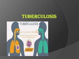
- 2. DEFINATION Tuberculosis is an infectious bacterial disease characterized by growth of nodules(tubercles) in the tissue, especially in lungs.
- 3. TYPES Primary Tuberculosis: This is when a person is infected by M. tuberculosis for the first time. Secondary Tuberculosis: The dormant bacteria gets reactivated mostly in people who are immune compromised leading to secondary TB. E.g. AIDS
- 4. CAUSITIVE ORGANISM (the culprit) M. tuberculosis is a slender, straight or slightly curved, aerobic acid fast bacilli. These bacilli cannot be seen properly in gram staining so the Ziehl- Neelsen staining is done. (The tuberculosis bacilli, it stains red in Ziehl- Neelsen staining)
- 5. PATHOGENESIS There are mainly two important events taking place in TB. 1.Facaltative intracellular growth- The bacilli grows within the macrophage. 2. Cell mediated immunity- Granuloma formation and many more things
- 6. The bacilli- local invasion Macrophages Engulfs the bacilli 1.The intracellular growth The tubercular Bacilli multiplies within The macrophage Now some of the macrophages Succeed in killing the bacteria. These then run towards the T- cells and present the remains of the bacilli to the T- cells
- 7. The Granuloma Formation T- cell The macrophages presenting the Remnants of the bacilli to T-cells Sensitized t- cells The t-cells get sensitized and these sensitized t- cells multiply. These t-cells Then enter the circulation in search of bacilli
- 8. Lymphokines Attracts and activates macrophages ,neutrophils. The activated macrophages kill the organism This causes local caseous necrosis which gets surrounded by other immune cells GRANULOMA Most of the times after this process the bacilli are trapped and so do not spread. But sometimes (in immune compromised people) it can spread to other organs causing miliary TB
- 9. Granuloma Definition: Granuloma is defined as a circumscribed, tiny lesion, about 1mm in diameter composed predominantly of collection of modified macrophages called epitheliod cells and rimmed at the periphery by lymphoid cells. Cells present: Consists of centre caseous (cheese like) necrosis. Epitheliod cells-- Modified macrophages. With slipper shaped nucleus Langhans’ giant cells-- Formed by the fusion of 10-20 epitheliod cells. These are multinucleated cells with their nuclei arranged in horse shoe form. Lymphoid cells form the periphery.
- 10. The microscopic and diagrammatic representation of granuloma
- 11. CLINICAL MANIFESTATION 1. Asymptomatic primary infection: Once the granuloma is formed the foci of the bacteria is trapped in the caseous necrosis. These granulomas then heal by scar formation which is followed by calcification and fibrosis and form tiny tubercles. Ghon’s focus: Calcified tubercles in the upper or middle lobes of the lung. Ghon’s complex: Ghon’s focus accompanied by perihilar lymph node calcified granuloma.
- 12. 2. Secondary or reactivation tuberculosis: • The already trapped foci of the bacteria can be reactivated later in life • This happens mostly in immune compromised individuals. • The organ system involved in tuberculosis: Pulmonary Tuberculosis– Lungs are the most common site form of tuberculosis. • It normally reactivates at the upper part of the lower lobe and lower part of the upper lobe due to the rich oxygen as M. tuberculosis is a aerobic organism. • Slowly these areas of infection grow, caseate, liquify and cavitate. • Clinically patients present with chronic low grade fever, night sweats, weight loss, and productive cough that may have blood in it.
- 13. Liquid fluid cavities in the lungs In pulmonary TB
- 14. Lymph node infection: this is the most common form of extra pulmonary tuberculosis. The cervical lymph nodes are mostly affected . They become swollen and mat together and drain. Lymph node tuberculosis is called scrofula. Pleural and pericardial infection: Infection in the spaces results in infected fluid collection around the lungs or the heart respectively. Kidney: Patients may have red and white blood cells but no bacteria is seen in gram stain Skeletal: This involves thoracic and lumber spines. Joints: there is chronic arthritis of joint. Central nervous system: Tuberculosis causes sub acute meningitis and forms granulomas in the brain.
- 15. Miliary Tuberculosis: Tiny millet sized tubercles(granulomas) are disseminated all over the body like shotgun blast. lungs, liver and kidney and other organs are riddled with tubercles. this disease normally occurs in children and elderly.
- 16. TUBERCULIN TEST Principal: Tuberculin skin test is delayed or type I hypersensitivity reaction. Reagent: i. Old tuberculin- It was described by Robert Koch. This may lead to some serious complications in patients. It is rarely used. ii. Purified protein derivative (PPD)- It was formed growing M. tuberculosis in semi synthetic medium. The dosage of PPD is expressed in Tuberculin Unit (TU). Method: i. Mantoux method- 0.1ml of PPD is injected intradermally into the flexor aspect of the forearm. ii. Heaf test- this is done with multiple puncture apparatus that pricks the skin.
- 17. Results: In mantoux test the site of injection is examined after 48-72 hours and is interpreted as follows- i. Positive test- In a positive reaction, there is a induration ( local oedema) of 10mm diameter or more with erythemea at the site of inoculation. Positive test only confirms past infection with the tubercle bacilli but does not indicate active form of the disease. ii. False negative – the test may become negative in two conditions: miliary tuberculosis When anergy develops following overwhelming infection of measles, Hodgkin's disease, sarcodiosis, lepromatous leprosy,. iii. False positive – this is observed in presence of related mycobacteria like atypical mycobacteria
- 18. Uses: To measure prevalence of infection in a community. To diagnose active infection in young children. It is used as an indicator of successful BCG vaccination.
- 19. LABORATORY DIAGNOSIS 1. Specimen collection: Pulmonary tuberculosis – sputum is the most common specimen. A morning specimen may be collected in three consecutive days. If sputum is scanty, a 24hrs specimen may be collected. When sputum is not available, laryngeal swab or bronchial washings are collected. In children gastric washings may be done. Meningitis- The CSF is collected. Renal tuberculosis- the urine sample is collected. Bone and joint tuberculosis- aspirated fluid Tissue biopsy of tissue.
- 20. 2. Concentration of specimen: This is done to achieve- a. Homogenization of specimen b. decontamination i.e. to kill other bacteria present c. concentrate the bacilli in small volume without inactivation. Methods of concentration i. Petroff’s method – sputum is mixed with equal volume of 4% sodium hydroxide and is incubated at 37 C with frequent shaking for about 30 minutes. It is then centrifuged at 3000rpm for 30 minutes. The supernatant fluid is poured off and the deposit is neutralized by adding 8% of HCL in the presence of a drop of phenol red indicator. ii. Other methods – Dilute acids, mucolytic agents such as N- acetyl-L-cystiene with sodium hydroxide with sodium hydroxide and pancreatin are used for concentration of specimen.
- 21. 3. Directly microcopy Smear is made from the specimen and stained using the Ziehl- Neelsen technique. It is then examined under oil immersion lens. the acid fast bacilli appear bright red against a blue background. To detect the bacilli microscopically there should be atleast 1000 bacilli per ml of sputum.
- 22. 4. Culture: Selective medium – Lowenstein- Jensen medium. • The concentrated material is inoculated on two bottles of the LJ medium. • In case of gastric washing the specimen is first neutralized by adding alkali. • The culture medium is incubated at 37 C . • The tubercle bacilli grows usually within 2-8 weeks. • In positive culture – ROUGH, TOUGH and BUFF colonies are formed . • In liquid medium bacilli grow as a surface pellicle.
- 23. 5. Biochemical reaction: Tubercle bacilli is Niacin positive. In radiometric method such as BACTEC, the growth may be detected in about a week by using 14C labeled substrates. 6. Animal inoculation: 0.5ml of concentrated specimen is inoculated intramuscularly into the thigh of two tuberculin negative guinea pig. The animals are weighed prior to inoculation and therefore after weakly interval. Tuberculin test is conducted with 2 weeks and animals infected show a positive result. Animal is killed after 6 weeks. Autopsy shows- • Caseous necrosis at the site of inoculation. • Enlarged caseous ingual lymph nodes. • Tubercles may be seen in many organs e.g. lungs, liver, spleen. • Kidneys are infected.
- 24. 7.Molecular methods: Polymerase chain reaction (PCR) is a rapid method in diagnosis of tuberculosis. It is based on DNA amplification and has been used to detect M. tuberculosis in clinical specimens. 8. Serology: Serology includes detection of antimycobacterial antibodies in patient serum. Various methods like- • Enzyme linked immuno sorbent assay (ELISA) • Radio- immunoassay (RIA) • Latex agglutination assay.
- 25. PROPHYLAXIS BCG- Bacille Calmette Guerin is a vaccine prepared by accentuated M. bovis by growing it on a potato medium. Dose and administration: BCG is available in liquid and freeze dry form. The freeze dry form is normally used. The freeze – dried vaccine is reconstituted by sterile physiological saline to make a final concentration of 0.1 mg in 0.1ml of the vaccine. After reconstitution the vaccine should be used within 3-6 hours. Vaccine is given intradermally with the dose of 0.1ml soon after birth failing which it can be administered later in life. A small nodule develops at the site of injection within 2-3 weeks.
- 26. It increases slowly and attains a diameter of 4-8 diameters. It then subsides and breaks into a small ulcer which heals forming a scar of 4-8 mm diameter. Such individual become tuberculin positive with 4-6 weeks. Contraindications: BCG is contraindicated in patients with AIDS , eczema, petrusis, measles and patients on steroids .
- 27. TREATMENT AND MDR-TB The antitubercular drugs include Rifampicin (R), isoniazid (H), Pyrazinamide (Z), streptomycin, ethanbutol (E), ethomanide, amino salicylic acid and cycloserine. A combination of four drugs (HRZE) is given three times a week initially for 2 months followed by only two drugs (HR) three times a week for 4- 6 months. MRD-TB: Also called as multidrug resistance tuberculosis refers to the resistance of the bacilli toward Rifampicin and isoniazid. When first line amino salicylic acid, ethanbutol and cycloserine are used. The directly observed therapy under supervision (DOTS) is being used to prevent detoriation of resistance.
- 28. XDR – TB Another serious condition extensively drug resistant tuberculosis (XDR – TB) has emerged recently. XDR – TB is due to M. tuberculosis strains which are resistant to any fluoroquinones and atleast one of the three injectable second line drugs, in addition to isoniazid and Rifampicin.
- 29. Bye