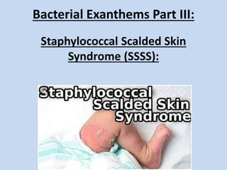
A brief description of a very common bacterial skin condition affecting children and adults. Characterized by fever, rash and peeling of the skin. Useful information for medical students, doctors especially dermatologists and pediatricians and nurses. Helpful information for exam preparation of USMLE, FCPS, MCPS, MRCP derma.
2. Definition and nomenclature:
Staphylococcal scalded skin syndrome is an
exfoliative dermatosis in which most of the body
surface becomes tender and erythematous and the
superficial epidermis strips off
Synonyms and inclusions
• Ritter disease
3. Epidemiology:
Age
• Children (< 6 years) and neonates most commonly
affected by the generalized form of SSSS,
• Adults may be affected by both generalized as well
as more commonly the localized form of the
disease.
5. Associated diseases
• Renal failure (as Exfoliative Toxins are usually
eliminated through the kidneys),
• Malignancy,
• Immunosuppression
• Alcohol abuse
6. Pathophysiology:
Predisposing factors
• Staphylococcus aureus strains produce an exfoliative toxin
(ETA, ETB).
• ETs target the cell adhesion protein desmoglein 1 (DG1)
resulting in separation of keratinocytes just beneath the
granular layer in the epidermis (intraepidermal).
• In bullous impetigo, the ETs remain local in the infected skin
but in SSSS the ETs spread haematogenously resulting in
widespread skin involvement.
7. Pathology
• Splitting of the epidermis between the granular and
spinous layers which does not usually contain
inflammatory cells.
• A few lymphocytes surround the superficial blood
vessels.
Pathogenesis:
• ETs either Type A or B are serine proteases that
selectively cleave the cellular adhesion molecule DG1
found on epidermal keratinocytes.
• Antibodies to desmoglein may develop in some
patients with SSSS
• ETA producing strains are more commonly isolated
from patients with bullous impetigo and ETB from
patients with SSSS
8. Split in
the
granular
layer
Histology of skin biopsy specimen shows an
epidermal detachment in the uppermost layer.
Histological findings are consistent with
staphylococcal scalded skin syndrome (SSSS)
9. Clinical features:
Presentation
• The initial source of infection may be an impetigo on
the face/ umbilicus, or a staphylococcal throat
/gastrointestinal tract infection.
• A few days later, patients develop fever, irritability and
skin tenderness.
• A widespread erythematous eruption follows which is
usually accentuated in the flexures
• Rapid progression to superficial blister formation
(Nikolsky positive).
• The tender skin becomes gathered into folds and, as it
shrinks, leaves raw areas which are extremely painful.
• The condition usually heals within 7–14 days.
10.
11. Clinical variants
• Localized SSSS:
– Favours the flexures, especially axillae, groin and limb flexures.
– Healing of localized form of disease leaves wrinkled desquamating
skin with hyperpigmentation.
– Localized form is considered to be less dangerous than the
generalized form of the disease.
Staphylococcal scalded skin
syndrome in an adult.
Localized staphyloccocal scalded skin syndrome
(SSSS) which subsequently healed with wrinkling
desquamation and hyperpigmentation.
12.
13. Differential diagnosis:
Toxic Epidermal Necrolysis: SSSS
Cell Necrosis prominent Absent
Epidermis separates from dermis Split between granular and spinous layer
Tzank smear shows only a few rounded
epithelial cells and many inflammatory cell
Tzanck smear shows a number of epithelial
cells with large nuclei but no inflammatory
cells.
14. Toxic epidermal necrolysis:
Parakeratosis overlying the epidermis which
has separated from the papillary dermis. On
the left, there are multiple Civatte bodies
affecting the full thickness of the epidermis
in a ‘gunshot’ distribution; on the right, there
is fullthickness epidermal necrosis.
Within the dermis, there is a superficial,
predominantly lymphocytic perivascular
infiltrate, vascular telangiectasiae and red
cell extravasation.
Staphylococcal Scalded Skin Syndrome:
Histology shows an epidermal
detachment in the granular layer; no cell
necrosis; no inflammatory infiltrate.
15. Disease course and prognosis:
• SSSS usually settles within a few weeks when
treated with appropriate systemic antibiotics.
• Localized form of the disease may be prolonged
with episodes of relapse over several months.
• If antibiotics are administered early, children usually
recover within 7 days and the mortality rate is 4%.
• In adults, the overall mortality rate is higher, around
60%.
• Those patients without underlying disease recover
more rapidly.
16. Investigations:
• Swabs and cultures of blister fluids
do not grow the staphylococci, as the blisters are mediated
by the toxins which are disseminated haematogenously.
• Staphylococci may be isolated from the original septic site if
identified.
• Typing of S. aureus strains
• PCR for toxin production
• In adults with generalized SSSS, blood cultures are often
positive for the staphylococci, whereas not in children.
• Erythrocyte sedimentation rate may be elevated.
17. Management:
First Line Treatment:
– Flucloxacillin,
– Clindamycin: may be given orally or parenterally either
alone (or in combination with rifampicin or
tetracyclines),
– Temocillin,
– Tigecycline
– Daptomycin
If MRSA is suspected:
– vancomycin or
– tobramycin