
Recommended
More Related Content
What's hot
What's hot (20)
Similar to Kawasaki Disease: Causes, Symptoms and Treatment
Similar to Kawasaki Disease: Causes, Symptoms and Treatment (20)
More from DrYusraShabbir
More from DrYusraShabbir (16)
Recently uploaded
Recently uploaded (20)
Kawasaki Disease: Causes, Symptoms and Treatment
- 2. Definition and nomenclature: Kawasaki disease (KD), also known as “Mucocutaneous lymph node syndrome” or “Kawasaki syndrome”, is an acute febrile illness of early childhood; characterized by vasculitis of the medium-sized arteries.
- 3. Introduction and general description: • Affects infants and children less than 5 years of age. • Associated with coronary arteritis • The commonest cause of acquired heart disease in children. • Prompt diagnosis, subsequent treatment with aspirin and intravenous immunoglobulins reduces heart complications
- 4. Epidemiology: Incidence and prevalence • The annual incidence per 100 000 children aged under 5 years is 8.4 in the UK • The incidence is rising in Japan every year. Age • Almost always occurs in children. Sex • There is a mild male predilection. Ethnicity • Asians, particularly Japanese. Associated diseases • Coronary vessel aneurysms • Myocardial infarction.
- 5. Pathophysiology: • Unknown infectious etiology, or environmental trigger or genetic susceptibility all contribute to activation of immune mechanisms. • Subsequent release of cytokines causes other cell types, including vascular endothelium, to undergo an uncontrolled immunological reaction. Epicardial coronary artery (right) and epicardial vein (left) from a 19-month-old child who died 10 months after Kawasaki disease onset. The epicardial vein contains blood and shows mild thickening of the wall, while the coronary artery shows almost complete occlusion by luminal myo- fibroblastic proliferation with a fine slit-like lumen.
- 7. Pathology: • The angiitis of Kawasaki disease affects nearly all organs, especially the heart. • It is predominantly a vasculitis of medium‐sized arteries but can involve any smaller and larger calibre blood vessels. • Initially, there is medial oedema associated with neutrophilic infiltration. • The inflammatory processes result in the breakdown of internal and external elastic laminae, resulting in aneurysms and thrombosis. • The inflammation heals with scarring and resultant stenosis of the affected blood vessel.
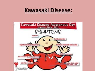
- 8. Clinical Features: • There are two forms of KD: – Complete – Incomplete. Complete KD: • Diagnosis of Complete KD requires fever of at least 5 days' duration along with 4 or 5 of the principal clinical features. • The principal clinical features are as under: (details will follow) – Extremity changes – Polymorphous rash – Oropharyngeal changes – Bilateral, nonexudative, limbic sparing, painless bulbar conjunctival injection – Acute unilateral nonpurulent cervical lymphadenopathy with lymph node diameter greater than 1.5 cm
- 9. The acronym "FEBRILE" is used to remember the criteria as follows: – Fever – Enanthem (mucous membrane changes) – Bulbar conjunctivitis – Rash – Internal organ involvement (not part of the criteria) – Lymphadenopathy – Extremity changes
- 10. Incomplete Kawasaki Disease: • Incomplete KD is diagnosed when a patient presents with fever for 5 days or longer, 2 or 3 of the principal clinical features, and laboratory findings suggestive of the disease or echocardiographic abnormalities. Suggestive laboratory findings include: • Elevated erythrocyte sedimentation rate (ESR), • Elevated C-reactive protein (CRP), • Hypoalbuminemia, • Anemia, • Elevated alanine aminotransferase (ALT), • Thrombocytosis, • Leukocytosis, and • Pyuria.
- 12. Chronic Phase: aneurysm rupture in late life in those with established heart disease. Lasts 2 weeks Lasts 4-6 weeks Increased risk of myocardial infarction in large aneurysms. Lasts 3 months Clinical Features Contd: The disease typically has the following stages:
- 13. Clinical Features Contd: The principal clinical features are as follows: • Extremity changes: – Erythema of the palms and soles, sometimes accompanied by firm and painful induration of the hands or feet impeding ambulation – Desquamation of the fingers and toes beginning in the periungual region, within 2-3 weeks after the onset of fever – Beau's lines (deep transverse grooves across the nails) may be present 1-2 months after fever onset • Polymorphous rash: – Typically diffuse and maculopapular, within 5 days of fever – The rash is usually extensive, primarily involving the trunk and extremities, and accentuation in the groin with early desquamation. – Scarlatiniform or erythema- multiforme like rash also common • Oropharyngeal changes: – Erythema, fissuring, bleeding, and/or crusting of the lips; – Strawberry tongue with prominent fungiform papillae; – Diffuse erythema of the oropharyngeal mucosa • Conjunctivitis: – Bilateral, nonexudative, limbic sparing, painless bulbar conjunctival injection – Anterior uveitis • Lymphadenopathy: – Acute unilateral nonpurulent cervical lymphadenopathy with lymph node diameter of at least 1.5 cm (least common criteria present)
- 14. (A) Diffuse Maculopapular rash: (B) Conjunctivitis: Bulbar conjunctival injection without exudate (C) Erythema and cracking of lips and strawberry tongue; (D) and (E): Palmar and plantar erythema accompanied by swelling;
- 15. Huge cervical lymphadenopathy: Strawberry Tongue: Peeling and erythema of the fingertips
- 16. (G) Magnetic resonance image of the left ventricular outflow tract showing a giant right coronary artery (RCA) aneurysm with nonocclusive thrombus (yellow arrow) and a giant left main coronary artery (LMCA) aneurysm. Ao indicates aorta; AoV, aortic valve; LV, left ventricle; and RV, right ventricle. (H) Peripheral artery aneurysms: Magnetic resonance image showing aneurysms in the axillary and subclavian arteries and the iliac and femoral arteries (yellow arrows).
- 17. Figure 6: (a) Beau lines; (b) Induration at BCG inoculation site:
- 19. Differential Diagnosis of Kawasaki Disease:
- 22. Disease course and prognosis: • Deaths are due to: – Myocarditis, – Dysrhythmias, – Pericarditis, – Rupture of aneurysms and – Occlusion of coronary arteries; • Increased risk of atherosclerosis due to endothelial cell dysfunction. • Coronary aneurysms seen in around 20% of patients (and in 90% of those who die); • Some aneurysms will regress potentially with stenosis • Giant aneurysms (>80 mm) may require bypass surgery. • Clinical factors that predict a higher risk of coronary artery arteritic lesions or aneurysms, or that predict a poor response to treatment: – Age below 1 year, – Low serum albumin, – Low haemoglobin, – High CRP, – Abnormal liver function – Duration of fever before treatment. – Peripheral blood eosinophilia (>4%) after treatment • Early intravenous immunoglobulin (IVIg) reduces the coronary aneurysm risk from around 25% to less than 5%. • Delaying IVIg beyond day 10 of fever increases the risk of death, particularly in boys under 1 year old.
- 23. Investigations: • Complete KD is a clinical diagnosis; no laboratory or imaging evaluations are required aside from echocardiography once the diagnosis is made. • Pre-diagnosis laboratory and imaging evaluations are helpful for cases of incomplete KD, when the diagnosis is suspected but the patient does not meet criteria for complete KD. • Labs Include: – Acute stage: • Increased ESR ≥ 40mm//hr • Increased C-Reactive protein ≥ 3mg/dL • Leukocytosis >15000/mm3 with neutrophilic predominance • Normocytic, normochromic anemia. • Sterile pyuria : WBC ≥ 10/HPF • Elevated serum transaminase levels, with or without mild hyperbilirubinemia. • Albumin ≤3g/dL • Echocardiogram: at baseline and at 6 weeks • ECG
- 24. Investigations Contd: • Subacute Phase: – Thrombocytosis which, in severe cases, may reach levels as high as 100,0000/mm – Elevated Cholesterol – Elevated Triglycerides – Decreased HDL • Convalesent Phase: – ESR and CRP begin to return to normal – Platelet levels begin to normalize
- 25. Management: • Patients need to be treated in a specialist paediatric unit. • Aspirin and Intravenous Immunoglobulins (IVIg) are the mainstay of treatment. First line Treatment: • Intravenous immunoglobulin and aspirin should be given early. • Aspirin, 100 mg/kg/day is given initially until the fever has settled and is then reduced to 3–5 mg/kg/day for 6–8 weeks in those with no cardiac abnormality, but longer in those with coronary aneurysms. • All children should receive IVIg, usually given as a single dose of 2 g/kg over 12 h.
- 26. The Harada score is used as an indication for intravenous immunoglobulin therapy: Four of following seven criteria are needed: • White blood count >12 000/ mm; • Platelet count <35x10 4 /mm; • CRP >3; • Haematocrit <35%; • Albumin <3.5 g/dL; • Age <12 months; and • Male sex
- 27. Second line Treatment: • For children who remain febrile 36 h after the first dose of IVIg, a further dose of 2 g/kg can be given. • Patients who are unresponsive to IVIg can be treated with high‐dose prednisolone 2 mg/kg/day, which should be tapered after normalization of the CRP.
- 28. Topics To Be Discussed In Future: Palmoplantar Keratoderma Previously Discussed Topics: Viral Diseases: 1. Viral warts 2. Epidermodysplasia verruciformis 3. Measle 4. Rubella 5. Erythema Infectiosum 6. Roseola Infantum 7. Cytomegalovirus 8. Infectious Mononucleosis Bacterial Diseases: 1. Toxic Shock Syndrome 2. Scarlet Fever 3. Staphylococcal Scalded Skin Syndrome