2. Chest Cavity
• The chest cavity is bounded by the chest wall
on each side and below by the diaphragm.
• It extends upward into the root of the neck
about one fingerbreadth above the clavicle on
each side
• The chest cavity can be divided into a median
partition, called the mediastinum, and the
laterally placed pleurae and lungs
3. Mediastinum
• The mediastinum is a partition
between the two pleural cavities and
two lungs
• It extends;
• Superiorly to the thoracic outlet and
the root of the neck
• Inferiorly to the diaphragm.
• Anteriorly to the sternum
• Posteriorly to the vertebral column
4.
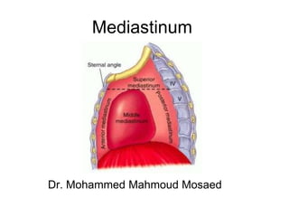
5. Divisions of mediastinum
Mediastinum divided by an imaginary plane
(passing from the sternal angle anteriorly to the
lower border of the body of the fourth thoracic
vertebra posteriorly) into:
The superior mediastinum
The inferior mediastinum which is subdivided
into;
the middle mediastinum, which consists of the
pericardium and heart;
the anterior mediastinum, which is a space
between the pericardium and the sternum;
the posterior mediastinum, which lies between
the pericardium and the vertebral column.
6.
7.
8. SUPERIOR MEDIASTINUM
• The superior mediastinum lies between the manubrium sterni
and the upper four thoracic vertebrae.
• It is bounded below by the sternal plane, above by the plane of
the thoracic inlet and laterally by the mediastinal pleurae.
Contents
1. Large arteries Internal thoracic arteries, the aortic arch and its
branches (the brachiocephalic, left common carotid and
subclavian arteries)
2. Large veins; Internal thoracic veins, brachiocephalic vein, the
upper half of the superior vena cava, and the left superior
intercostal vein.
3. Nerves; the vagus, cardiac, phrenic and left recurrent
laryngeal nerves and sympathetic trunks
4. Lymph node; the paratracheal, brachiocephalic and
tracheobronchial lymph nodes and the thoracic duct
5. The trachea, oesophagus and remnants of the thymus
gland
9.
10.
11. Inferior Mediastinum
• Anterior mediastinum
• It contains loose connective tissue, the sternopericardial ligaments
and the remnants of thymus gland
• Posterior mediastinum
Contents:
The descending thoracic aorta.
The azygos, hemiazygos and accessory azygos veins.
The vagus and splanchnic nerves.
The oesophagus, thoracic duct and posterior mediastinal lymph nodes
• Middle mediastinum
Contents:
The pericardium, heart and ascending aorta.
The lower half of the superior vena cava.
The pulmonary trunk and the right and left pulmonary arteries.
12.
13. Clinical Notes
• Mediastinitis
Causes
• deep infection of the neck spread readily into the thorax through the loose
connective tissue that is continuous with that of the mediastinum
• Penetrating wounds of the chest involving the esophagus may produce a
mediastinitis
• In esophageal perforations, air escapes into the connective tissue spaces
and ascends beneath the fascia to the root of the neck, producing
subcutaneous emphysema.
• Mediastinal Tumors or Cysts
• A tumor of the left lung can rapidly spread to involve the mediastinal lymph
nodes, which on enlargement may compress the left recurrent laryngeal
nerve, producing paralysis of the left vocal fold.
• An expanding cyst or tumor can partially occlude the superior vena cava,
causing severe congestion of the veins of the upper part of the body.
• Other pressure effects can be seen on the sympathetic trunks, phrenic
nerves, and sometimes the trachea, main bronchi, and esophagus.