

Recommended
More Related Content
What's hot
What's hot (20)
Similar to Lung anatomy.
Similar to Lung anatomy. (20)
More from siti hamidah
More from siti hamidah (20)
Recently uploaded
Recently uploaded (20)
Lung anatomy.
- 1. ANATOMY OF LUNGS -
- 2. 1. Gross Anatomy of Lungs 6. Histopathology of Alveoli 2. Surfaces and Borders of Lungs 7. Surfactant 3. Hilum and Root of Lungs 8. Blood supply of lungs 4. Fissures and Lobes of 9. Lymphatics of Lungs Lungs 10. Nerve supply of Lungs 5. Bronchopulmonary 11. Pleura segments 12. Mediastinum
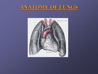
- 3. GROSS ANATOMY OF LUNGS Lungs are a pair of respiratory organs situated in a thoracic cavity. Right and left lung are separated by the mediastinum. Texture -- Spongy Color – Young – brown Adults -- mottled black due to deposition of carbon particles Weight- Right lung - 600 gms Left lung - 550 gms
- 5. SHAPE - Conical Apex (apex pulmonis) Base (basis pulmonis) 3 Borders - anterior (margo anterior) - posterior (margo posterior) - Inferior (margo inferior) 2 Surfaces - costal (facies costalis) - medial (facies mediastinus) - anterior (mediastinal) - posterior (vertebral)
- 6. APEX Blunt Grooved by- b Lies above the level of Subclavian artery anterior end of 1st Rib. Subclavian vein Reaches 1-2 cm above medial 1/3rd of clavicle. Coverings – cervical pleura. suprapleural membane
- 8. BASE Semilunar and concave. Rests on dome of Diaphragm. Right sided dome is higher than left.
- 9. BORDERS ANTERIOR BORDER – 1. Corresponds to the anterior (Costomediastinal) line of pleural reflection. 2. It is deeply notched in the left lung posterior to 5th costal cartilage by the pericardium and extends vertically downwards to form Lingula. This is called cardiac notch(percussion in this area gives a dull note as compared to dull note obtained over lung).
- 10. INFERIOR BORDER Thin and sharp It seperates the base of lung from the costal surface and extends into phrenicocostal sinus.
- 11. POSTERIOR BORDER Thick and ill defined Fits into deep paravertebral gutter. Extends from C7 to T10.
- 12. SURFACES OF THE LUNG 1. Costal Surface - It is in contact with costal pleura and overlying thoracic wall. 2. Medial Surface - Posterior / Vertebral Part - Anterior / Mediastinal Part
- 13. Relations of Posterior Part 1. Vertebral Part 2. Intervertebral Discs 3. Posterior Intercostal Vessels 4. Splanchic Nerves
- 14. RELATIONS OF ANTERIOR PART RIGHT SIDE LEFT SIDE 1. Right atrium 1. Left ventricle 2. Small part of RV 2. Pulmonary trunk 3. SVC 3. Arch of Aorta 4. Right brachiocephalic 4. Descending thoracic vein(lower part) aorta 5. Azygos vein 5. Left Subclavian Artery 6. Esophagus 6. Thoracic duct 7. IVC 7. Left Brachiocephalic 8. Trachea Vein 9. Right vagus nerve 8. Left vagus nerve 10. Right phrenic nerve 9. Left phrenic nerve 10. Left recurrent laryngeal nerve
- 17. HILUM It is a large depressed area that lies near the centre of the medial surface. Various structures enter and leave the lung via its root.
- 18. ROOT OF THE LUNG The root is enclosed in a short tubular sheet of pleura that joins the pulmonary and mediastinal parts of pleura . It extends inferiorly as a narrow fold - The pulmonary ligament. It lies opposite of the bodies of 5th, 6th and 7th thoracic vertebra
- 19. STRUCTURES OF THE ROOT Principal Bronchus on the left side. Eparterial and Hyparterial on the right side. One pulmonary artery . Two pulmonary veins - Superior Inferior Bronchial arteries One on right side Two on left side
- 20. Bronchial veins Anterior and posterior pulmonary plexus of nerves. Lymphatics Bronchopulmonary Lymphnodes Areolar tissue.
- 21. ARRANGEMENT OF STRUCTURES IN THE ROOT BEFORE BACKWARDS 1. Superior pulmonary vein. 2. Pulmonary artery. 3. Bronchus.
- 22. ARRANGEMENT OF STRUCTURES IN THE ROOT ABOVE DOWNWARDS A. Right Side 1. Eparterial Bronchus. 2. Pulmonary Artery. 3. Hyparterial Bronchus. 4. Inferior Pulmonary Vein. .
- 23. ARRANGEMENT OF STRUCTURES IN THE ROOT ABOVE DOWNWARDS B. Left Side 1. Pulmonary artery. 2. Bronchus. 3. Inferior pulmonary vein
- 24. FISSURES AND LOBES OF LUNGS
- 25. OBLIQUE FISSURE It begins posteriorly at the level of 5th thoracic vertebra. Passes antero-inferiorly in a spiral course to meet the inferior margin close to 6th costochondral junction.
- 26. HORIZONTAL FISSURE It extends from anterior margin at the level of 4th costal cartilage. Runs horizontally backwards to meet the oblique fissure in the mid-axillary line. Pulmonary pleura extends into the fissures of the lungs so that the lobes can move on each other during respiration.
- 28. BRONCHOPULMONARY SEGMENTS These are well defined areas of the lungs, each of which is aerated by a segmental / tertiary bronchus.
- 30. Trachea Right and Left Principal Bronchus Lobar Bronchi(Secondary)[2L,3R] Segmental Bronchi(Tertiary)[8L,10R] Terminal Bronchioles(25000 in no.) Respiratory Bronchioles Alveolar ducts ACINUS Alveolar sacs Alveoli
- 32. The ultimate pulmonary unit from respiratory brochiole to alveoli is called Acinus. There are about 28 orders of division of tracheo-bronchial tree. Total no. of alveoli has been estimated to be between 200 - 600 million, with a total surface area of 40 - 80 meter square.
- 34. BRONCHOPULMONARY SEGMENTS Right main bronchus Left main bronchus 1. Shorter 1. Longer 2. Wider. 2. Narrower. 3. More in line with 3. More oblique than trachea. the right.
- 35. BRONCHOPULMONARY SEGMENTS Right Main Bronchus Right upper lobe Bronchus Right Middle lobe Bronchus Right Lower Lobe Bronchus Segmental Bronchi Segmental Bronchi Segmental Bronchi Apical Apical Medial Anterior Anterior Lateral Posterior Posterior Medial and Lateral
- 36. BRONCHOPULMONARY SEGMENTS Left Main Bronchus Left upper lobe Bronchus Left lower lobe bronchus Upper Branch Lower Branch Segmental Bronchi Apical Anterior Superior Lingular Anterior Apico-posterior Inferior Lingular Posterior Lateral
- 38. These segments are pyramidal in shape with apex towards the root of lung. Each segment is an independent respiratory unit. Each segment has its own separate artery(branches of pulmonary artery). Pulmonary Veins run in inter-segmental planes between adjoining segments. Thus a bronchopulmonary segment is not a bronchovascular segment as it does not have its own vein.
- 40. CLINICAL SIGNIFICANCE Segmental resection with minimal destruction to the surrounding lung tissue. To visualize the interior of a bronchi through a bronchoscope when diseases process is limited in a segment.
- 41. HISTOPATHOLOGY OF ALVEOLI ALVEOLAR WALL 1. Alveolar epithelial cells- Type I pneumocytes Type II pneumocytes 2. Basement Membrane 3. Interstitial Space- Collagen Elastin Unmyelinated Nerves Macrophages 4. Capillary Basement Membrane 5. Capillary Endothelial Cells. .
- 42. Type I Pneumocyte Pavement epithelial cells of alveoli . Less in no. than type II. More surface area(flattened) Contain pinocytic vesicles. Specialized for diffusion of gases.
- 43. Type II Pneumocytes More numerous than type I. Cuboidal in shape. Rich in mitochondria, ER and vacuoles containing osmiophillic lamellar bodies. Type I are precursors of type II.
- 44. ENDOTHELIAL CELLS Most numerous . Presence of pinocytic vacuoles that meet the luminal surface to form caveolae. Walls of caveolae has, ACE. Source of NO, natural pulmonary vasodilator.
- 45. ALVEOLAR MACROPHAGES Primary defence mechanism. Takes part in inflammatory and immunological reactions. Activates lysosomes , proteases,complement , thromboplastin, cytokines - IF-α, TNF- α, IL-1, IL-8.
- 46. SURFACTANT Lines the inner layer of alveolar epithelium. Synthesized by SER of type II pneumocytes. Function – 1. To reduce the surface tension of alveoli mainly during expiration, thus reduces the work of lung inflation. 2. Waterproofing. Surfactant synthesis starts after 26 weeks of fetal life. Therefore premature infants,with insufficient surfactant suffer from HMD.
- 47. All the best…