
Recommended
More Related Content
Similar to Nervous System Anatomy and Functions in 40 Characters
Similar to Nervous System Anatomy and Functions in 40 Characters (20)
More from DivyaPant16
More from DivyaPant16 (15)
Recently uploaded
Recently uploaded (20)
Nervous System Anatomy and Functions in 40 Characters
- 2. The Nervous System • The nervous system coordinates all body functions, enabling a person to adapt changes in internal and external environment • The nervous system is composed mainly of the nerve cells (neurons) and supporting cells (neuroglia)
- 3. Structural Classification of the Nervous System ∙ Central nervous system (CNS) ∙ Brain ∙ Spinal cord ∙ Peripheral nervous system (PNS) ∙ Nerve outside the brain and spinal cord
- 4. Functional Classification of the Peripheral Nervous System ∙ Sensory (afferent) division ∙ Nerve fibers that carry information to the central nervous system
- 5. Functional Classification of the Peripheral Nervous System ∙ Motor (efferent) division ∙ Nerve fibers that carry impulses away from the central nervous system
- 6. Functional Classification of the Peripheral Nervous System ∙ Motor (efferent) division ∙ Two subdivisions ∙ Somatic nervous system = voluntary ∙ Autonomic nervous system = involuntary
- 7. Organization of the Nervous System Slide 7.4
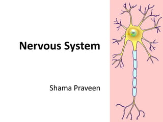
- 10. The basic unit of nervous tissue is the cell called the Neuron. Neuro n
- 11. Neuro ns consi st Nucleus ofa main part called the cell body, whichcontains the nucleus and various Cell Body Nucleol us • 4 to 100 micrometers in diameter • (Soma)
- 12. Neurons also contain cell body “extensions” called processes, which carry impulses to and from the cell body. axo n dendrit es Two different types of processes can come from the cell body. The first is called the dendrite and the other the axon.
- 13. Dendrites, also called afferent processes, carry impulses TOWARDS the cell body. Axons, also called efferent processes, carry impulses AWAY from the cell body.
- 14. ▪ Some axons are enveloped by Schwan Cells which provide structural and metabolic support. ▪ These are classified on Basis of Presence/Absence of Myeline Sheath. -> Myelinated -> Non-Myelinated
- 15. Axons and Nerve Impulses ∙ Axons end in axonal terminals ∙ Axonal terminals contain vesicles with neurotransmitters ∙ Axonal terminals are separated from the next neuron by a gap ∙ Synaptic cleft – gap between adjacent neurons ∙ Synapse – junction between nerves
- 17. Myelin • Myelin is a fatty covering which envelops many axons and permits action potentials to be propagated Myeli n
- 18. Nodes of Ranvier Nodes of Ranvier are short fragments of unmyelinated segments of the axon, which are found periodically in between the cells of the myelin sheath. • These nodes are areas where the action potential is amplified using a high density of sodium (Na+) ions and is subsequently passed along the axon. • The points between segments of myelin are called nodes of Ranvier. Nodes of Ranvier
- 20. There are actually three different types of neurons in the human body. They can be classified either according totheir function or structure: Functional Classification: Sensory Neurons Motor Neurons Interneurons (Association Neurons) Structural Classification: 1. Unipolar Neurons 2. Bipolar Neurons 3. Multipolar Neurons
- 21. Structural Classification of Neurons ∙ Multipolar neurons – many extensions from the cell body
- 22. Structural Classification of Neurons ∙ Bipolar neurons – one axon and one dendrite
- 23. Structural Classification of Neurons ∙ Unipolar neurons – have a short single process leaving the cell body
- 25. Sensory neurons or Afferent neurons– These neuro ns transmit impulses from the periphery of the body to the central nervous system. They are described as unipolar meaning they have only a single process. • This process is actually an axon which branches into two parts and spread in opposite directions. • In this way, one branch acts as the dendrite, while the other acts as the axon.
- 26. Motor neurons or Efferent neurons – These neurons carry impulses AWAY from the cell body and thus the central nervous system to muscles, gland, or some other “effector” to produce a certain action. This action can be the contraction of a muscle or the secretion of a gland. Motor neurons are classified as multipolar, which means they have numerous branching dendrites leading into the cell body and a single long axon leading out. Most of the neurons in the spinal cord and many of those in the brain are motor neurons.
- 28. Interneurons or Association neurons- This type of neuron is restricted to the central nervous system. They are also called connector neurons. These neurons act as bridges between sensory and motor neurons or relay impulses to various functional centers of the brain or spinal cord. They resemble motor neurons in that they have one axon with multiple dendrites, however, their function is much different.
- 30. Neuroglia
- 32. Neurogli a • Neuroglia do not generate or conduct nerve impulses. • However, unlike other cells can regenerate if injured.
- 33. • Astrocytes - Provide energy and other metabolic needs of neurons as well as giving nervous tissue structural support. • Abundant, star-shaped cell • Brace neuron • Form barrier between capillaries and neurons • Control the chemical environment of the brain (CNS)
- 34. Microglia - Phagocytic cells, similar to macrophages, that perform a housekeeping function by removing dead cellular material and bacteria from the CNS. Spider-like
- 35. ∙ Ependymal Cells - Cells that line the cerebral spinal fluid (CSF) containing cavities of the brain - the ventricles. Line cavities of the brain and spinal cord ∙ Circulate cerebrospinal fluid
- 36. Oligodendrocytes - Produce myelin sheath around nerve fibers in the central nervous system
- 37. Support Cells of the PNS ∙ Satellite cells ∙ Protect neuron cell bodies ∙ Schwann cells ∙ Form myelin sheath in the peripheral nervous system Figure 7.3e
- 38. Thank you