SlideShare a Scribd company logo
1 of 53
FETAL MEMBRANE & TWIN PREGNANCY
DR LOVELY JAIN
MD ANATOMY
N.S.C.B MEDICAL COLLEGE
JABALPUR
EarlyHumanDevelopment
Cardiac swelling
Umbilical cord
Fetal membrane
• The term fetal membrane is applied to those structures
derived from the blastocyst which do not contribute to
the embryo.
The amnion,
the chorion,
the yolk sac
Allantois
Umbilical cord
AMNION It is a thin, transparent &tough fluid-
filled, membranoussac surrounding the
embryo
 form by--- amniotic epi. +extraembryonic
mesoderm
 At term amnion is a tough, tenacious &
pliable membrane.
 Inner most ,avescular,&provide tensile
strength.
• It lacks smooth muscles,lymphatics,nervs&
bld vesscles.
Bourne (1962) described five separate layers of
amnion –
1)innermost single layer of cuboidal epithelium
derived from embryonic ectoderm.basement
membrane
2)acellular compact layer, which is composed
primarily of interstitial collagens
3)fibroblast-like mesenchymal cells, derived from
embryonic
4)disc mesoderm
5)acellular zona spongiosa, contiguous with second
fetal membrane, the chorion laeve
• a
Devlopment
Early during implantation:- a space develops between the embryonic cell mass and
adjacent trophoblasts.
 Small cells that line this inner surface of trophoblasts have been called amniogenic
cells—precursors of amnionic epithelium.
 At First : It is seen as a small cavity lying dorsal to the embryonic plate.
 At Stage of Chorionic Vesicle: The amnion becomes separated from the
chorion by chorionic cavity or extra embryonic coelom.
 After Folding: the amnion expands greatly and is becomes on the ventral
surface of the embryo.
 As a result of expansion of the amnion, the extra embryonic coelom is
gradually obliterated and amnion forms the epithelial covering of umbilical
cord & Reflected amnion is fused to the chorion laeve.
• Placental amnion covers placental surface & thereby is in contact with adventitial
surface of chorionic vessels.
• Umbilical amnion covers the umbilical cord.
• Diamniotic- dichorionic the conjoined portion of membranes of twin placenta, fused
amnions are separated by fused chorion laeve.
• Diamniotic-monochorionic placenta, there is no intervening tissue between the
fused amnions.
Amniotic Fluid
Produce by: 1)amniotic cells
2)infusion of fluid from maternal blood
3)urine output from the fetus
4)pulmonary secretions
Output: 1) absorbed by amniotic cells
• 2) fetus swallow
• Plays a major role in fetal growth and development.
• Daily contribution of fluid from respiratory tract is 300-400 ml.
• 500 ml of urine is added daily during the late pregnancy.
• Amniotic fluid volume is 30 ml at 10 weeks, 350 ml at 20 weeks, 700-1000 ml
at 37 weeks.
• Composition:99 % is water
• Desquamated fetal epithelial cells
• Organic & inorganic salts
• Protein, carbohydrates, fats, enzymes, hormones
• Meconium & urine in the late stage
function
• Helps maintain the body temperatureEnables the fetus to move freelyProvides
symmetrical external growth of the embryo.
• During labor it help dilatation of the cervix of the uterus and It wash birth
canal and protect the fetus against infections.
• Cushions & protects the embryo and fetus
Acts as a barrier to infection (it is an aseptic medium)
Permits normal fetal lung development
Prevents adherence of embryo to amnion
It protects embryo against external injuries.
Allows the embryo to move freely, aiding muscular development
in the limbs
It is involved in maintaining homeostasis of fluids & electrolytes
It permits studies on fetal enzymes, hormones and diagnosis of fetal sex and
chromosomal abnormalities
ABNORMALATIES OF AMNIOTIC FLUID VOLUME
: Oligo-hydramnios:The
volume is less than ½ liters
Causes :
Placental insufficiency
with low placental blood
flow
Preterm rupture of amnio-
chorionic membrane occurs
in 10% of pregnancies
Renal Agenesis (failure of
kidney
• development)
Obstructive Uropathy (urinary
tract obstruction) lead to
absence of fetal urine (the main
source)
Complications :
Fetal abnormalities
(pulmonary,
• facial & limb defects)
Polyhydra
mnios:(Hy
dramnios):
The volume is
more than 2liters,
it is diagnosed by
Ultrasonography.
Causes
Fetal ( 1-20% ) :
Esophageal atresia.
Maternal
(2-20%) :
defects in
maternal
circulation.
Idiopathic (3-
60%)
Abnormility Definition & causes Clinical significance
 Lined by typical amnionic
epithelium
 Fusion of amnionic folds with
subsequent fluid retention
Amnion nodosum  Tiny, creamy nodules in the
amnion made up of vernix
caseosa with hair, degenerated
squames and sebum
 Oligohydramnios
Found in
 fetuses with renal agenesis
 Prolonged preterm ruptured
Membranes
 The placenta of the donor
fetus with twin-to-twin
transfusion syndrome
Amnionic band Caused when disruption of the
amnion
leads to formation of bands or
strings
Intrauterine amputation
Amnionic
cyst
YOLK SAC
• It is essential in the transfer of
nutrients to the embryo during 2nd
& 3rd weeks, when the
uteroplacental circulation is not
established.
• It is large at 32 days
• Shrinks to 5mm pear shaped
remnant by 10th week & connected
to the midgut by a narrow yolk stalk
• Becomes very small at 20 weeks
Usually not visible thereafter
• It does not contain any yolk.
Its development passes
through three stages:
Primary yolk sac.
Secondary yolk sac.
Definitive yolk sac.
• a
Primary yolk sac
• a
Appears in the
Blastocyst stage at 10-
days, it lies ventral to
the embryonic
plate.
Its roof is formed by
hypoblast (primary
endoderm),
Its wall is formed by
exocoelomic membrane,
it lines the inner surface of
the cytotrophoblast, and
separated from it by the
extraembryonic
mesoderm
Secondary yolk sac
• a
Appears in the chorionic
vesicle stage
Its roof is formed by
hypoblast (embryonic
endoderm), its wall is
formed by exocoelomic
membrane + inner layer
(splanchnic layer) of the
extraembryonic
mesoderm.
At day 16: a diverticulum
appears from its
dorsocaudal
end (Allantois) into the
substance of the connecting
stalk
Definitive yolk sac
• a
After folding, part of
Yolk Sac is enclosed
within the embryo to
form the Gut (Foregut,
Midgut & Hindgut).
The remainder of Yolk
Sac that remains outside
the embryo becomes the
Definitive Yolk Sac
 The midgut is
temporarily connected to
Definitive Yolk Sac by a
narrow duct Vitello-
intestinal duct (Yolk
stalk), which is
incorporated inside the
umbilical cord.
 This is fibrosed and
degenerated by
• the end of (6th week)
Function of yolk sac
a
3rd week:
 (a) Blood formationt
• First formed in the extra-
embryonic mesoderm covering the
wall of the
yolk sac, until hemopoietic activity
begins in the liver during 6th week
 4th week: endoderm of yolk
sac is incorporated into the
embryo to form primordial gut
Epithelium of Respiratory system
&G.I.T
b)Primordial germ cells:- in the endodermal lining of the wall of caudal end of the
yolk sac migrate into the developing sex glands to differentiate into germ cells
(spermatogonia or oogonia)
Fate of yolk sac
• a
 Yolk stalk detached from
midgutby the end of 6th
week. In (2%) of adults, its
proximal intra-abdominal
part persists as ileal
diverticulum (Meckel
diverticulum).
 At 10 week, small definitive
yolk sac lies in the chorionic
cavity between amniotic &
chorionic sacs
 At 20 weeks, as pregnancy
advances, definitive yolk sac
atrophies and becomes a very
small cyst.
 In unusual cases, it
persists under the amnion
near the attachment of
Umbilical cord, on the fetal
surface of the placenta. Its
persistence is of no
significance
ALLANTOIS
3rd week:Appears as a
diverticulum from caudal wall of Y.S.
that extends into connecting stalk.
2nd month: Its extra- embryonic part
degenerates.
3rd month: Its intra-
embryonicpart extends from UB to
UC as thick tube , ‘(urachus) ’
 After birth: the urachus is
obliterated and fibrosed to form
median umbilical ligament, that
extendsfrom apex of UB to
umbilicus.
 Function:Blood formation in its
wall during 3rd to 5th week.
 Its blood vessels persist as the
umbilical vein & arteries.
• a
CHORION
• The extraembryonic somatic mesoderm and the two
layers of trophoblast form the chorion
• Chorion forms the wall of chorionic sac
• Embryo and its amniotic and yolk sacs are suspended into
it by connecting stalk
• Growth of these extensions are caused by underlying
extraembryonic somatic mesoderm
• The cellular projections form primary chorionic villi.
Chorionic villi
• As this sac grows, the villi associated with decidua
capsularis are compressed, reducing the blood supply to
them
• These villi soon degenerates producing an avascular bare
area smooth chorion (chorion laeve).
Primary chorionic vilii
• At the end of 2nd
week, finger-like
processes formed
of outer
syncytiotrophoblast
& inner
cytotrophoblast
appear & cover the
entire chorionic sac
until the beginning
of 8th week
Secondary chorionic villi
• Early in 3rd :-
week,
extraembryoni
c mesoderm
extends inside
the villi
• a
Tertiary Chorionic villi
• During 3rd week,
arterioles, venules
& capillaries
develop in the
mesenchyme of
villi & join
umbilical vessels
• By the end of 3rd
week, embryonic
blood begins to
flow slowly
through capillaries
in chorionic villi
• a
UMBILICAL CORD/ funis
Extends from fetal umbilicus to fetal surface of placenta or
chorionic plate.
• Exterior is dull white, moist, & covered with amnion, through
which three umbilical vessels may be seen.
• Origin :-It develops from the connecting stalk.
• Length:--At term, it measures about 50 cm.
• Diameter:--2 cm.
DEVELOPMENT:--Cord develops in yolk sac & umbilical vesicle
which are prominent early in pregnancy.
Embryo, at first, is a flattened disc interposed between amnion &
yolk sac.
Its dorsal surface grows faster than the ventral surface.
Embryo bulges into amnionic sac in association with elongation of
neural tube.
Dorsal part of yolk sac is incorporated into the body of embryo to
form gut.
Allantois projects into base of body stalk from the caudal wall of the yolk sac & later, forms
anterior wall of hindgut.
As pregnancy advances, yolk sac becomes smaller & its pedicle relatively longer.
Middle of 3rd month:- expanding amnion obliterates exocoelom, fuses with the chorion
laeve, & covers the bulging placental disc & lateral surface of the body stalk.Latter is then
called the umbilical cord—or funis.
Insertion:
The cord is inserted in the foetal surface of the placenta
near the center "eccentric insertion" (70%) Or
at the center "central insertion" (30%).
Structure: It consists of mesodermal connective tissue called Wharton's jelly, covered by
amnion.
It contains:
1.One umbilical vein carries oxygenated blood from the placenta to the foetus
2.Two umbilical arteries carry deoxygenated blood from the foetus to the placenta,
3.Remnants of the yolk sac and allantois..
CORD AT TERM
• It normally has two arteries
and one vein .
• Right umbilical vein
disappears early during fetal
development, leaving only
the left vein.
• Intra-abdominal portion of
duct of umbilical vesicle,
extending from umbilicus to
intestine, usually atrophies
& disappears.
• If patent, it is known as
Meckel’s diverticulum.
• Most common vascular
anomaly - absence of one
umbilical artery which may
be associated with fetal
anomalies.
• A
• Length
• Cord Coiling
• Single Umbilical Artery
• Four-vessel cord
• Abnormalities of cord insertion
• Cord Abnormalities capable of impeding blood flow
• Hematoma
• Cysts
LENGTH:--Appreciable variation –Average length of 55 cm
range-- of 30 to 100 cm.
No cord---(acordia) ~
lengths up to 300cm
Excessively long cords : ≥ 70cm( ≥2 SD ) Associated with
-maternal systemic disease
-delivery complications -cord prolapse, cord entanglement
-fetal anomalies and respiratory distress Perinatal mortality : ↑ nearly
threefold
Short umbilical cord:-Generally, cord length less than 30 cm is
considered abnormally short.
Adverse perinatal outcomes –
-fetal growth restriction
congenital malformations
intrapartum distress & risk of death (doubled)
Umbilical vessels: in a spiraled manner
CORD COILING:--Umbilical vessels: in a spiraled manner
Hypocoiled cords:-↑ in various adverse outcome in fetuses
- meconium staining, preterm birth and fetal distress.
Hypercoiled cords --higher incidence of preterm delivery &
cocaine abuse.
SINGLE UMBLICAL ARTERY:--Umbilical cord –2-arteries & 1
vein
Risk factors –↑ incidence in women with GDM, PIH, APH,
epilepsy, oligohydramnios & hydramnios.
----Âź of all infants with only 1 artery have associated congenital
anomalies.
FOUR VESSCLE CORD:- Venous remnantin 5% & Significance :
unknown
ABNORMALITIS IN CORD INSERSATION
rmalities Definition Incidenc Significance
cate insertion
Umbilical vessels separate from
the cord substance before their
insertion into the placenta
RARE Prone to twisting &
thromboses as vessels lose
their cushioning
ginal Inserion Battledore placenta:
Cord insertion at the placental
margin
7% at
term
Cord being pulled off
during delivery of the
placenta
mentous Insertion Umbilical vessels separate in the
membranes at a distance from
the placental margin.
Reach surrounded only by a fold
1.1% More frequently with
twins
28% of triples
Vasa pervia
• Associated with velamentous insertion when some of the fetal vessels in the
membranes cross the region of the cervical os below the presenting fetal part
Associated with :
• Velamentous insertion (50%)
• Marginal cord insertion
• Bilobed or Succenturiate-lobed placentas (50%)
Risk factors :
• Bilobed , Succenturiate or low-lying placenta (80%)
• Multifetal pregnancy
• Pregnancy resulting from in vitro fertilization.
Diagnosis
• Color Doppler examination
• Perinatal diagnosis :-- associated with increased survival
• Antenatal diagnosis : --associated with decreased fetal mortality compared with
discovery at delivery
• Antepartum or intrapartum haemorrhage
• Detecting fetal blood ( Apt test)
• Wright stain : smear the blood on glass slides stain the smears with Wright stain
and examine for nucleated RBC normally are present in cord blood but not
maternal blood
TWIN/MULTIPILE PREGNANCY
Two or more fertilization events
Single fertilization followed by splitting of zygote
Combination of both
Incidence:-Global incidence: 4/1000 births
Hellin’s Law : Twins: 1/80 singleton births
• Triplets: 1:802
• Quadruplets: 1:803
Conjoined twins: 1 : 60,000
Typesof twins………
1)-DIZYGOTIC
2)-MONOZYGOTIC
Dizygotic/ non-identical twins (binovular ,
fraternal, 2 eggtwins)
~ two third of twins.
fertilization of two independently released ova by
two different sperm.
In all polyzygotic multiple pregnancies, each zygote
develops its own amnion, chorion and placental
circulation, and hence will be polychorionic.
not true twins
Always dichorionic & diamnionic
Monozygotic twins
( uniovular, identical orsingle egg twins
One third of twins.
arise from the splitting of a single fertilized egg
within the first 14 days after fertilization.
Always same sex (Identical)
does not necessarily result in equal sharing of
genetic material , so they may be discordant for
genetic mutations , or may have the same genetic
disease but with marked variability in expression.
teratogenic event
ETIOLOGY:-Maternal age
 Race and heredity : Black race
 Parity: Increasing parity (2.7% in 4th pregnancy)
 Heredity
 Pituitary Gonadotropin
 ART: Ovulation induction with FSH and gonadotropin /chlomiphine Greater the number of
embryos transfered, the greater the risk of multiple pregnancy.
Determination of zygosity/chorionicity :--Chorionicity can be identified in
the first trimester with sonography
Before 10 weeks sonographic findings to determine chorionicity.
Number of
1.gestational sacs
2.amniotic sacs within the chorionic cavity
3.yolk sacs.
1. Number of Gestational Sacs
Each gestational sac forms its own placenta and chorion:
2 gestational sacs: DC twin
1 gestational sac with 2 identified heartbeats: MC twin.
2. Number of Amniotic Sacs Within the ChorionicCavity
 Diamniotic twins: separate and distinct amnions
before 10w the separate amnions of a diamniotic pregnancy will not have enlarged
sufficiently to contact each other and create the inter-twinseptum.
TAS: Each single amnion is extremely thin and delicate: very difficult to see TVS: often
successful in differentiating separate amnions.
3. Number of YolkSacs
2 yolk sacs are seen in the extra-embryonal coelom: diamniotic
 1 yolk sac-in most cases indicate monoamniotic twins
when there are dual embryos: follow-up 1st T scan to definitively assign amnionicitya
.After 10 weeks:-These sonographic
signs are no longer present:
gestational sacs are no longer
distinctly separable, and the inter-twin
membrane is formed.
Findings:-
• 1)-Genitalia
• 2)-Placental number
• 3)-Chorionic peak sign( Lambda sign
& T sign)
• 4)-Membranecharacteristics.
Inter-Twin Membrane :Characteristics
1)DC : -2 layers of amnion and 2 layers
of chorion.Thicker > 2 mm more
reflective
2)MC:- ≤ 2mm
In 2nd T: Number of membranes may be
counted, and if there are > 2, then
dichorionicity is strongly suggested
• .
Pregnancy complications
2 to3 fold increased than singletons
Threatened and spontaneous abortions (vanishing twin)
7.3 % risk in multiple
• pregnancy versus 0.9 % in singleton (Joo, 2012)
Hyperemesis
Severe anemia
Hypertensive disorders of pregnancy: 3 to 4 fold increase
Gestational diabetes
Antepartum hemorrhage: abruption
Preterm premature rupture of the membranes
Operative delivery
PPH : 3-4 fold increase
Increased maternal mortality
Fetal complications
Low birthweight- due to restricted fetal growth and preterm
delivery
Preterm birth
Monochorionic pregnancy complications
Perinatal asphyxia
Fetal death, Cord accidents
Increased perinatal mortality
• Congenital Malformations- 406/10000 in twins versus 238/10000
singletons .
Structural malformations
1)Conjoint twins,2)Acardiac fetus,3)Anencephaly
• 4)Talipes,5)Dislocation of hip etc.
Chromosomal anomalies
Down’ssyndrome
Aberrant twinningmechanisms
• a
Conjoined twins
• a
External parasitic twins- grossly defective fetus
or merely fetal parts attached externally to a
relatively normal twin
• Believed to result from demise of the defective
twin with its surviving tissues attached to and
vascularized by its normal twin
Fetus in fetu- early in development, one embryo
may be enfolded within its twin
• Classically vertebral or axial bones are found in
these fetiform mases, supported by their host by a
few large parasitic vessels
Monochorionic twins withvascular anastomoses
Two amniotic sacs and a common surrounding chorion
anatomical sharing of the two fetal circulations through
anastomoses of placental arteries and veins
Artery to artery anastomoses are most common and are identified on
the chorionic surface of the placenta- 75%
Vein to vein and artery to vein– approx. 50%.
Deep artery to vein connections can extend from capillary bed of a
given villus, creating a common villous compartment or third circulation
Depending on the degree to which they are hemodynamically
balanced, severity occurs
With significant pressure or flow gradients, a shunt will develop
between fetuses
Chorioinic feto fetal transfusion result in several clinical syndromes
Twin-Twin Transfusion syndrome
5 – 17 % of monochorionic twin
Mortality irrespective of gestational age is 60-70%
Mechanism: deep A-Vvascular anastomosis. Blood is
transfused from donor twin to its recipient sibling – donor is
anemic and growth may be restricted
Recipient becomes polycythemic, with circulatory overload
and may manifest as hydrops
Classic TTTS results from unidirectional flow through AV
anastomoses
Deoxygenated blood from donor placental artery- pumped into
a cotyledon shared by recipient. Once oxygen exchange is
completed in the chorionic villus, oxygenated blood leaves the
cotyledon via a placental vein of the recipient twin
.Clinically important
TTTS is frequently
chronic, results from
significant volume
differences
Presents in mid
pregnancy, donor fetus-
oliguric due to decreased
renal perfusion –
develops
oligohydramnios
Recipient-
polyhydramnios
Stuck twin,
polyhydramnios-
oligohydramnios –
syndrome (poly-oli
• .
THANK YOU
• .

More Related Content

What's hot

Placenta and amniotic fluid
Placenta and amniotic fluidPlacenta and amniotic fluid
Placenta and amniotic fluidShail Pandher
 
Chorionic villi copy
Chorionic villi   copyChorionic villi   copy
Chorionic villi copyanjalatchi
 
Implantation and placenta formation
Implantation and placenta formationImplantation and placenta formation
Implantation and placenta formationLucidante1
 
Umbilical Cord (General Embryology)
Umbilical Cord (General Embryology)Umbilical Cord (General Embryology)
Umbilical Cord (General Embryology)Dr. Sherif Fahmy
 
The placenta and fetal membranes
The placenta and fetal membranesThe placenta and fetal membranes
The placenta and fetal membranesSnigdha Gupta
 
Placenta development and its abnormalities
Placenta development and its abnormalitiesPlacenta development and its abnormalities
Placenta development and its abnormalitiesbhoomikasingh8
 
Amniotic fluid
Amniotic fluidAmniotic fluid
Amniotic fluidLa Lura White
 
Placenta yolk sac and amniotic_fluid_structure_function
Placenta yolk sac and amniotic_fluid_structure_functionPlacenta yolk sac and amniotic_fluid_structure_function
Placenta yolk sac and amniotic_fluid_structure_functionJitendra Kumar
 
Placenta & Amnion (General Embryology)
Placenta & Amnion (General Embryology)Placenta & Amnion (General Embryology)
Placenta & Amnion (General Embryology)Dr. Sherif Fahmy
 
Placenta development
Placenta developmentPlacenta development
Placenta developmentPrativa Dhakal
 
Placenta at term for nursing students
Placenta at term for nursing studentsPlacenta at term for nursing students
Placenta at term for nursing studentsNikita Barkat
 
Placenta types and grading
Placenta types and gradingPlacenta types and grading
Placenta types and gradingNISHANT RAJ
 
Unit 2
Unit 2Unit 2
Unit 2TUTH
 
Amnion & Umbilical Cord (General Embryology)
Amnion & Umbilical Cord (General Embryology)Amnion & Umbilical Cord (General Embryology)
Amnion & Umbilical Cord (General Embryology)Dr. Sherif Fahmy
 
Abnormalities of cord & placenta
Abnormalities of cord & placentaAbnormalities of cord & placenta
Abnormalities of cord & placentaRama Thakur
 
Revision on General Embryology 2
Revision on General Embryology 2Revision on General Embryology 2
Revision on General Embryology 2Dr. Sherif Fahmy
 

What's hot (20)

Placenta and amniotic fluid
Placenta and amniotic fluidPlacenta and amniotic fluid
Placenta and amniotic fluid
 
Placenta
PlacentaPlacenta
Placenta
 
Chorionic villi copy
Chorionic villi   copyChorionic villi   copy
Chorionic villi copy
 
Implantation and placenta formation
Implantation and placenta formationImplantation and placenta formation
Implantation and placenta formation
 
Placenta embryology
Placenta embryologyPlacenta embryology
Placenta embryology
 
Umbilical Cord (General Embryology)
Umbilical Cord (General Embryology)Umbilical Cord (General Embryology)
Umbilical Cord (General Embryology)
 
The placenta and fetal membranes
The placenta and fetal membranesThe placenta and fetal membranes
The placenta and fetal membranes
 
Placenta development and its abnormalities
Placenta development and its abnormalitiesPlacenta development and its abnormalities
Placenta development and its abnormalities
 
PLACENTA
PLACENTA PLACENTA
PLACENTA
 
Amniotic fluid
Amniotic fluidAmniotic fluid
Amniotic fluid
 
Placenta yolk sac and amniotic_fluid_structure_function
Placenta yolk sac and amniotic_fluid_structure_functionPlacenta yolk sac and amniotic_fluid_structure_function
Placenta yolk sac and amniotic_fluid_structure_function
 
Placenta & Amnion (General Embryology)
Placenta & Amnion (General Embryology)Placenta & Amnion (General Embryology)
Placenta & Amnion (General Embryology)
 
Placenta development
Placenta developmentPlacenta development
Placenta development
 
Fetal membranes
Fetal membranesFetal membranes
Fetal membranes
 
Placenta at term for nursing students
Placenta at term for nursing studentsPlacenta at term for nursing students
Placenta at term for nursing students
 
Placenta types and grading
Placenta types and gradingPlacenta types and grading
Placenta types and grading
 
Unit 2
Unit 2Unit 2
Unit 2
 
Amnion & Umbilical Cord (General Embryology)
Amnion & Umbilical Cord (General Embryology)Amnion & Umbilical Cord (General Embryology)
Amnion & Umbilical Cord (General Embryology)
 
Abnormalities of cord & placenta
Abnormalities of cord & placentaAbnormalities of cord & placenta
Abnormalities of cord & placenta
 
Revision on General Embryology 2
Revision on General Embryology 2Revision on General Embryology 2
Revision on General Embryology 2
 

Similar to Fetal mem&twin

fetal membranes and placenta.pdf
fetal membranes and placenta.pdffetal membranes and placenta.pdf
fetal membranes and placenta.pdfDr. Faiza Munir Ch
 
Human reproduction-III
Human reproduction-IIIHuman reproduction-III
Human reproduction-IIIChethan Kumar
 
PLACENTA & MEMBRANES.pptx
PLACENTA & MEMBRANES.pptxPLACENTA & MEMBRANES.pptx
PLACENTA & MEMBRANES.pptxDr. Rahul Shah
 
Embryology part 11
Embryology part 11Embryology part 11
Embryology part 11Amirrasa Kateb
 
placenta%20MD.pptx
placenta%20MD.pptxplacenta%20MD.pptx
placenta%20MD.pptxMonaMony6
 
2nd week of development.pdf
2nd week of development.pdf2nd week of development.pdf
2nd week of development.pdfDr. Faiza Munir Ch
 
Fertilization, implantaion and embryology
Fertilization, implantaion and embryologyFertilization, implantaion and embryology
Fertilization, implantaion and embryologyobgymgmcri
 
Placental pathology
Placental pathologyPlacental pathology
Placental pathologyAshwini Gowda
 
Conception, placenta, af, umbilical cord
Conception, placenta, af, umbilical cordConception, placenta, af, umbilical cord
Conception, placenta, af, umbilical cordDr.P.L Sankhwar
 
Breast - Anatomy and Phsiology with Congenital anomalies - Dr. Vijayandra.pptx
Breast - Anatomy  and Phsiology  with Congenital anomalies - Dr. Vijayandra.pptxBreast - Anatomy  and Phsiology  with Congenital anomalies - Dr. Vijayandra.pptx
Breast - Anatomy and Phsiology with Congenital anomalies - Dr. Vijayandra.pptxJhansi897032
 
8.Formation & features of umbilical cord, amniotic.ppt
8.Formation & features of umbilical cord, amniotic.ppt8.Formation & features of umbilical cord, amniotic.ppt
8.Formation & features of umbilical cord, amniotic.pptSomesh Shivaprasad
 
CH 5- 2nd Week development.pptx
CH 5- 2nd Week development.pptxCH 5- 2nd Week development.pptx
CH 5- 2nd Week development.pptxduremarkastu
 
Embryology of female genital organs
Embryology of female genital organsEmbryology of female genital organs
Embryology of female genital organsJ.Dieu SINGI
 
Embryology01-FertilizationToGastrulation(1).ppt
Embryology01-FertilizationToGastrulation(1).pptEmbryology01-FertilizationToGastrulation(1).ppt
Embryology01-FertilizationToGastrulation(1).pptahmedsohail44
 

Similar to Fetal mem&twin (20)

fetal membranes and placenta.pdf
fetal membranes and placenta.pdffetal membranes and placenta.pdf
fetal membranes and placenta.pdf
 
Human reproduction-III
Human reproduction-IIIHuman reproduction-III
Human reproduction-III
 
PLACENTA & MEMBRANES.pptx
PLACENTA & MEMBRANES.pptxPLACENTA & MEMBRANES.pptx
PLACENTA & MEMBRANES.pptx
 
Embryology part 11
Embryology part 11Embryology part 11
Embryology part 11
 
Physiological changes during pregnancy
Physiological changes during pregnancyPhysiological changes during pregnancy
Physiological changes during pregnancy
 
Gametogenesis
GametogenesisGametogenesis
Gametogenesis
 
placenta%20MD.pptx
placenta%20MD.pptxplacenta%20MD.pptx
placenta%20MD.pptx
 
2nd week of development.pdf
2nd week of development.pdf2nd week of development.pdf
2nd week of development.pdf
 
Fertilization, implantaion and embryology
Fertilization, implantaion and embryologyFertilization, implantaion and embryology
Fertilization, implantaion and embryology
 
Placental pathology
Placental pathologyPlacental pathology
Placental pathology
 
General embryo
General embryoGeneral embryo
General embryo
 
Conception, placenta, af, umbilical cord
Conception, placenta, af, umbilical cordConception, placenta, af, umbilical cord
Conception, placenta, af, umbilical cord
 
Breast - Anatomy and Phsiology with Congenital anomalies - Dr. Vijayandra.pptx
Breast - Anatomy  and Phsiology  with Congenital anomalies - Dr. Vijayandra.pptxBreast - Anatomy  and Phsiology  with Congenital anomalies - Dr. Vijayandra.pptx
Breast - Anatomy and Phsiology with Congenital anomalies - Dr. Vijayandra.pptx
 
8.Formation & features of umbilical cord, amniotic.ppt
8.Formation & features of umbilical cord, amniotic.ppt8.Formation & features of umbilical cord, amniotic.ppt
8.Formation & features of umbilical cord, amniotic.ppt
 
GENERAL EMBRYOLOGY
GENERAL EMBRYOLOGYGENERAL EMBRYOLOGY
GENERAL EMBRYOLOGY
 
Female2
Female2Female2
Female2
 
Extra embryonic structures
Extra embryonic structuresExtra embryonic structures
Extra embryonic structures
 
CH 5- 2nd Week development.pptx
CH 5- 2nd Week development.pptxCH 5- 2nd Week development.pptx
CH 5- 2nd Week development.pptx
 
Embryology of female genital organs
Embryology of female genital organsEmbryology of female genital organs
Embryology of female genital organs
 
Embryology01-FertilizationToGastrulation(1).ppt
Embryology01-FertilizationToGastrulation(1).pptEmbryology01-FertilizationToGastrulation(1).ppt
Embryology01-FertilizationToGastrulation(1).ppt
 

More from Dr Lovely Jain

White matter of cerebral hemisphere corpus callosum
White matter of cerebral hemisphere corpus callosum White matter of cerebral hemisphere corpus callosum
White matter of cerebral hemisphere corpus callosum Dr Lovely Jain
 
Rectum & anal canal
Rectum & anal canal Rectum & anal canal
Rectum & anal canal Dr Lovely Jain
 
Comparetive anatomy & kinesiology of large important synovial joint of h...
Comparetive  anatomy & kinesiology of large important synovial joint of h...Comparetive  anatomy & kinesiology of large important synovial joint of h...
Comparetive anatomy & kinesiology of large important synovial joint of h...Dr Lovely Jain
 
Cervical vertebrae
Cervical vertebraeCervical vertebrae
Cervical vertebraeDr Lovely Jain
 
Classification of joints
Classification of jointsClassification of joints
Classification of jointsDr Lovely Jain
 
External &;middle ear anatomy
External &;middle ear anatomyExternal &;middle ear anatomy
External &;middle ear anatomyDr Lovely Jain
 

More from Dr Lovely Jain (6)

White matter of cerebral hemisphere corpus callosum
White matter of cerebral hemisphere corpus callosum White matter of cerebral hemisphere corpus callosum
White matter of cerebral hemisphere corpus callosum
 
Rectum & anal canal
Rectum & anal canal Rectum & anal canal
Rectum & anal canal
 
Comparetive anatomy & kinesiology of large important synovial joint of h...
Comparetive  anatomy & kinesiology of large important synovial joint of h...Comparetive  anatomy & kinesiology of large important synovial joint of h...
Comparetive anatomy & kinesiology of large important synovial joint of h...
 
Cervical vertebrae
Cervical vertebraeCervical vertebrae
Cervical vertebrae
 
Classification of joints
Classification of jointsClassification of joints
Classification of joints
 
External &;middle ear anatomy
External &;middle ear anatomyExternal &;middle ear anatomy
External &;middle ear anatomy
 

Recently uploaded

pOOJA sexy Call Girls In Sector 49,9999965857 Young Female Escorts Service In...
pOOJA sexy Call Girls In Sector 49,9999965857 Young Female Escorts Service In...pOOJA sexy Call Girls In Sector 49,9999965857 Young Female Escorts Service In...
pOOJA sexy Call Girls In Sector 49,9999965857 Young Female Escorts Service In...Call Girls Noida
 
Russian Call Girls in Goa Samaira 7001305949 Independent Escort Service Goa
Russian Call Girls in Goa Samaira 7001305949 Independent Escort Service GoaRussian Call Girls in Goa Samaira 7001305949 Independent Escort Service Goa
Russian Call Girls in Goa Samaira 7001305949 Independent Escort Service Goanarwatsonia7
 
Call Girls Kukatpally 7001305949 all area service COD available Any Time
Call Girls Kukatpally 7001305949 all area service COD available Any TimeCall Girls Kukatpally 7001305949 all area service COD available Any Time
Call Girls Kukatpally 7001305949 all area service COD available Any Timedelhimodelshub1
 
Call Girls in Hyderabad Lavanya 9907093804 Independent Escort Service Hyderabad
Call Girls in Hyderabad Lavanya 9907093804 Independent Escort Service HyderabadCall Girls in Hyderabad Lavanya 9907093804 Independent Escort Service Hyderabad
Call Girls in Hyderabad Lavanya 9907093804 Independent Escort Service Hyderabaddelhimodelshub1
 
Escorts in Gurgaon Aarohi 9711199171 VIP Call Girl in Gurgaon Personal Number
Escorts in Gurgaon Aarohi 9711199171 VIP Call Girl in Gurgaon Personal NumberEscorts in Gurgaon Aarohi 9711199171 VIP Call Girl in Gurgaon Personal Number
Escorts in Gurgaon Aarohi 9711199171 VIP Call Girl in Gurgaon Personal NumberCall Girls Service Gurgaon
 
VIP Call Girls Sector 67 Gurgaon Just Call Me 9711199012
VIP Call Girls Sector 67 Gurgaon Just Call Me 9711199012VIP Call Girls Sector 67 Gurgaon Just Call Me 9711199012
VIP Call Girls Sector 67 Gurgaon Just Call Me 9711199012Call Girls Service Gurgaon
 
Gurgaon iffco chowk 🔝 Call Girls Service 🔝 ( 8264348440 ) unlimited hard sex ...
Gurgaon iffco chowk 🔝 Call Girls Service 🔝 ( 8264348440 ) unlimited hard sex ...Gurgaon iffco chowk 🔝 Call Girls Service 🔝 ( 8264348440 ) unlimited hard sex ...
Gurgaon iffco chowk 🔝 Call Girls Service 🔝 ( 8264348440 ) unlimited hard sex ...soniya singh
 
VIP Call Girls Hyderabad Megha 9907093804 Independent Escort Service Hyderabad
VIP Call Girls Hyderabad Megha 9907093804 Independent Escort Service HyderabadVIP Call Girls Hyderabad Megha 9907093804 Independent Escort Service Hyderabad
VIP Call Girls Hyderabad Megha 9907093804 Independent Escort Service Hyderabaddelhimodelshub1
 
Call Girls Secunderabad 7001305949 all area service COD available Any Time
Call Girls Secunderabad 7001305949 all area service COD available Any TimeCall Girls Secunderabad 7001305949 all area service COD available Any Time
Call Girls Secunderabad 7001305949 all area service COD available Any Timedelhimodelshub1
 
Call Girl Gurgaon Saloni 9711199012 Independent Escort Service Gurgaon
Call Girl Gurgaon Saloni 9711199012 Independent Escort Service GurgaonCall Girl Gurgaon Saloni 9711199012 Independent Escort Service Gurgaon
Call Girl Gurgaon Saloni 9711199012 Independent Escort Service GurgaonCall Girls Service Gurgaon
 
Dehradun Call Girls Service ❤️🍑 9675010100 👄🫦Independent Escort Service Dehradun
Dehradun Call Girls Service ❤️🍑 9675010100 👄🫦Independent Escort Service DehradunDehradun Call Girls Service ❤️🍑 9675010100 👄🫦Independent Escort Service Dehradun
Dehradun Call Girls Service ❤️🍑 9675010100 👄🫦Independent Escort Service DehradunNiamh verma
 
hyderabad call girl.pdfRussian Call Girls in Hyderabad Amrita 9907093804 Inde...
hyderabad call girl.pdfRussian Call Girls in Hyderabad Amrita 9907093804 Inde...hyderabad call girl.pdfRussian Call Girls in Hyderabad Amrita 9907093804 Inde...
hyderabad call girl.pdfRussian Call Girls in Hyderabad Amrita 9907093804 Inde...delhimodelshub1
 
Call Girls Madhapur 7001305949 all area service COD available Any Time
Call Girls Madhapur 7001305949 all area service COD available Any TimeCall Girls Madhapur 7001305949 all area service COD available Any Time
Call Girls Madhapur 7001305949 all area service COD available Any Timedelhimodelshub1
 
Russian Call Girls in Hyderabad Ishita 9907093804 Independent Escort Service ...
Russian Call Girls in Hyderabad Ishita 9907093804 Independent Escort Service ...Russian Call Girls in Hyderabad Ishita 9907093804 Independent Escort Service ...
Russian Call Girls in Hyderabad Ishita 9907093804 Independent Escort Service ...delhimodelshub1
 
College Call Girls Mumbai Alia 9910780858 Independent Escort Service Mumbai
College Call Girls Mumbai Alia 9910780858 Independent Escort Service MumbaiCollege Call Girls Mumbai Alia 9910780858 Independent Escort Service Mumbai
College Call Girls Mumbai Alia 9910780858 Independent Escort Service Mumbaisonalikaur4
 
Russian Call Girls in Chandigarh Ojaswi ❤️🍑 9907093804 👄🫦 Independent Escort ...
Russian Call Girls in Chandigarh Ojaswi ❤️🍑 9907093804 👄🫦 Independent Escort ...Russian Call Girls in Chandigarh Ojaswi ❤️🍑 9907093804 👄🫦 Independent Escort ...
Russian Call Girls in Chandigarh Ojaswi ❤️🍑 9907093804 👄🫦 Independent Escort ...High Profile Call Girls Chandigarh Aarushi
 
Call Girl Hyderabad Madhuri 9907093804 Independent Escort Service Hyderabad
Call Girl Hyderabad Madhuri 9907093804 Independent Escort Service HyderabadCall Girl Hyderabad Madhuri 9907093804 Independent Escort Service Hyderabad
Call Girl Hyderabad Madhuri 9907093804 Independent Escort Service Hyderabaddelhimodelshub1
 

Recently uploaded (20)

pOOJA sexy Call Girls In Sector 49,9999965857 Young Female Escorts Service In...
pOOJA sexy Call Girls In Sector 49,9999965857 Young Female Escorts Service In...pOOJA sexy Call Girls In Sector 49,9999965857 Young Female Escorts Service In...
pOOJA sexy Call Girls In Sector 49,9999965857 Young Female Escorts Service In...
 
Russian Call Girls in Goa Samaira 7001305949 Independent Escort Service Goa
Russian Call Girls in Goa Samaira 7001305949 Independent Escort Service GoaRussian Call Girls in Goa Samaira 7001305949 Independent Escort Service Goa
Russian Call Girls in Goa Samaira 7001305949 Independent Escort Service Goa
 
Call Girls Kukatpally 7001305949 all area service COD available Any Time
Call Girls Kukatpally 7001305949 all area service COD available Any TimeCall Girls Kukatpally 7001305949 all area service COD available Any Time
Call Girls Kukatpally 7001305949 all area service COD available Any Time
 
Call Girls in Hyderabad Lavanya 9907093804 Independent Escort Service Hyderabad
Call Girls in Hyderabad Lavanya 9907093804 Independent Escort Service HyderabadCall Girls in Hyderabad Lavanya 9907093804 Independent Escort Service Hyderabad
Call Girls in Hyderabad Lavanya 9907093804 Independent Escort Service Hyderabad
 
Escorts in Gurgaon Aarohi 9711199171 VIP Call Girl in Gurgaon Personal Number
Escorts in Gurgaon Aarohi 9711199171 VIP Call Girl in Gurgaon Personal NumberEscorts in Gurgaon Aarohi 9711199171 VIP Call Girl in Gurgaon Personal Number
Escorts in Gurgaon Aarohi 9711199171 VIP Call Girl in Gurgaon Personal Number
 
VIP Call Girls Sector 67 Gurgaon Just Call Me 9711199012
VIP Call Girls Sector 67 Gurgaon Just Call Me 9711199012VIP Call Girls Sector 67 Gurgaon Just Call Me 9711199012
VIP Call Girls Sector 67 Gurgaon Just Call Me 9711199012
 
Russian Call Girls in Dehradun Komal 🔝 7001305949 🔝 📍 Independent Escort Serv...
Russian Call Girls in Dehradun Komal 🔝 7001305949 🔝 📍 Independent Escort Serv...Russian Call Girls in Dehradun Komal 🔝 7001305949 🔝 📍 Independent Escort Serv...
Russian Call Girls in Dehradun Komal 🔝 7001305949 🔝 📍 Independent Escort Serv...
 
Gurgaon iffco chowk 🔝 Call Girls Service 🔝 ( 8264348440 ) unlimited hard sex ...
Gurgaon iffco chowk 🔝 Call Girls Service 🔝 ( 8264348440 ) unlimited hard sex ...Gurgaon iffco chowk 🔝 Call Girls Service 🔝 ( 8264348440 ) unlimited hard sex ...
Gurgaon iffco chowk 🔝 Call Girls Service 🔝 ( 8264348440 ) unlimited hard sex ...
 
VIP Call Girls Hyderabad Megha 9907093804 Independent Escort Service Hyderabad
VIP Call Girls Hyderabad Megha 9907093804 Independent Escort Service HyderabadVIP Call Girls Hyderabad Megha 9907093804 Independent Escort Service Hyderabad
VIP Call Girls Hyderabad Megha 9907093804 Independent Escort Service Hyderabad
 
Call Girls Secunderabad 7001305949 all area service COD available Any Time
Call Girls Secunderabad 7001305949 all area service COD available Any TimeCall Girls Secunderabad 7001305949 all area service COD available Any Time
Call Girls Secunderabad 7001305949 all area service COD available Any Time
 
Call Girl Gurgaon Saloni 9711199012 Independent Escort Service Gurgaon
Call Girl Gurgaon Saloni 9711199012 Independent Escort Service GurgaonCall Girl Gurgaon Saloni 9711199012 Independent Escort Service Gurgaon
Call Girl Gurgaon Saloni 9711199012 Independent Escort Service Gurgaon
 
Dehradun Call Girls Service ❤️🍑 9675010100 👄🫦Independent Escort Service Dehradun
Dehradun Call Girls Service ❤️🍑 9675010100 👄🫦Independent Escort Service DehradunDehradun Call Girls Service ❤️🍑 9675010100 👄🫦Independent Escort Service Dehradun
Dehradun Call Girls Service ❤️🍑 9675010100 👄🫦Independent Escort Service Dehradun
 
hyderabad call girl.pdfRussian Call Girls in Hyderabad Amrita 9907093804 Inde...
hyderabad call girl.pdfRussian Call Girls in Hyderabad Amrita 9907093804 Inde...hyderabad call girl.pdfRussian Call Girls in Hyderabad Amrita 9907093804 Inde...
hyderabad call girl.pdfRussian Call Girls in Hyderabad Amrita 9907093804 Inde...
 
Call Girls Madhapur 7001305949 all area service COD available Any Time
Call Girls Madhapur 7001305949 all area service COD available Any TimeCall Girls Madhapur 7001305949 all area service COD available Any Time
Call Girls Madhapur 7001305949 all area service COD available Any Time
 
Russian Call Girls in Hyderabad Ishita 9907093804 Independent Escort Service ...
Russian Call Girls in Hyderabad Ishita 9907093804 Independent Escort Service ...Russian Call Girls in Hyderabad Ishita 9907093804 Independent Escort Service ...
Russian Call Girls in Hyderabad Ishita 9907093804 Independent Escort Service ...
 
College Call Girls Mumbai Alia 9910780858 Independent Escort Service Mumbai
College Call Girls Mumbai Alia 9910780858 Independent Escort Service MumbaiCollege Call Girls Mumbai Alia 9910780858 Independent Escort Service Mumbai
College Call Girls Mumbai Alia 9910780858 Independent Escort Service Mumbai
 
Call Girl Guwahati Aashi 👉 7001305949 👈 🔝 Independent Escort Service Guwahati
Call Girl Guwahati Aashi 👉 7001305949 👈 🔝 Independent Escort Service GuwahatiCall Girl Guwahati Aashi 👉 7001305949 👈 🔝 Independent Escort Service Guwahati
Call Girl Guwahati Aashi 👉 7001305949 👈 🔝 Independent Escort Service Guwahati
 
Russian Call Girls in Chandigarh Ojaswi ❤️🍑 9907093804 👄🫦 Independent Escort ...
Russian Call Girls in Chandigarh Ojaswi ❤️🍑 9907093804 👄🫦 Independent Escort ...Russian Call Girls in Chandigarh Ojaswi ❤️🍑 9907093804 👄🫦 Independent Escort ...
Russian Call Girls in Chandigarh Ojaswi ❤️🍑 9907093804 👄🫦 Independent Escort ...
 
Call Girl Hyderabad Madhuri 9907093804 Independent Escort Service Hyderabad
Call Girl Hyderabad Madhuri 9907093804 Independent Escort Service HyderabadCall Girl Hyderabad Madhuri 9907093804 Independent Escort Service Hyderabad
Call Girl Hyderabad Madhuri 9907093804 Independent Escort Service Hyderabad
 
Model Call Girl in Subhash Nagar Delhi reach out to us at 🔝9953056974🔝
Model Call Girl in Subhash Nagar Delhi reach out to us at 🔝9953056974🔝Model Call Girl in Subhash Nagar Delhi reach out to us at 🔝9953056974🔝
Model Call Girl in Subhash Nagar Delhi reach out to us at 🔝9953056974🔝
 

Fetal mem&twin

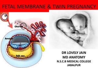
  • 1. FETAL MEMBRANE & TWIN PREGNANCY DR LOVELY JAIN MD ANATOMY N.S.C.B MEDICAL COLLEGE JABALPUR
  • 4.
  • 6. Fetal membrane • The term fetal membrane is applied to those structures derived from the blastocyst which do not contribute to the embryo. The amnion, the chorion, the yolk sac Allantois Umbilical cord
  • 7. AMNION It is a thin, transparent &tough fluid- filled, membranoussac surrounding the embryo  form by--- amniotic epi. +extraembryonic mesoderm  At term amnion is a tough, tenacious & pliable membrane.  Inner most ,avescular,&provide tensile strength. • It lacks smooth muscles,lymphatics,nervs& bld vesscles. Bourne (1962) described five separate layers of amnion – 1)innermost single layer of cuboidal epithelium derived from embryonic ectoderm.basement membrane 2)acellular compact layer, which is composed primarily of interstitial collagens 3)fibroblast-like mesenchymal cells, derived from embryonic 4)disc mesoderm 5)acellular zona spongiosa, contiguous with second fetal membrane, the chorion laeve • a
  • 8. Devlopment Early during implantation:- a space develops between the embryonic cell mass and adjacent trophoblasts.  Small cells that line this inner surface of trophoblasts have been called amniogenic cells—precursors of amnionic epithelium.  At First : It is seen as a small cavity lying dorsal to the embryonic plate.  At Stage of Chorionic Vesicle: The amnion becomes separated from the chorion by chorionic cavity or extra embryonic coelom.  After Folding: the amnion expands greatly and is becomes on the ventral surface of the embryo.  As a result of expansion of the amnion, the extra embryonic coelom is gradually obliterated and amnion forms the epithelial covering of umbilical cord & Reflected amnion is fused to the chorion laeve. • Placental amnion covers placental surface & thereby is in contact with adventitial surface of chorionic vessels. • Umbilical amnion covers the umbilical cord. • Diamniotic- dichorionic the conjoined portion of membranes of twin placenta, fused amnions are separated by fused chorion laeve. • Diamniotic-monochorionic placenta, there is no intervening tissue between the fused amnions.
  • 9. Amniotic Fluid Produce by: 1)amniotic cells 2)infusion of fluid from maternal blood 3)urine output from the fetus 4)pulmonary secretions Output: 1) absorbed by amniotic cells • 2) fetus swallow • Plays a major role in fetal growth and development. • Daily contribution of fluid from respiratory tract is 300-400 ml. • 500 ml of urine is added daily during the late pregnancy. • Amniotic fluid volume is 30 ml at 10 weeks, 350 ml at 20 weeks, 700-1000 ml at 37 weeks. • Composition:99 % is water • Desquamated fetal epithelial cells • Organic & inorganic salts • Protein, carbohydrates, fats, enzymes, hormones • Meconium & urine in the late stage
  • 10. function • Helps maintain the body temperatureEnables the fetus to move freelyProvides symmetrical external growth of the embryo. • During labor it help dilatation of the cervix of the uterus and It wash birth canal and protect the fetus against infections. • Cushions & protects the embryo and fetus Acts as a barrier to infection (it is an aseptic medium) Permits normal fetal lung development Prevents adherence of embryo to amnion It protects embryo against external injuries. Allows the embryo to move freely, aiding muscular development in the limbs It is involved in maintaining homeostasis of fluids & electrolytes It permits studies on fetal enzymes, hormones and diagnosis of fetal sex and chromosomal abnormalities
  • 11. ABNORMALATIES OF AMNIOTIC FLUID VOLUME : Oligo-hydramnios:The volume is less than ½ liters Causes : Placental insufficiency with low placental blood flow Preterm rupture of amnio- chorionic membrane occurs in 10% of pregnancies Renal Agenesis (failure of kidney • development) Obstructive Uropathy (urinary tract obstruction) lead to absence of fetal urine (the main source) Complications : Fetal abnormalities (pulmonary, • facial & limb defects) Polyhydra mnios:(Hy dramnios): The volume is more than 2liters, it is diagnosed by Ultrasonography. Causes Fetal ( 1-20% ) : Esophageal atresia. Maternal (2-20%) : defects in maternal circulation. Idiopathic (3- 60%)
  • 12. Abnormility Definition & causes Clinical significance  Lined by typical amnionic epithelium  Fusion of amnionic folds with subsequent fluid retention Amnion nodosum  Tiny, creamy nodules in the amnion made up of vernix caseosa with hair, degenerated squames and sebum  Oligohydramnios Found in  fetuses with renal agenesis  Prolonged preterm ruptured Membranes  The placenta of the donor fetus with twin-to-twin transfusion syndrome Amnionic band Caused when disruption of the amnion leads to formation of bands or strings Intrauterine amputation Amnionic cyst
  • 13. YOLK SAC • It is essential in the transfer of nutrients to the embryo during 2nd & 3rd weeks, when the uteroplacental circulation is not established. • It is large at 32 days • Shrinks to 5mm pear shaped remnant by 10th week & connected to the midgut by a narrow yolk stalk • Becomes very small at 20 weeks Usually not visible thereafter • It does not contain any yolk. Its development passes through three stages: Primary yolk sac. Secondary yolk sac. Definitive yolk sac. • a
  • 14. Primary yolk sac • a Appears in the Blastocyst stage at 10- days, it lies ventral to the embryonic plate. Its roof is formed by hypoblast (primary endoderm), Its wall is formed by exocoelomic membrane, it lines the inner surface of the cytotrophoblast, and separated from it by the extraembryonic mesoderm
  • 15. Secondary yolk sac • a Appears in the chorionic vesicle stage Its roof is formed by hypoblast (embryonic endoderm), its wall is formed by exocoelomic membrane + inner layer (splanchnic layer) of the extraembryonic mesoderm. At day 16: a diverticulum appears from its dorsocaudal end (Allantois) into the substance of the connecting stalk
  • 16. Definitive yolk sac • a After folding, part of Yolk Sac is enclosed within the embryo to form the Gut (Foregut, Midgut & Hindgut). The remainder of Yolk Sac that remains outside the embryo becomes the Definitive Yolk Sac  The midgut is temporarily connected to Definitive Yolk Sac by a narrow duct Vitello- intestinal duct (Yolk stalk), which is incorporated inside the umbilical cord.  This is fibrosed and degenerated by • the end of (6th week)
  • 17. Function of yolk sac a 3rd week:  (a) Blood formationt • First formed in the extra- embryonic mesoderm covering the wall of the yolk sac, until hemopoietic activity begins in the liver during 6th week  4th week: endoderm of yolk sac is incorporated into the embryo to form primordial gut Epithelium of Respiratory system &G.I.T b)Primordial germ cells:- in the endodermal lining of the wall of caudal end of the yolk sac migrate into the developing sex glands to differentiate into germ cells (spermatogonia or oogonia)
  • 18. Fate of yolk sac • a  Yolk stalk detached from midgutby the end of 6th week. In (2%) of adults, its proximal intra-abdominal part persists as ileal diverticulum (Meckel diverticulum).  At 10 week, small definitive yolk sac lies in the chorionic cavity between amniotic & chorionic sacs  At 20 weeks, as pregnancy advances, definitive yolk sac atrophies and becomes a very small cyst.  In unusual cases, it persists under the amnion near the attachment of Umbilical cord, on the fetal surface of the placenta. Its persistence is of no significance
  • 19.
  • 20. ALLANTOIS 3rd week:Appears as a diverticulum from caudal wall of Y.S. that extends into connecting stalk. 2nd month: Its extra- embryonic part degenerates. 3rd month: Its intra- embryonicpart extends from UB to UC as thick tube , ‘(urachus) ’  After birth: the urachus is obliterated and fibrosed to form median umbilical ligament, that extendsfrom apex of UB to umbilicus.  Function:Blood formation in its wall during 3rd to 5th week.  Its blood vessels persist as the umbilical vein & arteries. • a
  • 21. CHORION • The extraembryonic somatic mesoderm and the two layers of trophoblast form the chorion • Chorion forms the wall of chorionic sac • Embryo and its amniotic and yolk sacs are suspended into it by connecting stalk • Growth of these extensions are caused by underlying extraembryonic somatic mesoderm • The cellular projections form primary chorionic villi. Chorionic villi • As this sac grows, the villi associated with decidua capsularis are compressed, reducing the blood supply to them • These villi soon degenerates producing an avascular bare area smooth chorion (chorion laeve).
  • 22.
  • 23. Primary chorionic vilii • At the end of 2nd week, finger-like processes formed of outer syncytiotrophoblast & inner cytotrophoblast appear & cover the entire chorionic sac until the beginning of 8th week
  • 24. Secondary chorionic villi • Early in 3rd :- week, extraembryoni c mesoderm extends inside the villi • a
  • 25. Tertiary Chorionic villi • During 3rd week, arterioles, venules & capillaries develop in the mesenchyme of villi & join umbilical vessels • By the end of 3rd week, embryonic blood begins to flow slowly through capillaries in chorionic villi • a
  • 26. UMBILICAL CORD/ funis Extends from fetal umbilicus to fetal surface of placenta or chorionic plate. • Exterior is dull white, moist, & covered with amnion, through which three umbilical vessels may be seen. • Origin :-It develops from the connecting stalk. • Length:--At term, it measures about 50 cm. • Diameter:--2 cm. DEVELOPMENT:--Cord develops in yolk sac & umbilical vesicle which are prominent early in pregnancy. Embryo, at first, is a flattened disc interposed between amnion & yolk sac. Its dorsal surface grows faster than the ventral surface. Embryo bulges into amnionic sac in association with elongation of neural tube. Dorsal part of yolk sac is incorporated into the body of embryo to form gut.
  • 27. Allantois projects into base of body stalk from the caudal wall of the yolk sac & later, forms anterior wall of hindgut. As pregnancy advances, yolk sac becomes smaller & its pedicle relatively longer. Middle of 3rd month:- expanding amnion obliterates exocoelom, fuses with the chorion laeve, & covers the bulging placental disc & lateral surface of the body stalk.Latter is then called the umbilical cord—or funis. Insertion: The cord is inserted in the foetal surface of the placenta near the center "eccentric insertion" (70%) Or at the center "central insertion" (30%). Structure: It consists of mesodermal connective tissue called Wharton's jelly, covered by amnion. It contains: 1.One umbilical vein carries oxygenated blood from the placenta to the foetus 2.Two umbilical arteries carry deoxygenated blood from the foetus to the placenta, 3.Remnants of the yolk sac and allantois..
  • 28.
  • 29. CORD AT TERM • It normally has two arteries and one vein . • Right umbilical vein disappears early during fetal development, leaving only the left vein. • Intra-abdominal portion of duct of umbilical vesicle, extending from umbilicus to intestine, usually atrophies & disappears. • If patent, it is known as Meckel’s diverticulum. • Most common vascular anomaly - absence of one umbilical artery which may be associated with fetal anomalies. • A
  • 30. • Length • Cord Coiling • Single Umbilical Artery • Four-vessel cord • Abnormalities of cord insertion • Cord Abnormalities capable of impeding blood flow • Hematoma • Cysts
  • 31. LENGTH:--Appreciable variation –Average length of 55 cm range-- of 30 to 100 cm. No cord---(acordia) ~ lengths up to 300cm Excessively long cords : ≥ 70cm( ≥2 SD ) Associated with -maternal systemic disease -delivery complications -cord prolapse, cord entanglement -fetal anomalies and respiratory distress Perinatal mortality : ↑ nearly threefold Short umbilical cord:-Generally, cord length less than 30 cm is considered abnormally short. Adverse perinatal outcomes – -fetal growth restriction congenital malformations intrapartum distress & risk of death (doubled)
  • 32. Umbilical vessels: in a spiraled manner CORD COILING:--Umbilical vessels: in a spiraled manner Hypocoiled cords:-↑ in various adverse outcome in fetuses - meconium staining, preterm birth and fetal distress. Hypercoiled cords --higher incidence of preterm delivery & cocaine abuse. SINGLE UMBLICAL ARTERY:--Umbilical cord –2-arteries & 1 vein Risk factors –↑ incidence in women with GDM, PIH, APH, epilepsy, oligohydramnios & hydramnios. ----Âź of all infants with only 1 artery have associated congenital anomalies. FOUR VESSCLE CORD:- Venous remnantin 5% & Significance : unknown
  • 33. ABNORMALITIS IN CORD INSERSATION rmalities Definition Incidenc Significance cate insertion Umbilical vessels separate from the cord substance before their insertion into the placenta RARE Prone to twisting & thromboses as vessels lose their cushioning ginal Inserion Battledore placenta: Cord insertion at the placental margin 7% at term Cord being pulled off during delivery of the placenta mentous Insertion Umbilical vessels separate in the membranes at a distance from the placental margin. Reach surrounded only by a fold 1.1% More frequently with twins 28% of triples
  • 34. Vasa pervia • Associated with velamentous insertion when some of the fetal vessels in the membranes cross the region of the cervical os below the presenting fetal part Associated with : • Velamentous insertion (50%) • Marginal cord insertion • Bilobed or Succenturiate-lobed placentas (50%) Risk factors : • Bilobed , Succenturiate or low-lying placenta (80%) • Multifetal pregnancy • Pregnancy resulting from in vitro fertilization. Diagnosis • Color Doppler examination • Perinatal diagnosis :-- associated with increased survival • Antenatal diagnosis : --associated with decreased fetal mortality compared with discovery at delivery • Antepartum or intrapartum haemorrhage • Detecting fetal blood ( Apt test) • Wright stain : smear the blood on glass slides stain the smears with Wright stain and examine for nucleated RBC normally are present in cord blood but not maternal blood
  • 35. TWIN/MULTIPILE PREGNANCY Two or more fertilization events Single fertilization followed by splitting of zygote Combination of both Incidence:-Global incidence: 4/1000 births Hellin’s Law : Twins: 1/80 singleton births • Triplets: 1:802 • Quadruplets: 1:803 Conjoined twins: 1 : 60,000 Typesof twins……… 1)-DIZYGOTIC 2)-MONOZYGOTIC
  • 36.
  • 37. Dizygotic/ non-identical twins (binovular , fraternal, 2 eggtwins) ~ two third of twins. fertilization of two independently released ova by two different sperm. In all polyzygotic multiple pregnancies, each zygote develops its own amnion, chorion and placental circulation, and hence will be polychorionic. not true twins
  • 38. Always dichorionic & diamnionic
  • 39. Monozygotic twins ( uniovular, identical orsingle egg twins One third of twins. arise from the splitting of a single fertilized egg within the first 14 days after fertilization. Always same sex (Identical) does not necessarily result in equal sharing of genetic material , so they may be discordant for genetic mutations , or may have the same genetic disease but with marked variability in expression. teratogenic event
  • 40.
  • 41.
  • 42. ETIOLOGY:-Maternal age  Race and heredity : Black race  Parity: Increasing parity (2.7% in 4th pregnancy)  Heredity  Pituitary Gonadotropin  ART: Ovulation induction with FSH and gonadotropin /chlomiphine Greater the number of embryos transfered, the greater the risk of multiple pregnancy. Determination of zygosity/chorionicity :--Chorionicity can be identified in the first trimester with sonography Before 10 weeks sonographic findings to determine chorionicity. Number of 1.gestational sacs 2.amniotic sacs within the chorionic cavity 3.yolk sacs.
  • 43. 1. Number of Gestational Sacs Each gestational sac forms its own placenta and chorion: 2 gestational sacs: DC twin 1 gestational sac with 2 identified heartbeats: MC twin. 2. Number of Amniotic Sacs Within the ChorionicCavity  Diamniotic twins: separate and distinct amnions before 10w the separate amnions of a diamniotic pregnancy will not have enlarged sufficiently to contact each other and create the inter-twinseptum. TAS: Each single amnion is extremely thin and delicate: very difficult to see TVS: often successful in differentiating separate amnions. 3. Number of YolkSacs 2 yolk sacs are seen in the extra-embryonal coelom: diamniotic  1 yolk sac-in most cases indicate monoamniotic twins when there are dual embryos: follow-up 1st T scan to definitively assign amnionicitya
  • 44. .After 10 weeks:-These sonographic signs are no longer present: gestational sacs are no longer distinctly separable, and the inter-twin membrane is formed. Findings:- • 1)-Genitalia • 2)-Placental number • 3)-Chorionic peak sign( Lambda sign & T sign) • 4)-Membranecharacteristics. Inter-Twin Membrane :Characteristics 1)DC : -2 layers of amnion and 2 layers of chorion.Thicker > 2 mm more reflective 2)MC:- ≤ 2mm In 2nd T: Number of membranes may be counted, and if there are > 2, then dichorionicity is strongly suggested • .
  • 45. Pregnancy complications 2 to3 fold increased than singletons Threatened and spontaneous abortions (vanishing twin) 7.3 % risk in multiple • pregnancy versus 0.9 % in singleton (Joo, 2012) Hyperemesis Severe anemia Hypertensive disorders of pregnancy: 3 to 4 fold increase Gestational diabetes Antepartum hemorrhage: abruption Preterm premature rupture of the membranes Operative delivery PPH : 3-4 fold increase Increased maternal mortality
  • 46. Fetal complications Low birthweight- due to restricted fetal growth and preterm delivery Preterm birth Monochorionic pregnancy complications Perinatal asphyxia Fetal death, Cord accidents Increased perinatal mortality • Congenital Malformations- 406/10000 in twins versus 238/10000 singletons . Structural malformations 1)Conjoint twins,2)Acardiac fetus,3)Anencephaly • 4)Talipes,5)Dislocation of hip etc. Chromosomal anomalies Down’ssyndrome
  • 49. External parasitic twins- grossly defective fetus or merely fetal parts attached externally to a relatively normal twin • Believed to result from demise of the defective twin with its surviving tissues attached to and vascularized by its normal twin Fetus in fetu- early in development, one embryo may be enfolded within its twin • Classically vertebral or axial bones are found in these fetiform mases, supported by their host by a few large parasitic vessels
  • 50. Monochorionic twins withvascular anastomoses Two amniotic sacs and a common surrounding chorion anatomical sharing of the two fetal circulations through anastomoses of placental arteries and veins Artery to artery anastomoses are most common and are identified on the chorionic surface of the placenta- 75% Vein to vein and artery to vein– approx. 50%. Deep artery to vein connections can extend from capillary bed of a given villus, creating a common villous compartment or third circulation Depending on the degree to which they are hemodynamically balanced, severity occurs With significant pressure or flow gradients, a shunt will develop between fetuses Chorioinic feto fetal transfusion result in several clinical syndromes
  • 51. Twin-Twin Transfusion syndrome 5 – 17 % of monochorionic twin Mortality irrespective of gestational age is 60-70% Mechanism: deep A-Vvascular anastomosis. Blood is transfused from donor twin to its recipient sibling – donor is anemic and growth may be restricted Recipient becomes polycythemic, with circulatory overload and may manifest as hydrops Classic TTTS results from unidirectional flow through AV anastomoses Deoxygenated blood from donor placental artery- pumped into a cotyledon shared by recipient. Once oxygen exchange is completed in the chorionic villus, oxygenated blood leaves the cotyledon via a placental vein of the recipient twin
  • 52. .Clinically important TTTS is frequently chronic, results from significant volume differences Presents in mid pregnancy, donor fetus- oliguric due to decreased renal perfusion – develops oligohydramnios Recipient- polyhydramnios Stuck twin, polyhydramnios- oligohydramnios – syndrome (poly-oli • .