SlideShare a Scribd company logo
1 of 22
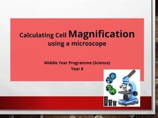
Calculating Cell Magnification
using a microscope
Middle Year Programme (Science)
Year 8
• To label the parts of a microscope.
• To safely use a microscope to magnify objects.
• To make scientific drawings of objects under the microscope.
• To learn how to calculate cell magnification
Learning Objectives
Put the following objects in order of size. You can use your device to research their
size.
bacteria red
blood cell
DNA
paperclip
grain of
salt
Extension:
Which of these objects can only be seen using a microscope?
bacteria red
blood cell
DNA paperclip
grain of
salt
Extension:
Which of these objects can only be seen using a microscope?
increasing size
bacteria, red blood cell and DNA
MICROSCOPES
• MICROSCOPES HAVE BEEN USED FOR MANY YEARS TO
OBSERVE OBJECTS THAT ARE TOO SMALL TO SEE WITH THE
NAKED EYE. THE FIRST MICROSCOPE WAS INVENTED IN THE
1500S.
• OVER TIME, THE MAGNIFICATION AND RESOLUTION OF
MICROSCOPES HAS SIGNIFICANTLY IMPROVED DUE TO
DEVELOPMENTS IN TECHNOLOGY. WE NOW HAVE
MICROSCOPES THAT CAN EXAMINE SPECIMENS AT AN
ATOMIC LEVEL.
• MANY IMPORTANT SCIENTIFIC DISCOVERIES HAVE BEEN
MADE USING MICROSCOPES.
PARTS OF A MICROSCOPE
You are given a diagram of the microscope. Stick the paper onto your
copybook. Can you label any parts of the light microscope?
PARTS OF A MICROSCOPE
Can you label any parts of the light microscope?
arm
stage clips
eyepiece lens
objective lens
stage
coarse adjustment knob
fine adjustment knob
light source
base
diaphragm
USING A LIGHT MICROSCOPE
1. Plug in the microscope and turn on the light. If your microscope
has a mirror, you may need to adjust it so light is directed through
the diaphragm.
2. Place your specimen (the object you want to observe) on the stage and
secure it with the stage clips.
3. Turn the objective lens to the lowest magnification (usually ×4).
4. Turn the coarse adjustment knob until the objective lens is almost
touching the microscope slide. Look from the side of the microscope as
you do this, not through the eyepiece, so you do not damage the slide.
5. Looking through the eyepiece, turn the coarse adjustment knob to move
the stage away from the objective lens until the image comes into focus.
6. Use the fine adjustment knob to make the image clearer.
7. Turn to a higher power objective lens (×10 or ×40) and refocus the image
using the fine adjustment knob.
8. Make a scientific drawing of the specimen or write down any
observations.
FUN EXERCISE 1: USING A LIGHT MICROSCOPE
Have a go at looking at some objects under the microscope. These could be
prepared slides provided by your teacher, but you could also try looking at a
strand of your hair, the tip of a pencil or any other objects you can find in the
classroom.
Object Diagram/Observation Remember to start
on the lowest
magnification!
Use a sharp pencil
for drawing scientific
diagrams.
CALCULATING PLANT CELL MAGNIFICATION
• PLEASE WATCH THE VIDEO ON CALCULATION OF CELL
MAGNIFICATION
• HTTPS://WWW.YOUTUBE.COM/WATCH?V=FDALMKOHF2O
WHAT IS CELL
MAGNIFICATION ?
MAGNIFICATION IS H
OW MANY TIMES
BIGGER THE IMAGE
OF A SPECIMEN
OBSERVED IS IN
COMPARISON TO
THE ACTUAL (REAL-
LIFE) SIZE OF THE
SPECIMEN
Example 1
Red blood cells have an average diameter of 0.012mm. The image above shows a
red blood cell with a diameter of 100mm.
Calculate the magnification using the equation below.
diameter of image = real diameter magnification
×
Answer
diameter of image = real diameter magnification
×
Red blood cells have an average diameter of 0.012mm. The image above shows a
red blood cell with a diameter of 100mm.
Calculate the magnification using the equation below.
100mm = 0.012 magnification
×
Re-arrange the equation = 100 ÷ 0.012 = 8333.3
EXAMPLE 2
Use the formula
ANSWER
PLENARY
1. Which part of the microscope do you look through?
diaphragm
objective lens
eyepiece lens
eyepiece lens
2. Which part of the microscope is used to move the stage up and down?
coarse adjustment knob
stage clips
fine adjustment knob
coarse adjustment knob
3. What is the name of the object that you observe with a microscope?
sample
specimen
species
specimen
4. Which part of the microscope can be adjusted to control the amount of
light reaching the specimen.
arm
objective lens
diaphragm
diaphragm
5. A light microscope has three objective lenses: ×4, ×10 and ×40. Which
objective lens should be used first when viewing an object?
×4
×40
×10
×4
EXTENSION EXERCISES AT HOME
Task 1
Research the history of the
microscope and how it has
developed over time. Add
pictures and diagrams.
Task 2
Describe the differences
between a light microscope
and an electron microscope.
NEXT LESSON
PRACTICAL ON CELL MAGNIFICATION

More Related Content

Similar to Microscope and Cell Magnification.pptx

Week 08 - Leading for ChangeFollowership Objecti.docx
Week 08 - Leading for ChangeFollowership Objecti.docxWeek 08 - Leading for ChangeFollowership Objecti.docx
Week 08 - Leading for ChangeFollowership Objecti.docxcelenarouzie
 
1.0 - The Light Miscroscope (2).pptx0ppt
1.0 - The Light Miscroscope (2).pptx0ppt1.0 - The Light Miscroscope (2).pptx0ppt
1.0 - The Light Miscroscope (2).pptx0pptstephenopokuasante
 
1.0 - The Light Miscroscope.ppt microscopy
1.0 - The Light Miscroscope.ppt microscopy1.0 - The Light Miscroscope.ppt microscopy
1.0 - The Light Miscroscope.ppt microscopystephenopokuasante
 
BIOLOGY FORM 1 Notes.pptx
BIOLOGY FORM 1 Notes.pptxBIOLOGY FORM 1 Notes.pptx
BIOLOGY FORM 1 Notes.pptxMgungweFrank
 
Microscope Notes- Types & Use.ppt.pdf
Microscope Notes- Types & Use.ppt.pdfMicroscope Notes- Types & Use.ppt.pdf
Microscope Notes- Types & Use.ppt.pdfMunishGuleria4
 
Microscope introduction
Microscope introductionMicroscope introduction
Microscope introductionHaileybury
 
COMPOUND MICROSCOPE COT 2ND.pptx
COMPOUND MICROSCOPE COT 2ND.pptxCOMPOUND MICROSCOPE COT 2ND.pptx
COMPOUND MICROSCOPE COT 2ND.pptxjenniferatilano3
 
Lecture 3 Unit-I Bright-field & Dark-field Microscopy.pdf
Lecture 3 Unit-I Bright-field & Dark-field Microscopy.pdfLecture 3 Unit-I Bright-field & Dark-field Microscopy.pdf
Lecture 3 Unit-I Bright-field & Dark-field Microscopy.pdfSushmitaChoudhary16
 
Second (2nd) Quarter Topics in Science 7
Second (2nd) Quarter Topics in Science 7Second (2nd) Quarter Topics in Science 7
Second (2nd) Quarter Topics in Science 7Jady Claire Lullegao
 
Microscopes Applied
Microscopes AppliedMicroscopes Applied
Microscopes Appliedbeckydaw
 
Microscopy.ppt Microscopy.pptvMicroscopy.pptMicroscopy.pptMicroscopy.pptMicro...
Microscopy.ppt Microscopy.pptvMicroscopy.pptMicroscopy.pptMicroscopy.pptMicro...Microscopy.ppt Microscopy.pptvMicroscopy.pptMicroscopy.pptMicroscopy.pptMicro...
Microscopy.ppt Microscopy.pptvMicroscopy.pptMicroscopy.pptMicroscopy.pptMicro...KAOUKAAlaedin
 
Integrated Science M1 More tools used in Science - Microscope
Integrated Science M1 More tools used in Science - MicroscopeIntegrated Science M1 More tools used in Science - Microscope
Integrated Science M1 More tools used in Science - MicroscopeeLearningJa
 
Microscopy for Microbiology – Use and FunctionHands-On L.docx
Microscopy for Microbiology – Use and FunctionHands-On L.docxMicroscopy for Microbiology – Use and FunctionHands-On L.docx
Microscopy for Microbiology – Use and FunctionHands-On L.docxARIV4
 
The Microscope Zacharias Janssen is to be credited for i.docx
The Microscope Zacharias Janssen is to be credited for i.docxThe Microscope Zacharias Janssen is to be credited for i.docx
The Microscope Zacharias Janssen is to be credited for i.docxoreo10
 

Similar to Microscope and Cell Magnification.pptx (20)

Week 08 - Leading for ChangeFollowership Objecti.docx
Week 08 - Leading for ChangeFollowership Objecti.docxWeek 08 - Leading for ChangeFollowership Objecti.docx
Week 08 - Leading for ChangeFollowership Objecti.docx
 
1.0 - The Light Miscroscope (2).pptx0ppt
1.0 - The Light Miscroscope (2).pptx0ppt1.0 - The Light Miscroscope (2).pptx0ppt
1.0 - The Light Miscroscope (2).pptx0ppt
 
1.0 - The Light Miscroscope.ppt microscopy
1.0 - The Light Miscroscope.ppt microscopy1.0 - The Light Miscroscope.ppt microscopy
1.0 - The Light Miscroscope.ppt microscopy
 
BIOLOGY FORM 1 Notes.pptx
BIOLOGY FORM 1 Notes.pptxBIOLOGY FORM 1 Notes.pptx
BIOLOGY FORM 1 Notes.pptx
 
Lab 0-B Microscope Training
Lab 0-B Microscope TrainingLab 0-B Microscope Training
Lab 0-B Microscope Training
 
Microscope Notes- Types & Use.ppt.pdf
Microscope Notes- Types & Use.ppt.pdfMicroscope Notes- Types & Use.ppt.pdf
Microscope Notes- Types & Use.ppt.pdf
 
Microscope introduction
Microscope introductionMicroscope introduction
Microscope introduction
 
SCIENCE-7-QUARTER-2-LAS (1).pdf
SCIENCE-7-QUARTER-2-LAS (1).pdfSCIENCE-7-QUARTER-2-LAS (1).pdf
SCIENCE-7-QUARTER-2-LAS (1).pdf
 
COMPOUND MICROSCOPE COT 2ND.pptx
COMPOUND MICROSCOPE COT 2ND.pptxCOMPOUND MICROSCOPE COT 2ND.pptx
COMPOUND MICROSCOPE COT 2ND.pptx
 
Lecture 3 Unit-I Bright-field & Dark-field Microscopy.pdf
Lecture 3 Unit-I Bright-field & Dark-field Microscopy.pdfLecture 3 Unit-I Bright-field & Dark-field Microscopy.pdf
Lecture 3 Unit-I Bright-field & Dark-field Microscopy.pdf
 
Second (2nd) Quarter Topics in Science 7
Second (2nd) Quarter Topics in Science 7Second (2nd) Quarter Topics in Science 7
Second (2nd) Quarter Topics in Science 7
 
Microscopes Applied
Microscopes AppliedMicroscopes Applied
Microscopes Applied
 
Microscope.pptx
Microscope.pptxMicroscope.pptx
Microscope.pptx
 
MICROSCOPY1.pptx
MICROSCOPY1.pptxMICROSCOPY1.pptx
MICROSCOPY1.pptx
 
Microscope.pptx
Microscope.pptxMicroscope.pptx
Microscope.pptx
 
Microscopy.ppt Microscopy.pptvMicroscopy.pptMicroscopy.pptMicroscopy.pptMicro...
Microscopy.ppt Microscopy.pptvMicroscopy.pptMicroscopy.pptMicroscopy.pptMicro...Microscopy.ppt Microscopy.pptvMicroscopy.pptMicroscopy.pptMicroscopy.pptMicro...
Microscopy.ppt Microscopy.pptvMicroscopy.pptMicroscopy.pptMicroscopy.pptMicro...
 
Integrated Science M1 More tools used in Science - Microscope
Integrated Science M1 More tools used in Science - MicroscopeIntegrated Science M1 More tools used in Science - Microscope
Integrated Science M1 More tools used in Science - Microscope
 
Microscopy for Microbiology – Use and FunctionHands-On L.docx
Microscopy for Microbiology – Use and FunctionHands-On L.docxMicroscopy for Microbiology – Use and FunctionHands-On L.docx
Microscopy for Microbiology – Use and FunctionHands-On L.docx
 
The Microscope Zacharias Janssen is to be credited for i.docx
The Microscope Zacharias Janssen is to be credited for i.docxThe Microscope Zacharias Janssen is to be credited for i.docx
The Microscope Zacharias Janssen is to be credited for i.docx
 
Chapter 1 Microscopy
Chapter 1 MicroscopyChapter 1 Microscopy
Chapter 1 Microscopy
 

Recently uploaded

FAIRSpectra - Enabling the FAIRification of Spectroscopy and Spectrometry
FAIRSpectra - Enabling the FAIRification of Spectroscopy and SpectrometryFAIRSpectra - Enabling the FAIRification of Spectroscopy and Spectrometry
FAIRSpectra - Enabling the FAIRification of Spectroscopy and SpectrometryAlex Henderson
 
Porella : features, morphology, anatomy, reproduction etc.
Porella : features, morphology, anatomy, reproduction etc.Porella : features, morphology, anatomy, reproduction etc.
Porella : features, morphology, anatomy, reproduction etc.Cherry
 
The Mariana Trench remarkable geological features on Earth.pptx
The Mariana Trench remarkable geological features on Earth.pptxThe Mariana Trench remarkable geological features on Earth.pptx
The Mariana Trench remarkable geological features on Earth.pptxseri bangash
 
Understanding Partial Differential Equations: Types and Solution Methods
Understanding Partial Differential Equations: Types and Solution MethodsUnderstanding Partial Differential Equations: Types and Solution Methods
Understanding Partial Differential Equations: Types and Solution Methodsimroshankoirala
 
X-rays from a Central “Exhaust Vent” of the Galactic Center Chimney
X-rays from a Central “Exhaust Vent” of the Galactic Center ChimneyX-rays from a Central “Exhaust Vent” of the Galactic Center Chimney
X-rays from a Central “Exhaust Vent” of the Galactic Center ChimneySérgio Sacani
 
GBSN - Biochemistry (Unit 2) Basic concept of organic chemistry
GBSN - Biochemistry (Unit 2) Basic concept of organic chemistry GBSN - Biochemistry (Unit 2) Basic concept of organic chemistry
GBSN - Biochemistry (Unit 2) Basic concept of organic chemistry Areesha Ahmad
 
Concept of gene and Complementation test.pdf
Concept of gene and Complementation test.pdfConcept of gene and Complementation test.pdf
Concept of gene and Complementation test.pdfCherry
 
Use of mutants in understanding seedling development.pptx
Use of mutants in understanding seedling development.pptxUse of mutants in understanding seedling development.pptx
Use of mutants in understanding seedling development.pptxRenuJangid3
 
Daily Lesson Log in Science 9 Fourth Quarter Physics
Daily Lesson Log in Science 9 Fourth Quarter PhysicsDaily Lesson Log in Science 9 Fourth Quarter Physics
Daily Lesson Log in Science 9 Fourth Quarter PhysicsWILSONROMA4
 
POGONATUM : morphology, anatomy, reproduction etc.
POGONATUM : morphology, anatomy, reproduction etc.POGONATUM : morphology, anatomy, reproduction etc.
POGONATUM : morphology, anatomy, reproduction etc.Cherry
 
TransientOffsetin14CAftertheCarringtonEventRecordedbyPolarTreeRings
TransientOffsetin14CAftertheCarringtonEventRecordedbyPolarTreeRingsTransientOffsetin14CAftertheCarringtonEventRecordedbyPolarTreeRings
TransientOffsetin14CAftertheCarringtonEventRecordedbyPolarTreeRingsSérgio Sacani
 
LUNULARIA -features, morphology, anatomy ,reproduction etc.
LUNULARIA -features, morphology, anatomy ,reproduction etc.LUNULARIA -features, morphology, anatomy ,reproduction etc.
LUNULARIA -features, morphology, anatomy ,reproduction etc.Cherry
 
Reboulia: features, anatomy, morphology etc.
Reboulia: features, anatomy, morphology etc.Reboulia: features, anatomy, morphology etc.
Reboulia: features, anatomy, morphology etc.Cherry
 
Thyroid Physiology_Dr.E. Muralinath_ Associate Professor
Thyroid Physiology_Dr.E. Muralinath_ Associate ProfessorThyroid Physiology_Dr.E. Muralinath_ Associate Professor
Thyroid Physiology_Dr.E. Muralinath_ Associate Professormuralinath2
 
(May 9, 2024) Enhanced Ultrafast Vector Flow Imaging (VFI) Using Multi-Angle ...
(May 9, 2024) Enhanced Ultrafast Vector Flow Imaging (VFI) Using Multi-Angle ...(May 9, 2024) Enhanced Ultrafast Vector Flow Imaging (VFI) Using Multi-Angle ...
(May 9, 2024) Enhanced Ultrafast Vector Flow Imaging (VFI) Using Multi-Angle ...Scintica Instrumentation
 
Lipids: types, structure and important functions.
Lipids: types, structure and important functions.Lipids: types, structure and important functions.
Lipids: types, structure and important functions.Cherry
 
Module for Grade 9 for Asynchronous/Distance learning
Module for Grade 9 for Asynchronous/Distance learningModule for Grade 9 for Asynchronous/Distance learning
Module for Grade 9 for Asynchronous/Distance learninglevieagacer
 
Cyathodium bryophyte: morphology, anatomy, reproduction etc.
Cyathodium bryophyte: morphology, anatomy, reproduction etc.Cyathodium bryophyte: morphology, anatomy, reproduction etc.
Cyathodium bryophyte: morphology, anatomy, reproduction etc.Cherry
 
Genome sequencing,shotgun sequencing.pptx
Genome sequencing,shotgun sequencing.pptxGenome sequencing,shotgun sequencing.pptx
Genome sequencing,shotgun sequencing.pptxCherry
 
Genome Projects : Human, Rice,Wheat,E coli and Arabidopsis.
Genome Projects : Human, Rice,Wheat,E coli and Arabidopsis.Genome Projects : Human, Rice,Wheat,E coli and Arabidopsis.
Genome Projects : Human, Rice,Wheat,E coli and Arabidopsis.Cherry
 

Recently uploaded (20)

FAIRSpectra - Enabling the FAIRification of Spectroscopy and Spectrometry
FAIRSpectra - Enabling the FAIRification of Spectroscopy and SpectrometryFAIRSpectra - Enabling the FAIRification of Spectroscopy and Spectrometry
FAIRSpectra - Enabling the FAIRification of Spectroscopy and Spectrometry
 
Porella : features, morphology, anatomy, reproduction etc.
Porella : features, morphology, anatomy, reproduction etc.Porella : features, morphology, anatomy, reproduction etc.
Porella : features, morphology, anatomy, reproduction etc.
 
The Mariana Trench remarkable geological features on Earth.pptx
The Mariana Trench remarkable geological features on Earth.pptxThe Mariana Trench remarkable geological features on Earth.pptx
The Mariana Trench remarkable geological features on Earth.pptx
 
Understanding Partial Differential Equations: Types and Solution Methods
Understanding Partial Differential Equations: Types and Solution MethodsUnderstanding Partial Differential Equations: Types and Solution Methods
Understanding Partial Differential Equations: Types and Solution Methods
 
X-rays from a Central “Exhaust Vent” of the Galactic Center Chimney
X-rays from a Central “Exhaust Vent” of the Galactic Center ChimneyX-rays from a Central “Exhaust Vent” of the Galactic Center Chimney
X-rays from a Central “Exhaust Vent” of the Galactic Center Chimney
 
GBSN - Biochemistry (Unit 2) Basic concept of organic chemistry
GBSN - Biochemistry (Unit 2) Basic concept of organic chemistry GBSN - Biochemistry (Unit 2) Basic concept of organic chemistry
GBSN - Biochemistry (Unit 2) Basic concept of organic chemistry
 
Concept of gene and Complementation test.pdf
Concept of gene and Complementation test.pdfConcept of gene and Complementation test.pdf
Concept of gene and Complementation test.pdf
 
Use of mutants in understanding seedling development.pptx
Use of mutants in understanding seedling development.pptxUse of mutants in understanding seedling development.pptx
Use of mutants in understanding seedling development.pptx
 
Daily Lesson Log in Science 9 Fourth Quarter Physics
Daily Lesson Log in Science 9 Fourth Quarter PhysicsDaily Lesson Log in Science 9 Fourth Quarter Physics
Daily Lesson Log in Science 9 Fourth Quarter Physics
 
POGONATUM : morphology, anatomy, reproduction etc.
POGONATUM : morphology, anatomy, reproduction etc.POGONATUM : morphology, anatomy, reproduction etc.
POGONATUM : morphology, anatomy, reproduction etc.
 
TransientOffsetin14CAftertheCarringtonEventRecordedbyPolarTreeRings
TransientOffsetin14CAftertheCarringtonEventRecordedbyPolarTreeRingsTransientOffsetin14CAftertheCarringtonEventRecordedbyPolarTreeRings
TransientOffsetin14CAftertheCarringtonEventRecordedbyPolarTreeRings
 
LUNULARIA -features, morphology, anatomy ,reproduction etc.
LUNULARIA -features, morphology, anatomy ,reproduction etc.LUNULARIA -features, morphology, anatomy ,reproduction etc.
LUNULARIA -features, morphology, anatomy ,reproduction etc.
 
Reboulia: features, anatomy, morphology etc.
Reboulia: features, anatomy, morphology etc.Reboulia: features, anatomy, morphology etc.
Reboulia: features, anatomy, morphology etc.
 
Thyroid Physiology_Dr.E. Muralinath_ Associate Professor
Thyroid Physiology_Dr.E. Muralinath_ Associate ProfessorThyroid Physiology_Dr.E. Muralinath_ Associate Professor
Thyroid Physiology_Dr.E. Muralinath_ Associate Professor
 
(May 9, 2024) Enhanced Ultrafast Vector Flow Imaging (VFI) Using Multi-Angle ...
(May 9, 2024) Enhanced Ultrafast Vector Flow Imaging (VFI) Using Multi-Angle ...(May 9, 2024) Enhanced Ultrafast Vector Flow Imaging (VFI) Using Multi-Angle ...
(May 9, 2024) Enhanced Ultrafast Vector Flow Imaging (VFI) Using Multi-Angle ...
 
Lipids: types, structure and important functions.
Lipids: types, structure and important functions.Lipids: types, structure and important functions.
Lipids: types, structure and important functions.
 
Module for Grade 9 for Asynchronous/Distance learning
Module for Grade 9 for Asynchronous/Distance learningModule for Grade 9 for Asynchronous/Distance learning
Module for Grade 9 for Asynchronous/Distance learning
 
Cyathodium bryophyte: morphology, anatomy, reproduction etc.
Cyathodium bryophyte: morphology, anatomy, reproduction etc.Cyathodium bryophyte: morphology, anatomy, reproduction etc.
Cyathodium bryophyte: morphology, anatomy, reproduction etc.
 
Genome sequencing,shotgun sequencing.pptx
Genome sequencing,shotgun sequencing.pptxGenome sequencing,shotgun sequencing.pptx
Genome sequencing,shotgun sequencing.pptx
 
Genome Projects : Human, Rice,Wheat,E coli and Arabidopsis.
Genome Projects : Human, Rice,Wheat,E coli and Arabidopsis.Genome Projects : Human, Rice,Wheat,E coli and Arabidopsis.
Genome Projects : Human, Rice,Wheat,E coli and Arabidopsis.
 

Microscope and Cell Magnification.pptx

  • 1. Calculating Cell Magnification using a microscope Middle Year Programme (Science) Year 8
  • 2. • To label the parts of a microscope. • To safely use a microscope to magnify objects. • To make scientific drawings of objects under the microscope. • To learn how to calculate cell magnification Learning Objectives
  • 3. Put the following objects in order of size. You can use your device to research their size. bacteria red blood cell DNA paperclip grain of salt Extension: Which of these objects can only be seen using a microscope?
  • 4. bacteria red blood cell DNA paperclip grain of salt Extension: Which of these objects can only be seen using a microscope? increasing size bacteria, red blood cell and DNA
  • 5. MICROSCOPES • MICROSCOPES HAVE BEEN USED FOR MANY YEARS TO OBSERVE OBJECTS THAT ARE TOO SMALL TO SEE WITH THE NAKED EYE. THE FIRST MICROSCOPE WAS INVENTED IN THE 1500S. • OVER TIME, THE MAGNIFICATION AND RESOLUTION OF MICROSCOPES HAS SIGNIFICANTLY IMPROVED DUE TO DEVELOPMENTS IN TECHNOLOGY. WE NOW HAVE MICROSCOPES THAT CAN EXAMINE SPECIMENS AT AN ATOMIC LEVEL. • MANY IMPORTANT SCIENTIFIC DISCOVERIES HAVE BEEN MADE USING MICROSCOPES.
  • 6. PARTS OF A MICROSCOPE You are given a diagram of the microscope. Stick the paper onto your copybook. Can you label any parts of the light microscope?
  • 7. PARTS OF A MICROSCOPE Can you label any parts of the light microscope? arm stage clips eyepiece lens objective lens stage coarse adjustment knob fine adjustment knob light source base diaphragm
  • 8. USING A LIGHT MICROSCOPE 1. Plug in the microscope and turn on the light. If your microscope has a mirror, you may need to adjust it so light is directed through the diaphragm. 2. Place your specimen (the object you want to observe) on the stage and secure it with the stage clips. 3. Turn the objective lens to the lowest magnification (usually ×4). 4. Turn the coarse adjustment knob until the objective lens is almost touching the microscope slide. Look from the side of the microscope as you do this, not through the eyepiece, so you do not damage the slide. 5. Looking through the eyepiece, turn the coarse adjustment knob to move the stage away from the objective lens until the image comes into focus. 6. Use the fine adjustment knob to make the image clearer. 7. Turn to a higher power objective lens (×10 or ×40) and refocus the image using the fine adjustment knob. 8. Make a scientific drawing of the specimen or write down any observations.
  • 9. FUN EXERCISE 1: USING A LIGHT MICROSCOPE Have a go at looking at some objects under the microscope. These could be prepared slides provided by your teacher, but you could also try looking at a strand of your hair, the tip of a pencil or any other objects you can find in the classroom. Object Diagram/Observation Remember to start on the lowest magnification! Use a sharp pencil for drawing scientific diagrams.
  • 10. CALCULATING PLANT CELL MAGNIFICATION • PLEASE WATCH THE VIDEO ON CALCULATION OF CELL MAGNIFICATION • HTTPS://WWW.YOUTUBE.COM/WATCH?V=FDALMKOHF2O
  • 11. WHAT IS CELL MAGNIFICATION ? MAGNIFICATION IS H OW MANY TIMES BIGGER THE IMAGE OF A SPECIMEN OBSERVED IS IN COMPARISON TO THE ACTUAL (REAL- LIFE) SIZE OF THE SPECIMEN
  • 12. Example 1 Red blood cells have an average diameter of 0.012mm. The image above shows a red blood cell with a diameter of 100mm. Calculate the magnification using the equation below. diameter of image = real diameter magnification ×
  • 13. Answer diameter of image = real diameter magnification × Red blood cells have an average diameter of 0.012mm. The image above shows a red blood cell with a diameter of 100mm. Calculate the magnification using the equation below. 100mm = 0.012 magnification × Re-arrange the equation = 100 ÷ 0.012 = 8333.3
  • 14. EXAMPLE 2 Use the formula
  • 16. PLENARY 1. Which part of the microscope do you look through? diaphragm objective lens eyepiece lens eyepiece lens
  • 17. 2. Which part of the microscope is used to move the stage up and down? coarse adjustment knob stage clips fine adjustment knob coarse adjustment knob
  • 18. 3. What is the name of the object that you observe with a microscope? sample specimen species specimen
  • 19. 4. Which part of the microscope can be adjusted to control the amount of light reaching the specimen. arm objective lens diaphragm diaphragm
  • 20. 5. A light microscope has three objective lenses: ×4, ×10 and ×40. Which objective lens should be used first when viewing an object? ×4 ×40 ×10 ×4
  • 21. EXTENSION EXERCISES AT HOME Task 1 Research the history of the microscope and how it has developed over time. Add pictures and diagrams. Task 2 Describe the differences between a light microscope and an electron microscope.
  • 22. NEXT LESSON PRACTICAL ON CELL MAGNIFICATION