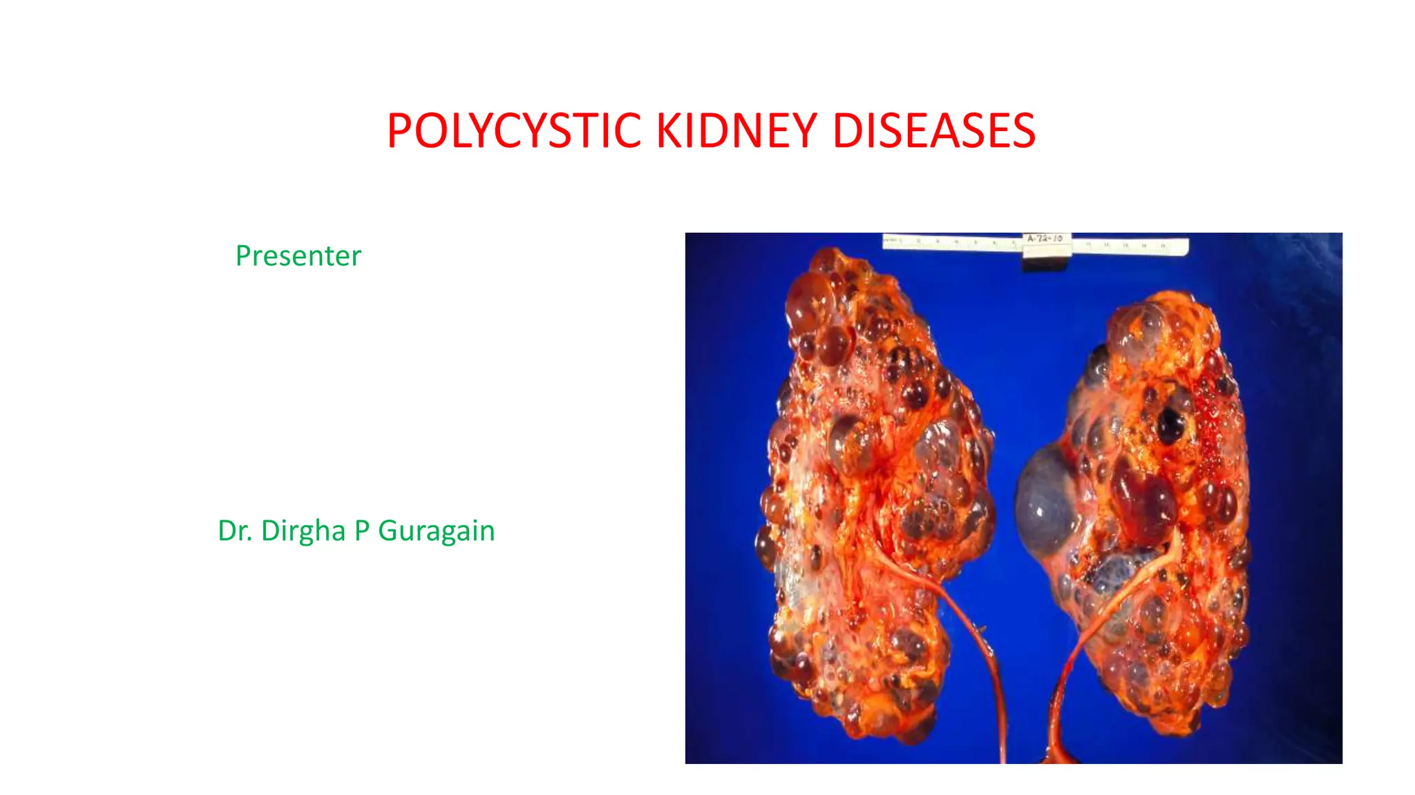
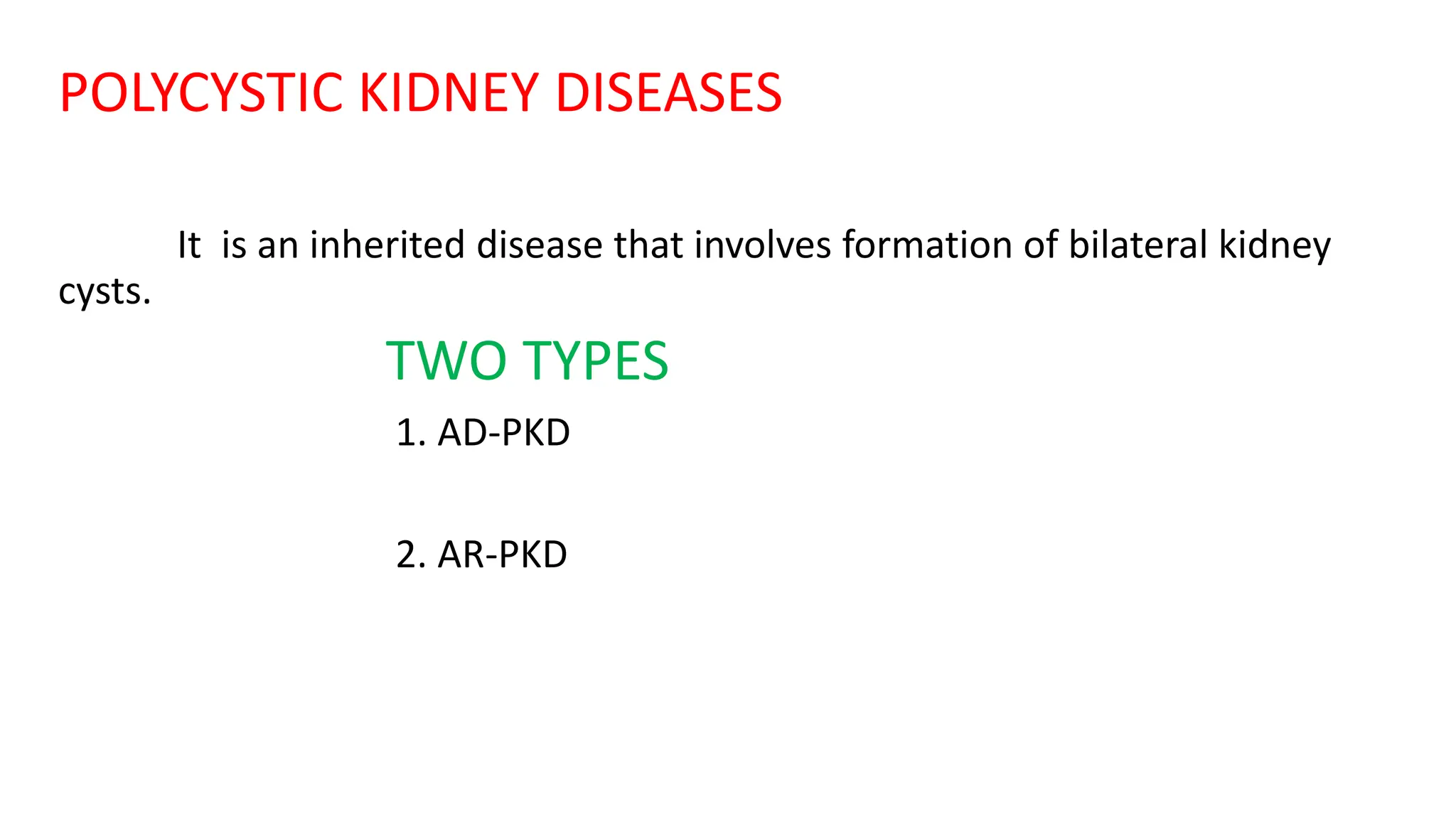
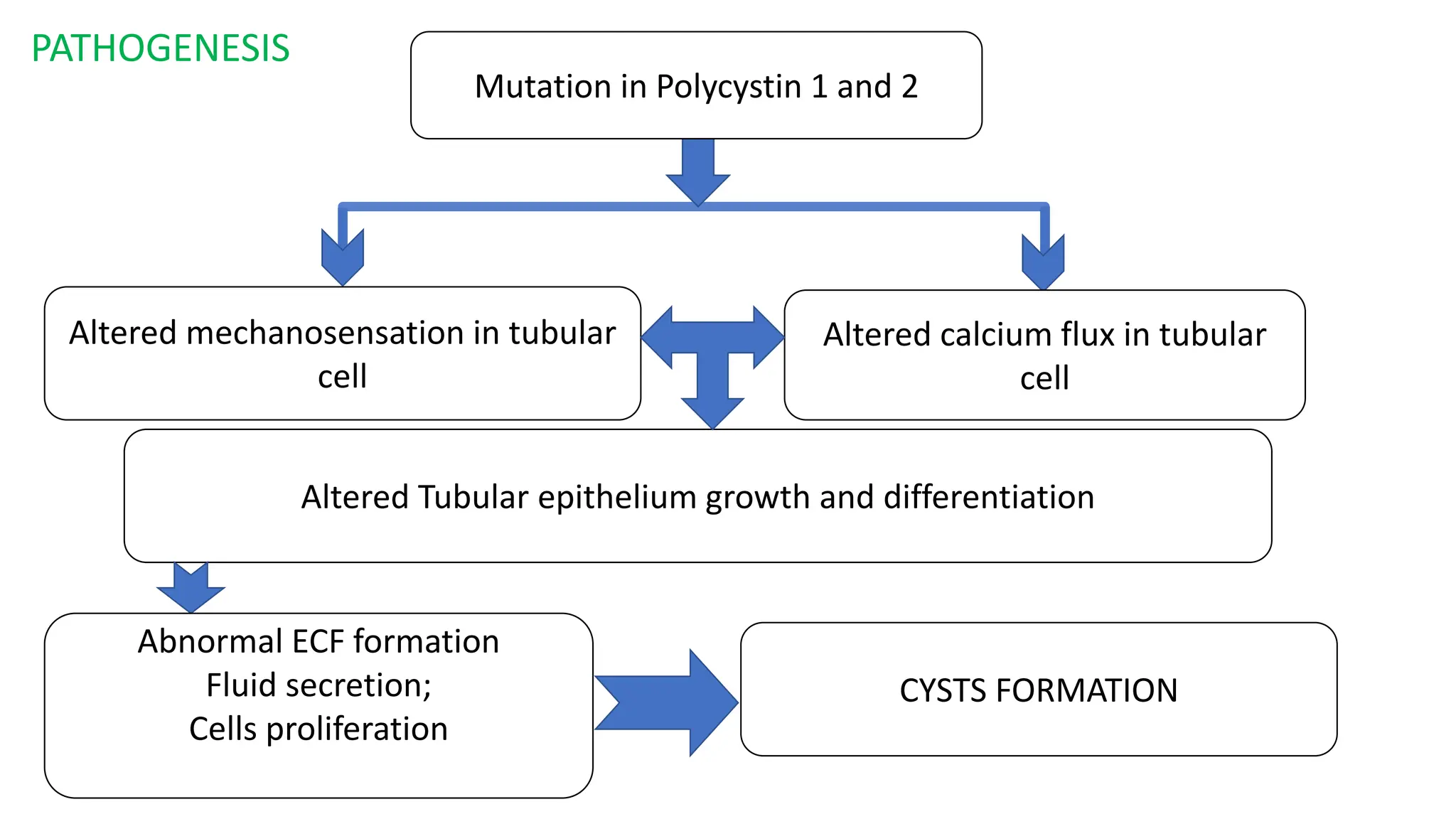
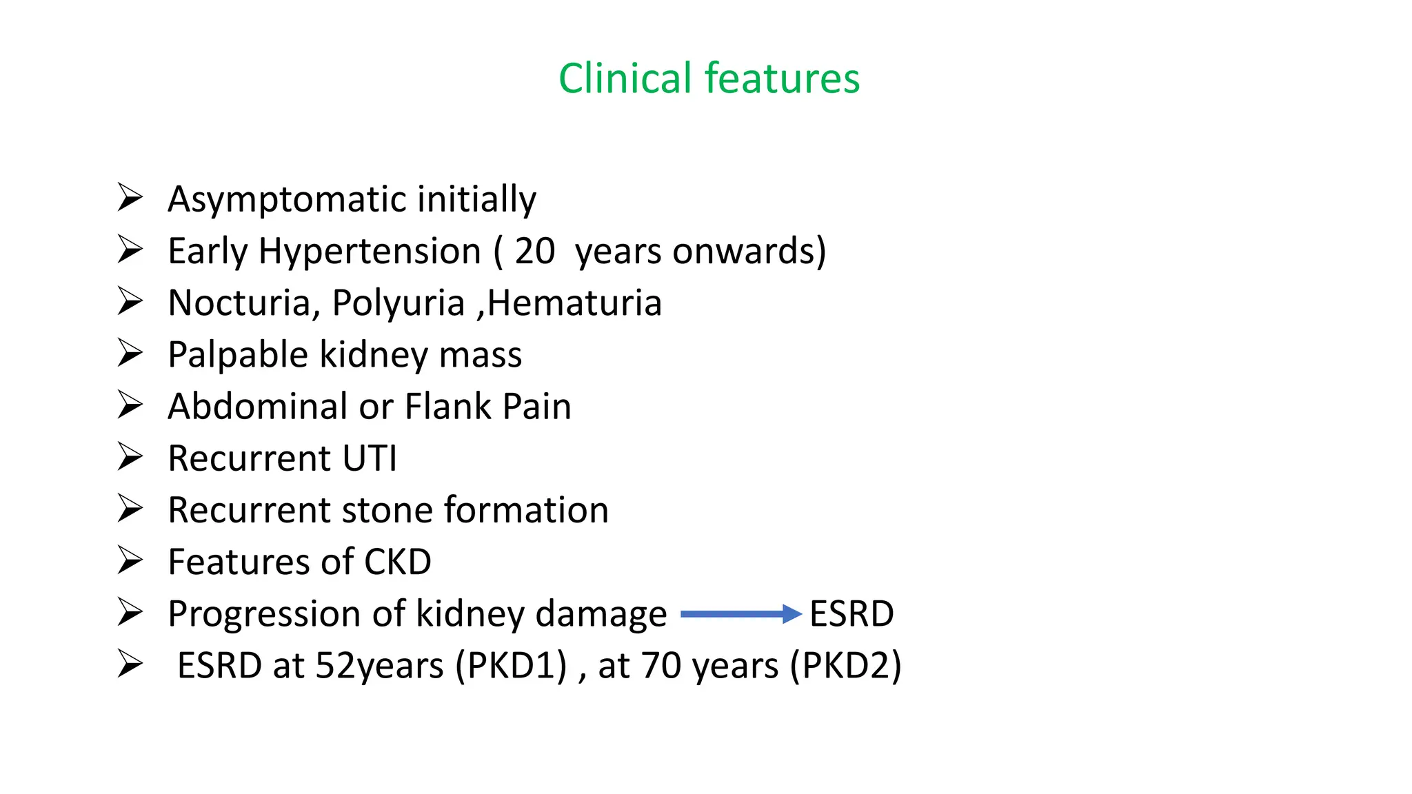
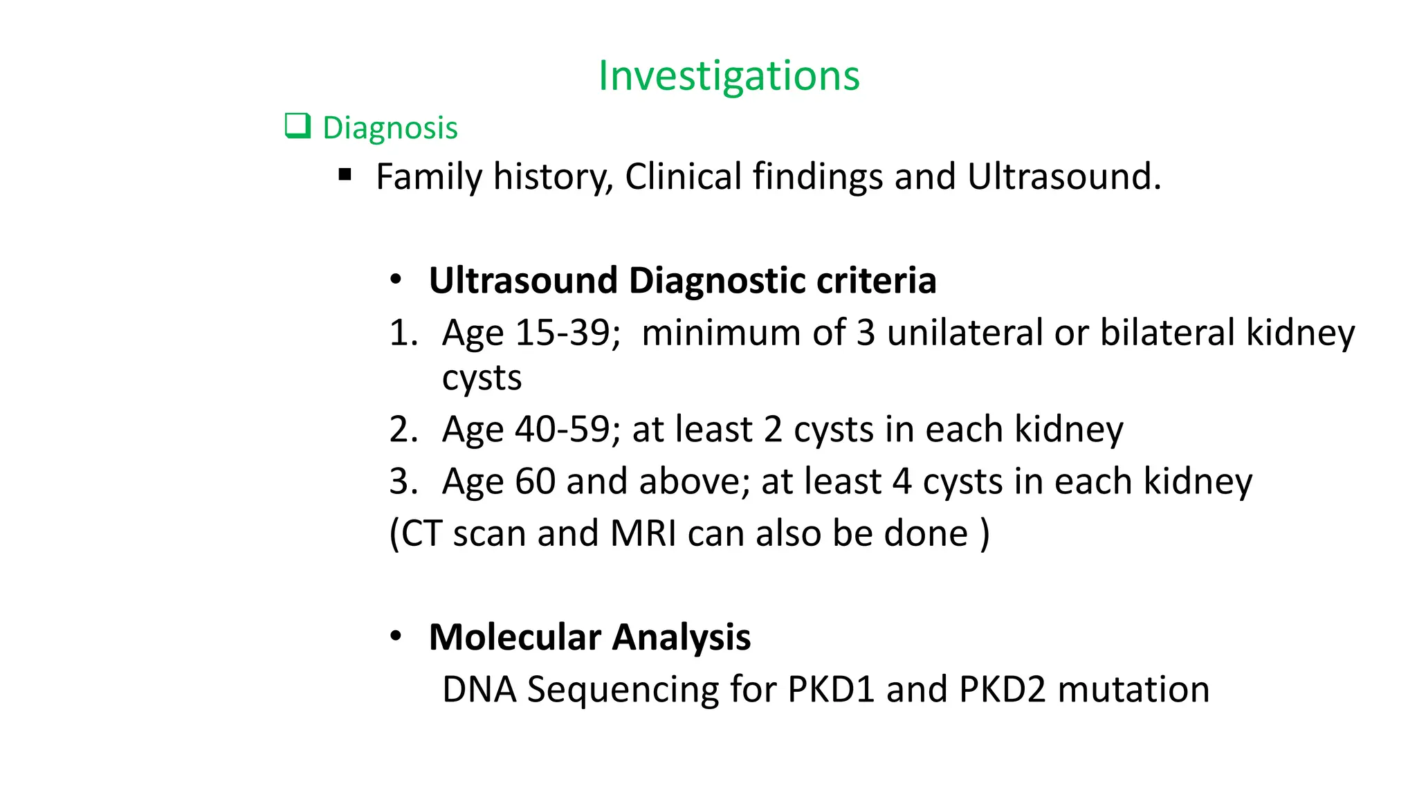
POLYCYSTIC KIDNEY DISEASES
Presenter
Dr. Dirgha P Guragain
POLYCYSTIC KIDNEY DISEASES
It is an inherited disease that involves formation of bilateral kidney
cysts.
TWO TYPES
1. AD-PKD
2. AR-PKD
AUTOSOMAL DOMINANT PKD
ADPKD is a multisystem disorder characterized by multiple ,
bilateral renal cysts associated with cysts in other organs such
as liver, pancreas and arachnoid membrane.
Prevalence 1:1000
ETIOLOGY
Genetic mutation in PKD1 and PKDK2 genes that encodes
Polycystin 1 and Polycyctin 2 proteins.
PATHOGENESIS
Altered calcium flux in tubular
cell
Altered mechanosensation in tubular
cell
Altered Tubular epithelium growth and differentiation
Mutation in Polycystin 1 and 2
Abnormal ECF formation
Fluid secretion;
Cells proliferation
CYSTS FORMATION
Pathogenesis
Multiple Renal cysts
compression of kidney parenchyma Vascular compression and ischemia
Damage of remaining normal kidney Increase in renin production
Progression of Kidney damage Increase in angiotensin
End stage renal disease Early Hypertension
Clinical features
 Asymptomatic initially
 Early Hypertension ( 20 years onwards)
 Nocturia, Polyuria ,Hematuria
 Palpable kidney mass
 Abdominal or Flank Pain
 Recurrent UTI
 Recurrent stone formation
 Features of CKD
 Progression of kidney damage ESRD
 ESRD at 52years (PKD1) , at 70 years (PKD2)
Associated Features
 Hepatic cysts ( m/c association) usually asymptomatic
 Pancreatic and Spleenic cysts
 Colonic diverticulas
 Abdominal wall hernias
 Mitral/aortic regurgitation
 Mitral Prolapse
 Berry aneurysms
Investigations
 Diagnosis
 Family history, Clinical findings and Ultrasound.
• Ultrasound Diagnostic criteria
1. Age 15-39; minimum of 3 unilateral or bilateral kidney
cysts
2. Age 40-59; at least 2 cysts in each kidney
3. Age 60 and above; at least 4 cysts in each kidney
(CT scan and MRI can also be done )
• Molecular Analysis
DNA Sequencing for PKD1 and PKD2 mutation
Investigation for disease manifestation
o Blood tests
• CBC, PBS, ESR, CRP
• RFT: Urea, Creatinine
• Electrolytes: Sodium, Potassium, Calcium,
phosphate
• Serum renin
o Urine examination
• Dipstick for hematuria, proteinuria, leukocytes
esterase’s test.
• Microscopy : cells , casts and crystals
o CT scan KUB, for stones
o Renal scan using (MAG3, DMSA)
Complementary Investigations
CT , MRI abdomen for liver , pancreatic cysts
Echocardiography for MR,AR, mitral prolapse
Barium enema and colonoscopy for diverticula
CTA ,MRA for Berry’s aneurysms
MANAGEMENT
• Blood Pressure Control
Target : <130/80mmhg
 Salt restriction
 ACE inhibitors, ARB
 CCB
• Slow progression of disease
 Vasopressin V2 receptor antagonist ( Tolvaptan)
• Treatment of renal complications
 Electrolytes balance, Fluid balance
 Correction of acid base balance
 Correction of anemia
• RRT and Renal transplantation.

Autosomal Dominant Polycystic Kidney Disease (ADPKD).pptx

  • 1.
  • 2.
    POLYCYSTIC KIDNEY DISEASES Itis an inherited disease that involves formation of bilateral kidney cysts. TWO TYPES 1. AD-PKD 2. AR-PKD
  • 3.
    AUTOSOMAL DOMINANT PKD ADPKDis a multisystem disorder characterized by multiple , bilateral renal cysts associated with cysts in other organs such as liver, pancreas and arachnoid membrane. Prevalence 1:1000 ETIOLOGY Genetic mutation in PKD1 and PKDK2 genes that encodes Polycystin 1 and Polycyctin 2 proteins.
  • 4.
    PATHOGENESIS Altered calcium fluxin tubular cell Altered mechanosensation in tubular cell Altered Tubular epithelium growth and differentiation Mutation in Polycystin 1 and 2 Abnormal ECF formation Fluid secretion; Cells proliferation CYSTS FORMATION
  • 5.
    Pathogenesis Multiple Renal cysts compressionof kidney parenchyma Vascular compression and ischemia Damage of remaining normal kidney Increase in renin production Progression of Kidney damage Increase in angiotensin End stage renal disease Early Hypertension
  • 6.
    Clinical features  Asymptomaticinitially  Early Hypertension ( 20 years onwards)  Nocturia, Polyuria ,Hematuria  Palpable kidney mass  Abdominal or Flank Pain  Recurrent UTI  Recurrent stone formation  Features of CKD  Progression of kidney damage ESRD  ESRD at 52years (PKD1) , at 70 years (PKD2)
  • 7.
    Associated Features  Hepaticcysts ( m/c association) usually asymptomatic  Pancreatic and Spleenic cysts  Colonic diverticulas  Abdominal wall hernias  Mitral/aortic regurgitation  Mitral Prolapse  Berry aneurysms
  • 8.
    Investigations  Diagnosis  Familyhistory, Clinical findings and Ultrasound. • Ultrasound Diagnostic criteria 1. Age 15-39; minimum of 3 unilateral or bilateral kidney cysts 2. Age 40-59; at least 2 cysts in each kidney 3. Age 60 and above; at least 4 cysts in each kidney (CT scan and MRI can also be done ) • Molecular Analysis DNA Sequencing for PKD1 and PKD2 mutation
  • 9.
    Investigation for diseasemanifestation o Blood tests • CBC, PBS, ESR, CRP • RFT: Urea, Creatinine • Electrolytes: Sodium, Potassium, Calcium, phosphate • Serum renin o Urine examination • Dipstick for hematuria, proteinuria, leukocytes esterase’s test. • Microscopy : cells , casts and crystals o CT scan KUB, for stones o Renal scan using (MAG3, DMSA)
  • 10.
    Complementary Investigations CT ,MRI abdomen for liver , pancreatic cysts Echocardiography for MR,AR, mitral prolapse Barium enema and colonoscopy for diverticula CTA ,MRA for Berry’s aneurysms
  • 11.
    MANAGEMENT • Blood PressureControl Target : <130/80mmhg  Salt restriction  ACE inhibitors, ARB  CCB • Slow progression of disease  Vasopressin V2 receptor antagonist ( Tolvaptan) • Treatment of renal complications  Electrolytes balance, Fluid balance  Correction of acid base balance  Correction of anemia • RRT and Renal transplantation.