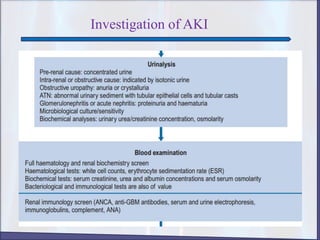
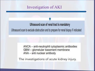
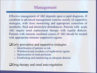
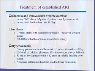
RAHAD AHAMED
M. Pharm , Jagannath University, Dhaka
ACUTE KIDNEY INJURY
Acute kidney injury
(AKI), previously
called acute renal
failure (ARF), is an
abrupt loss of kidney
function that develops
within 7 days. This
results in an
accumulation of
nitrogenous waste
products and other
toxins.
Classification and Causes of AKI
The most useful practical classification
comprises three main groups:
1. Pre-renal ( functional/ hypoperfusion)
2. Renal ( Structural/ intrinsic)
3. Post-renal (obstructive)
Classification and Causes
1. Pre-renal acute kidney injury
a) Volume depletion resulting from:
i. Hemorrhage
ii. Renal losses(diuretics)
iii. GI losses (vomiting, diarrhea)
b) Impaired cardiac efficiency resulting from:
i. MI
ii. Heart failure
iii. Dysrhythmias
iv. Cardiogenic shock
c) Vasodilation resulting from:
i. Sepsis
ii. Anaphylaxis
iii. Antihypertensive medications or other medication that
cause vasodilation
Classification and Causes
2. Renal ( Structural/ intrinsic)
a) Prolonged renal ischemia resulting from:
i. Pigment nephropathy
ii. Myoglobinuria( trauma, crush injury, burns)
iii. Hemoglobinuria( transfusion reaction, hemolytic anemia)
b) Nephrotoxic agents such as:
i. Aminoglycosides antibiotics
ii. Radiopaque contrast media
iii. Heavy metals
iv. NSAIDS, ACE inhibitors
c) Infectious processes such as:
i. Acute pylonephritis
ii. Acute GN
Classification and Causes
3. Post-renal (obstructive)
Urinary tract obstruction including:
i. Calculi( stones)
ii. Bladder tumor
iii. BPH
iv. Stricture
v. Blood clots
vi. Medications
Comparing categories of AKI
Categories Pre-renal Renal Post-renal
Etiology hypoperfusion parenchymal Obstruction
BUN value Increased Increased Increased
Creatinine increased increased increased
Urine output Decreased Varies, often
decreased
Varies, may be
decreased or
sudden anura
Urine sodium Decreased to <20
mEq/L
Increased to >
40mEq/L
Varies, decreased to
<20 mEq/L
Urinary
sedimentation
Normal, few hyaline
cast
Abnormal casts and
debris
Usually normal
Urine osmolarity Increased to 500
mOsm
About 350 mOsm
Similar to serum
Varies, increased or
equal to serum
Urine specific gravity Increased Low normal Varies
Diagrammatic Presentation
Pre-renal AKI Post-renal AKI
Clinical Manifestations
The patient may exhibit signs and symptoms of volume
depletion or overload, depending upon the precipitating
conditions, course of the disease and prior treatment.
 Acute kidney injury with volume depletion:
o Fatigue
o Loss of appetite
o Headache
o Nausea and vomiting
o Gastrointestinal hemorrhage
o Muscle cramp
o Tachycardia
o Blood pressure
o Postural hypotension
o Cold extremities
o Reduced skin turgor
Clinical Manifestations
Acute kidney injury with volume overload:
o Weight increased
o Orthopnoea/nocturnal dyspnoea
o Ankle swelling
o Oedema
o Jugular venous pressure distension
o Pulmonary crackles
Diagnosis
 History collection
 Physical examination
o Asterixis and myoclonus
o Peripheral edema( if volume overload is present)
o Pulmonary rales( if volume overload is present)
o Elevated right atrial pressure ( if volume overlaod is
present)
o Identification of precipitating cause
o Serum creatinine and BUN level
o Urine analysis
o Renal bladder ultra sound
o Renal scan
o CT scan and Mri
o The urine will be examined under a microscope
o Biopsy
Investigation of AKI
Management
Effective management of AKI depends upon a rapid diagnosis. If
condition is advanced management consists mainly of supportive
strategies, with close monitoring and appropriate correction of
metabolic, fluid and electrolyte disturbances. Patients with acute
AKI require renal replacement therapy with regular dialysis.
Patients with immune mediated causes of AKI should be treated
with appropriate immune suppressive agents.
Early preventive and supportive strategies
o Identification of patients at risk
o Withdrawal and avoidance of nephrotoxic agents
o Optimization of renal perfusion
o Establishing and maintaining an adequate diuresis
Drug therapy and renal auto-regulation
Treatment of established AKI
Uraemia and intravascular volume overload
o Intake NaCl about 1-2g/day if patient is not hyponatraemic.
o Intake total fluid to less than 1L/day
Acidosis
o Treated orally with sodium bicarbonate 1-6g/day in divided
doses
o 50-100mmol of bicarbonate ions intravenously.
Hyperkalaemia
o Dietary potassium should be restricted to less than 40mmol/day
o 10-30mL of calcium gluconate 10% intravenously over 5-10 min
o 50 mL of 50% glucose with 8-12 units of soluble insuline over
10min
o Nebulised salbutamol has been used to lower potassium.
Treatment of established AKI
Hypocalcaemia
o Oral calcium supplementation with calcium carbonate is usually
adequate.
o Vit. D may be used to treat the hypocalcaemia.
Hypophosphatemia
o Phosphate binding agents may be used to retain phosphate ions
in the gut.
o Most commonly used agents are calcium carbonate or acetate
and are given with food.
Infection
o Broad spectrum antibiotics are used.
Treatment of established AKI
Uraemic gastro-intestinal erosions
o Proton pump inhibitors can be efficient.
o H2 antagonists are an appropriate alternative.
Nutrition
o Electrolyte free amino acid solutions is usually supplied as 12-30
g/day.
o Mg and Zn supplementation may be required.
o Parenteral glucose may be required in AKI patients.
Aki updated

Aki updated

  • 1.
    RAHAD AHAMED M. Pharm, Jagannath University, Dhaka
  • 2.
    ACUTE KIDNEY INJURY Acutekidney injury (AKI), previously called acute renal failure (ARF), is an abrupt loss of kidney function that develops within 7 days. This results in an accumulation of nitrogenous waste products and other toxins.
  • 3.
    Classification and Causesof AKI The most useful practical classification comprises three main groups: 1. Pre-renal ( functional/ hypoperfusion) 2. Renal ( Structural/ intrinsic) 3. Post-renal (obstructive)
  • 4.
    Classification and Causes 1.Pre-renal acute kidney injury a) Volume depletion resulting from: i. Hemorrhage ii. Renal losses(diuretics) iii. GI losses (vomiting, diarrhea) b) Impaired cardiac efficiency resulting from: i. MI ii. Heart failure iii. Dysrhythmias iv. Cardiogenic shock c) Vasodilation resulting from: i. Sepsis ii. Anaphylaxis iii. Antihypertensive medications or other medication that cause vasodilation
  • 5.
    Classification and Causes 2.Renal ( Structural/ intrinsic) a) Prolonged renal ischemia resulting from: i. Pigment nephropathy ii. Myoglobinuria( trauma, crush injury, burns) iii. Hemoglobinuria( transfusion reaction, hemolytic anemia) b) Nephrotoxic agents such as: i. Aminoglycosides antibiotics ii. Radiopaque contrast media iii. Heavy metals iv. NSAIDS, ACE inhibitors c) Infectious processes such as: i. Acute pylonephritis ii. Acute GN
  • 6.
    Classification and Causes 3.Post-renal (obstructive) Urinary tract obstruction including: i. Calculi( stones) ii. Bladder tumor iii. BPH iv. Stricture v. Blood clots vi. Medications
  • 7.
    Comparing categories ofAKI Categories Pre-renal Renal Post-renal Etiology hypoperfusion parenchymal Obstruction BUN value Increased Increased Increased Creatinine increased increased increased Urine output Decreased Varies, often decreased Varies, may be decreased or sudden anura Urine sodium Decreased to <20 mEq/L Increased to > 40mEq/L Varies, decreased to <20 mEq/L Urinary sedimentation Normal, few hyaline cast Abnormal casts and debris Usually normal Urine osmolarity Increased to 500 mOsm About 350 mOsm Similar to serum Varies, increased or equal to serum Urine specific gravity Increased Low normal Varies
  • 8.
  • 9.
    Clinical Manifestations The patientmay exhibit signs and symptoms of volume depletion or overload, depending upon the precipitating conditions, course of the disease and prior treatment.  Acute kidney injury with volume depletion: o Fatigue o Loss of appetite o Headache o Nausea and vomiting o Gastrointestinal hemorrhage o Muscle cramp o Tachycardia o Blood pressure o Postural hypotension o Cold extremities o Reduced skin turgor
  • 10.
    Clinical Manifestations Acute kidneyinjury with volume overload: o Weight increased o Orthopnoea/nocturnal dyspnoea o Ankle swelling o Oedema o Jugular venous pressure distension o Pulmonary crackles
  • 11.
    Diagnosis  History collection Physical examination o Asterixis and myoclonus o Peripheral edema( if volume overload is present) o Pulmonary rales( if volume overload is present) o Elevated right atrial pressure ( if volume overlaod is present) o Identification of precipitating cause o Serum creatinine and BUN level o Urine analysis o Renal bladder ultra sound o Renal scan o CT scan and Mri o The urine will be examined under a microscope o Biopsy
  • 12.
  • 13.
  • 14.
  • 15.
    Management Effective management ofAKI depends upon a rapid diagnosis. If condition is advanced management consists mainly of supportive strategies, with close monitoring and appropriate correction of metabolic, fluid and electrolyte disturbances. Patients with acute AKI require renal replacement therapy with regular dialysis. Patients with immune mediated causes of AKI should be treated with appropriate immune suppressive agents. Early preventive and supportive strategies o Identification of patients at risk o Withdrawal and avoidance of nephrotoxic agents o Optimization of renal perfusion o Establishing and maintaining an adequate diuresis Drug therapy and renal auto-regulation
  • 16.
    Treatment of establishedAKI Uraemia and intravascular volume overload o Intake NaCl about 1-2g/day if patient is not hyponatraemic. o Intake total fluid to less than 1L/day Acidosis o Treated orally with sodium bicarbonate 1-6g/day in divided doses o 50-100mmol of bicarbonate ions intravenously. Hyperkalaemia o Dietary potassium should be restricted to less than 40mmol/day o 10-30mL of calcium gluconate 10% intravenously over 5-10 min o 50 mL of 50% glucose with 8-12 units of soluble insuline over 10min o Nebulised salbutamol has been used to lower potassium.
  • 17.
    Treatment of establishedAKI Hypocalcaemia o Oral calcium supplementation with calcium carbonate is usually adequate. o Vit. D may be used to treat the hypocalcaemia. Hypophosphatemia o Phosphate binding agents may be used to retain phosphate ions in the gut. o Most commonly used agents are calcium carbonate or acetate and are given with food. Infection o Broad spectrum antibiotics are used.
  • 18.
    Treatment of establishedAKI Uraemic gastro-intestinal erosions o Proton pump inhibitors can be efficient. o H2 antagonists are an appropriate alternative. Nutrition o Electrolyte free amino acid solutions is usually supplied as 12-30 g/day. o Mg and Zn supplementation may be required. o Parenteral glucose may be required in AKI patients.