
Recommended
More Related Content
What's hot
What's hot (20)
Similar to Unit 11 nervous system
Similar to Unit 11 nervous system (20)
Recently uploaded
Recently uploaded (20)
Unit 11 nervous system
- 1. UNIT – 11 NERVOUS SYSTEM CHANDAN KUMARI SHAH M.SC NURSING 1ST YEAR
- 2. UNIT 11 INTRODUCTION TO NERVOUS SYSTEM 11.1 Introduction:- Nerve, neuroglia, synapse nerve transmission 11.2 Types of nerves 11.3 Mechanism of stimuli transmission 11.4 The central nervous system:- Covering membrane(meninges), Cerebrum, Mid brain, Cerebellum, Brain stem, Ventricles, Spinal cord 11.5 The peripheral nervous system:- Spinal nerve, cranial nerve, Autonomic nervous system 11.6 Neurones Mechanism of stimuli transmitted in the nervous system- Reflex action CNS- Brain and spinal cord PNS- Spinal cord and cranial nerves ANS-Sympathetic and parasympathetic system
- 3. INTRODUCTION The nervous system is the system of communication between the various parts of the body. It is the mechanism by which sensation of all kinds are received from the environment, from the tissues and ongans of the body itself. It is the system by which action are also carried out by the sending of impulse to other parts of the nervous system and organs of the body. Humans, like all living organisms, can respond to their environment. Humans have two complimentary control systems to do this: the nervous system and the endocrine (hormonal) system. The human nervous system controls everything from breathing and producing digestive enzymes, to memory and intelligence.
- 4. THIS NERVOUS SYSTEM CAN BE DIVIDED INTO TWO MAIN PARTS: 1. The central nervous system (CNS) that consists of : a) The brains b) Spinal cord 2). Three peripheral nervous system (PNS) that consists of : a) Spinal nerve – 31 pairs b) Cranial nerves – 12 pairs c) Autonomic nervous system (ANS) i) Sympathetic and ii) Parasympathetic nerves
- 7. FUNCTIONS OF THE NERVOUS SYSTEM Communication ♦ Monitors impressions and information from external stimuli ♦ Monitors information from internal stimuli ♦ Responds to danger; pain, and other situations ♦ Responds to internal and external changes ♦ Helps to maintain homeostasis ♦ Responds to conscious decisions and thoughts ♦ Coordinates processing of new learning ♦ Stores and retrieves memories, including previous learning ♦ Facilitates judgment, reasoning, and decision making Control ♦ Directs all body activities ♦ Maintains blood pressure, respiration, and other vital functions ♦ Regulates body systems (in coordination with endocrine system) ♦ Coordinates reflex actions ♦ Controls instinctual behaviors ♦ Controls conscious movement and activities ♦ Stores unconscious thoughts The basic functions of the nervous system are to receive sensory input (stimuli), to integrate and interpret stimuli, and to respond to the stimuli
- 8. FUNCTION Registration of information Regulation of body function. Transportation of sensations. Storage of information Formation of thoughts through learning and gaining experience.
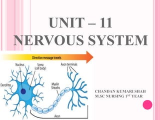
- 9. THE NERVE CELL (NEURONS) The nervous system composed of nerve cells, or neurons. which are structural and functional unit of nervous system. Neurons are supported by a special kinds of connective tissue called neuroglia. The basic properties of neuron are irritability or excitability and conductivity.
- 10. The neurone can be divided into three parts: I. A cell body (Soma) II. An axon III. Dendrites
- 11. Cell body:- cell body forms the grey matter of nervous system and found at the periphery of the brain & in the central of spinal cord. Group of cell bodies are called nuclei in CNS and ganglia in PNS. The cell body contains protoplasm and centrally placed nucleus. The protoplasm of cell has large granules called Nissil granules or bodies.
- 12. Axone:- Each nerve cell has only one axone , which begins at a tapered area of the cell body, the axon hillock. It carries impulses away from the cell body . The membrane of axon is called axalemma. The axon may be myelinated or non myelinated usually large axon. The axon has a uniform diameter. In additional to this difference in structure , There is a fundamental functional difference between dendrites and axon. The axon is sometimes about 100 cm long. Function of myelin sheath are:- To prevent axon from pressure & injury. To act as electrical insulator so that flow of impulses goes unrestricted along the nerves.
- 14. Dendrites:- Dendrites are the many short processes that receive and carry incoming impulses towards call bodies . In motor neurons dendrites from part of synapes and in sensory neurones they form the sensory receptors that respond to specific stimuli.
- 15. There are several differences between axons and dendrites: Axons Dendrites Take information away from the cell body Bring information to the cell body Smooth Surface Rough Surface (dendritic spines) Generally only 1 axon per cell Usually many dendrites per cell No ribosomes Have ribosomes Can have myelin No myelin insulation Branch further from the cell body Branch near the cell body
- 16. Neurons are similar to other cells in the body because: Neurons are surrounded by a cell membrane. Neurons have a nucleus that contains genes. Neurons contain cytoplasm, mitochondria and other organelles. Neurons carry out basic cellular processes such as protein synthesis and energy production. Neurons differ from other cells in the body because: Neurons have specialised extensions called dendrites and axons. Dendrites bring information to the cell body and axons take information away from the cell body. Neurons communicate with each other through an electrochemical process. Neurons contain some specialized structures (for example, synapses) and chemicals (for example, neurotransmitters).
- 17. HUMANS HAVE THREE TYPES OF NEURONES Sensory neurons have long axons and transmit nerve impulses from sensory receptors all over the body to the central nervous system. Motor neurons also have long axons and transmit nerve impulses from the central nervous system to effectors (muscles and glands) all over the body. Inter neurons (also called connector neurons or relay neurons) are usually much smaller cells, with many interconnections.
- 18. MOTOR NEURON: Efferent Neuron – Moving toward a central organ or point Relays messages from the brain or spinal cord to the muscles and organs.
- 19. SENSORY NEURON: Afferent Neuron – Moving away from a central organ or point Relays messages from receptors to the brain or spinal cord.
- 20. INTERNEURON (RELAY NEURON): Relays message from sensory neuron to motor neuron Make up the brain and spinal cord
- 21. Sensory neuron Interneuron Motor Neuron Length of Fibers Long dendrites and short axon Short dendrites and short or long anxon Short dendrites and long axons Location Cell body and dendrite are outside of the spinal cord; the cell body is located in a dorsal root ganglion Entirely within the spinal cord or CNS Dendrites and the cell body are located in the spinal cord; the axon is outside of the spinal cord Function Conduct impulse to the spinal cord Interconnect the sensory neuron with appropriate motor neuron Conduct impulse to an effector (muscle or gland)
- 23. THE REFLEX ARC The three types of neurons are arranged in circuits and networks, the simplest of which is the reflex arc. In a simple reflex arc, such as the knee jerk, a stimulus is detected by a receptor cell, which synapses with a sensory neuron. The sensory neuron carries the impulse from site of the stimulus to the central nervous system (the brain or spinal cord), where it synapses with an inter neuron. The inter neuron synapses with a motor neuron, which carries the nerve impulse out to an effectors, such as a muscle, which responds by contracting.
- 25. THE NERVES IMPULSES (ACTION POTENTIAL) Action potentials are rapidly developing electrochemical changes occurring in cell membranes of excitable cells. The transmission of impulses , is due to movement of ions across the nerves cell membrane. In resting state ,the nerve cell membrane is polarized due to differences in the concentration of ions across the plasma membrane which is called resting membrane potential(The RMP of a neurone is about -70mV(mV=millivolt) this means that the inside of the neurone is -70mV less than the outside. At rest, there are relatively more sodium ions outside the neurone and more potassium ions inside the neurone ) . Or, The potential difference between the two sides of the membrane of a nerve cell when the cell is not conducting impulses. At rest the charge on outside is positive & inside is negative . The ions involved are:- Sodium (Na+), Main extracellular cation Potassium(K+) ,Main intracellular cation.
- 26. In resting state there is continual tendency for these ion to diffuse along their concentration gradients ie K+ outsides & Na+ into cells through the ion leaky channels. Now, when stimulated ,the permeability of the nerve cell membrane to these ions changes . It produces an initial opening of voltage- gated sodium channel causing Na+ to flood inside the cell from extracellular fluid leading depolarization, creating a nerve impulses or action potential followed by opening of voltage-gated potassium channel leading to repolarization . Almost immediately following the entry of sodium, k+ floods out of the neuron & the movement of these ions returns the membrane potential to its resting state. This is called refractory period during which restimulation is not possible. As the neuron returns to its original resting state, the action of sodium – potassium pump (sodium – potassium ATPs ases) expel Na+ from the cell in exchange for K+.
- 27. In myelinated neurons ,the insulating properties of the myelin sheath prevent the movement of ions. Therefore electrical charges across the membrane occurs only at the nodes of Ranvier. When an impulses occure at one node, depolarization passes along the myelin sheath to the next node so that the flow of current appears to leap from one node to the next. This is called saltatory conduction(Quick propropagation of the action potential along a myelinated axon owing to voltage gated Na+ channela being present only at the node of ranvier ). The speed of conduction depends on the diameter of neuron; larger the diameter , faster the conduction. So , mylinated fibers conduct impulses faster than unmyelinated fibers because saltatory conduction is faster than simple propagation.
- 30. NEUROGLLA Neuroglia is the nervous system cell. They surround the neuron. These cells consists of specialized connective tissues. They perform a variety of supportive functions to impulses conduction . Neuroglia produces the myelin that insulates(protect from heat, cold or noice by surrounding with insulating material) and functionally isolates neurons. Varieties of neuroglia are:- a. Ependymal cells:- are the epithelial cells that line the neural canal and brain ventricles. b. Astrocytes:- Are present around the blood vessels in the brain parenchyma. c. Oligodendroglia:- Produce myelin in the CNS. d. Microglia :- is part of the mononuclear phagocyte to system in brain. e. Satellite cells:- Are present in PNS ganglia.
- 32. SYNAPES Synapse is the functional region between two neurons, where information from one neuron is transmitted or relayed (hand over) to another neuron ,but there is no protoplasmic connection between the two neurons. So there is space (gap) between the two neurons called synaptic cleft. A synapse = presynaptic knob + synaptic cleft + post synaptic membrane. Neural signals propagate along an axon in the form of electrochemical waves called action potentials, which produce cell-to-cell signals at points where axon terminals make synaptic contact with other cells.
- 34. Synapses may be electrical or chemical. Electrical synapses make direct electrical connections between neurons(impulses as heart muscles), but chemical synapses are much more common, and much more diverse in function.At a chemical synapse, the cell that sends signals is called presynaptic, and the cell that receives signals is called postsynaptic. Both the presynaptic and postsynaptic areas are full of molecular machinery that carries out the signalling process. The presynaptic area contains large numbers of tiny spherical vessels called synaptic vesicles, packed with neurotransmitter chemicals. When the presynaptic terminal is electrically stimulated, an carray of molecules embedded in the membrane are activated, and cause the contents of the vesicles to be released into the narrow space between the presynaptic and postsynaptic membranes, called the synaptic cleft.
- 35. The neurotransmitter then binds to receptors embedded in the postsynaptic membrane, causing them to enter an activated state. Depending on the type of receptor, the resulting effect on the postsynaptic cell may be excitatory, inhibitory, or modulatory in more complex ways. For example, release of the neurotransmitter acetylcholine at a synaptic contact between a motor neuron and a muscle cell induces rapid contraction of the muscle cell. The entire synaptic transmission process takes only a fraction of a millisecond, although the effects on the postsynaptic cell may last much longer
- 38. TYPES OF SYNAPSE There are three types of synapses:- a. Axo – dendritic:- Axone is terminating on a dendrone of another neuron b. Axo- somatic :- Axone is terminating on the nerve cell body (Soma) of another neuron c. Axo axonic , where an axone is terminating on another axon.
- 40. Neurotransmitter Acetylcholine (ACH) Motor Movement and. Dopamine Motor movement and Alertness. Endorphins Pain Control Serotonin Mood Control
- 41. NERVES TERMINATION Afferent (Sensory) termination:- Input of nerve impulses into the central nervous system results from stimulation of a wide variety of sensory receptors which may be special sense receptors, cutaneous receptor, proprioceptors and enteroceptors. For example, in the skin , the sensory nerve lose their myelin sheath and neurilemma(a memberanous sheath around a nerve fiber) and divide into small branches known as sensory receptors. The cutaneous receptors are stimulated by touch, pain , temperature. Then these impulses are transmitted to the brain where sensation is perceived.
- 42. Efferent (motor)termination:- Efferent (motor)nerve carry out the impulses from the brain to the various part of the body. as the axon supplying a skeletal muscle fiber approaches its termination , it loses its myelin sheath and divided into a number of terminal buttons or end feet . The end feet fit into depression in the motor end plate, the thickened portion of the muscle membrane of the junction. There is a tiny space between the nerve ending and the thickened muscle membrane is comparable to the synaptic cleft at synapses. The whole structure is known as the neuromuscular junction. Only nerve fiber end on each end plate.
- 43. THE CENTRAL NERVOUS SYSTEM The central nervous system consists of brain and spinal cord.
- 44. THE BRAIN AND ITS COVERINGS The brain lies in the cranial cavity . An average weight is about 1.5 kg. The brain consists of the following important parts:- Covering membranes Cerebrum Brain stem Cerebellum Ventricles
- 45. A) COVERING MEMBRANES The brain and spinal cord are very important but delicated organs. They are protected by the following coverings. i. The bony covering of the cranium and vertebras ii. Three membranous covering called meninges. The meninges:- Brain and spinal cord are completely surrounded by a membranous sheet called meninges. It has three layers:- i. Dura mater ii. Archnoid mater iii. Pia mater
- 47. Dura mater:- It consists of 2 layer of dense fibrous tissue. The outer layer takes place of peristeum which lies closest to skull bone and the inner layer (meningeal layer), which lies closest to the brain. The space between the skull & the dura mater called epidural space & the space between dura & archnoid mater is called subdural space. The dura mater has following folds:- Flax cerebri:- Tentorium cerebelli Flax cerebelli Diaphragma sellae:-
- 50. Flax cerebri:- Larger sickle shaped that separates the cerebral hemispheres(either half of the cerebrum). The superior sagittal sinus is formed by flax cerebri. Tentorium cerebelli:- the second largest; crescent shaped(moon in shaped) that separates the occipital lobes from cerebellum. It forms the straight & transverse sinuses. Flax cerebelli:- Vertical infolding that lies inferior to the tentorium cerebelli separating the cerebellum. It forms the straight & tranverse sinuses. Diaphragma sellae:- Smallest infolding covering the pituitary gland & sella turcica.
- 51. Arachnoid mater:- Is the middle layer of the meninges. The archnoid mater is a thin transparent membrane. The arachnoid mater also extends down like dura mater and ends at second sacral vertebra, The space between archnoid & pia matter is called sub archoid space which contain the cerebrospinal fluid(CSF) Pia mater:- the pia mater is the innermost layer of meninges.it is very thin , but it is thicker than the arachnoid mater. It is a very delicated layer(highly vascularized) of connective tissue containing many minute blood vessels and it adheres to the surface of the brain , spinal cord. It dips into all sulci and fissues. The spinal pia mater extends from the foramen magnum(large oval opening in occipital bone) to the lower border of the first lumber vertebrae and continues below as filum terminale.
- 54. VENTRICLES OF THE BRAIN AND CEREBROSPINAL FLUID The brain contain 4 irregular – shaped cavity,: ventricles, where CSF is produced within each ventricles is a region of choroid plexus(a vascular plexus of the cerebral ventricles that regulate intraventricular pressure), a network of ependymal cells(Thin epithelial membrane lining the ventricles of the brain and spinal cord canal) involved in production of CSF . The ventricles are lined up with ependyma a specialized form of ventricles. Right and left lateral ventricles Third ventricles Fourth ventricle
- 57. Right and left lateral ventricles:- These ventricles lie within the cerebral hemisphere one on each side just below the corpus callosum(is a wide, flat bundle of neural fibers about 10 cm long beneath the cortex). They communicate with 3rd ventricle by a inter ventricular foraming( foramina of Monro , are channels that connect the paired lateral ventricles with the third ventricle at the midline of the brain.). Third ventricles:- it is situated in the dienecephalon below lateral ventricles between the two parts of thalamus. It communicates with fourth ventricle by a canal the cerebral aqueduct. Fourth ventricle:- it is a diamond shaped cavity situated below & behind the 3rd ventricle between cerebellum & pons . It is continuous below the central canal of spinal cord & communicates with subarchnoid space via cisterna magna.
- 60. CEREBROSPINAL FLUIDS(CSF) CSF a clear , colourless & slightly alkaline fluids produced by choroid plexus in the ventricles of the brain. CSF is secreted continuously at a rate of 0.5 ml/min. The volume remains fairly constant at about 150ml as reabsorption takes place through arachnoid villi , which project into venous sinuses . The CSF constites:- Water Mineral salt Glucose Plasma protein: small amount of albumin & globulin Urea , creatinine- in small amount A few leukocytes
- 61. CHARACTERISTICS:- Color: crystal clear Cell count:-0-4 cell/mm3 Glucose; 2/3 of the blood glucose level(40- 60 mg/dl) Protein:- 20- 45mg/dl Specific gravity:- 1.003-1.008 pH ;-7.31- 7.34 Pressure:-10 - 18 cm of H2O (8- 15 mmHg in lying down position) : 20 - 30 cm of H2O (16 – 24 mmHg in sitting position) 60 – 150 mm of CSF in supine and 200 – 250 mm of CSF in sitting position. And newborn CSF pressure ranges from 8 to 10 cmH2O (4.4–7.3 mmHg) Circulating volume:- 120 – 150 ml of circulating volume it is formed at the rate of about 20 ml per hour or 1500ml/day. Rate of absorption:- As rapidly as it is produced leaving 120 to 150 ml of circulating volume, CSF turnover rate :-3.7 times/day 1 cmH2O = 0.76 mm Hg, 1 mm Hg = 1.36 cmH2O
- 62. FUNCTION To cushion the soft tissues of the brain and spinal cord. To act as the medium for exchange of nutrients and waste products in between the blood, brain and the spinal cord. Maintain intra cranial pressure around the brain & spinal cord & act as a cushion or shock absorber. It keeps the brain & spinal cord moist & there may be exchange of nutrient & waste products between CSF & nerve cells.
- 63. BRAIN The brain is an organ that serves as the center of the nervous system in all vertebrate and most invertebrate animals. The brain is located in the head, usually close to the sensory organs for senses such as vision. The brain is the most complex organ in a vertebrate's body. In a human, the cerebral cortex contains approximately 15–33 billion neurons, ach connected by synapses to several thousand other neurons. These neurons communicate with one another by means of long protoplasmic fibers called axons, which carry trains of signal pulses called action potentials to distant parts of the brain or body targeting specific recipient cells.
- 64. The brain consists of a. Cerebrum :- Cerebral cortex, Corpus callosus b. The diencephalon:- Thalamus, pineal body, hypothalamus c. The brain stem:- Mid brain, pons, Medulla oblongata d. Cerebellum The brain receives about 15% of the cardiac output per minute. The circle of willis or circulus arteriosus & its contributing arteries plays a vital role in maintaining supply of oxygen & glucose to the brain.
- 68. A) CEREBRUM It is the largest part of the brain & is divided by a deep cleft , the longitudinal cerebral fissure , into right & left cerebral hemispheres each containing one of the lateral ventricle . Deep within the brain ,the hemispheres are connected by a mass of white matter called the corpus callosum. The peripheral part of the cerebrum is composed of grey matter ,forming the cerebral cortex & the deeper layer consists of white matter . Cerebral cortex shows many infoldings. The exposed area of fold are the gyri & these are separated by sulci (fissures). It greatly increase the surface area of cerebrum . Each hemispheres of cerebrum is divided into frontal , parietal , temporal & occipital lobes.
- 71. The frontal :- These are the largest of all the lobe, and they form the anterior part of the cerebral hemisphares. They are located anterior to central sulci and superior to the lateral sulci. Parietal :- These lobes are related to the internal aspects of posterior and superior part of the parietal bones. Each lobes is bounded anteriorly by the central sulcus, posteriorly by the superior part of the parieto- occipital sulcus and inferiorly by the imaginary line extending from the posterior ramus of the lateral sulcus. Temporal ;- These lobes lie inferior to the lateral sulci. Occipital lobes:- These lobe are relatively small and are located posterior to the parieto occipital sulci although, these lobes are very important because they contain visual area.
- 72. THE FUNCTIONAL AREA OF CEREBRAL CORTEX i. The motor area ii. The premotor area iii. The motor speech area iv. The sensory area v. Visual area vi. The auditory area(of hearing) vii. The taste area
- 74. FUNCTION OF CEREBRAL CORTEX Mental activities involved in memory , intelligence, sense of responsibility, thinking , reasoning , moral sense & learning. Sensory perception , including the perception of pain , temperature, touch, slight , hearing, taste & smell. Initiation & control of skeletal muscle contraction & therefore voluntary movement. The motor impulses arrive in the frontal lobe immediately anterior to central sulcus & sensory impulses arrive at the posterior part behind the the central sulcus.
- 75. B) DIENCEPHALON Diencephalon connects the cerebrum and midbrain. It consists of mainly thalamus , hypothalamus & pineal body. Thalamus:- Thalamus is situated within cerebral hemispheres just below corpus collosum. The main function of thalamus is to relay motor & sensory signals to the cerebral cortex . It is also involved in processing of some emotional & complex reflexes & also regulates sleep, alertness & wakefulness. Hypothalamus:- It is a small but important structure situated below & in front of thalamus , immediately above pituitary gland. Hypothalamus control the output of hormones from both lobe of pituitary gland. The other functions of hypothalamus are:-
- 76. Control of autonomic nervous system. Control of appetite & satiety. Control of thirst & water balance. Control of body temperature. Control of emotional reactions – pleasure , fear. Control of sexual behaviour & child rearing. Sleeping & waking cycles.
- 77. C) BRAIN STEM It consists of mid brain, pons &medulla oblongata. Mid brain:- it is situated between the cerebrum & pons below . It consists of nuclei 7 nerve fibers(tracts), which connect the cerebrum with lower parts of the brain & the spinal cord. The nuclei act as relay stations for ascending & descending nerve fibers. Pons:- It is situated in front of cerebellum, below midbrain & above medulla oblongata. There are nuclei within the pons that act as relay station & some of these are associated with cranial nerves. Others form the pneumotaxic & apnoustic centre in the medulla oblongata. Medulla oblongata:- It extends from the pons above & is continuous with the spinal cord below . It lies just within the cranium above the foramen magnum. The outer aspect is composed of white mater & grey matter is located centrally . Some cells constitute relay stations for sensory nerve passing from spinal cord to cerebrum.
- 79. The vital center located are:- Cardiovascular center Respiratory center Reflex center of vomiting , coughing, sneezing & swallowing.
- 80. D) CEREBELLUM Cerebellum is situated behind the pons & immediately below posterior portion of cerebrum. It has 2 hemispheres seprated by a narrow median strip called the vermis . Grey matter forms the surface & white matter lies deep inside the cerebellum. The cerebellum is concerned with the co – ordination of voluntary muscular movement ,posture & balance , The sensory input for these function is derived from muscle & joints, eye & ears. Damage to cerebellum resulting in clumsy uncoordinated muscle movement, staggering gait & inability to carry out smooth , steady, precise movement.
- 81. SPINAL CORD The spinal cord consists of nerves that carry incoming and outgoing messages between the brain and the rest of the body. The spinal cord is elongated, almost cylindrical part of the CNS which is suspended in vertebral canal surrounded by the meanings & CSF . Spinal cord is continues above with the medulla oblongata & extends from the upper border of atlas to the lower border of 1st lumber vertebra. Spinal cord is composed of grey matter in the centre and white matter in the peripheral . Spinal cord links the brain with rest of the body. Nerves conveying impulses from brain to organ descends through spinal cord & leave the cord at appropriate level. Similarly , sensory nerve from organs & tissue enter & pass upwards in spinal cord to the brain.
- 82. Sensory nerve tracts in the spinal cord:- Nerves that transmit impulses towards the brain are called sensory . The main source of sensation are:- skin (Cutaneous receptors), tendons , muscles & joints(Proprioceptors) Motor nerve tract in the spinal cord:-Neurons that transmit nerve impulses away from the brain are motor neurons. Motor neurons stimulation results in :- contraction of skeletal muscle(Voluntary) - Contraction of smooth muscles (involuntary) like cardiac muscle , secretion by glands controlled by nerves of ANS
- 83. THE PERIPHERAL NERVOUS SYSTEM The peripheral nervous system consists of : 31 pairs of spinal nerves 12 pairs of cranial nerves The autonomic nervous system Sympathetic and parasympathetic
- 84. THE SPINAL NERVES A spinal nerve is a mixed nerve, which carries motor, sensory, and autonomic signals between the spinal cord and the body. There are 31 pairs of spinal nerves that leave the vertebral canal by passing through the intervertebral formina formed by adjacent vertebra. Spinal nerves are:- 8 cervical 12 thoracic 5 lumber 5 sacral 1 coccygeal
- 85. The lumber, sacral, coccygeal nerves leaves the spinal cord near its termination at the level of 1st lumber vertebra & extend downwards inside the vertebral canal in the subarchnoid space , forming a sheaf(bundle) of nerves which resembles a horses tail , the cauda equina. The nerves leave the vertebral canal at the appropriate lumber, sacral or coccygeal level.
- 88. PLEXUS:- In the cervical , lumber & sacral regions the anterior rami unit near their origins to form large mass of nerves or plexus where nerves fibers are regrouped & rearranged before proceding to supply particular area :- Cervical plexuses:- Serves head , neck & shoulder Brachial plexus:- Serve chest, shoulder, arms & hands Lumber plexus:- Serves the back, abdomen, groin, thigh, knee, & calves. Sacral plexus:- Serve the pelvic, buttocks, genitals, thigh, calves & feet. Coccygeal plexus:- serves a small region over the coccyx.
- 95. THORACIC NERVES:- These nerves do not form plexus. There are 12 pairs & the first 11 are intercostal nerves which pass between ribs & supply them , the intercostal muscle & skin overlying it. The 12th pair comprise of subcostal nerves. The 7th to 12th thoracic nerves also supply the mucus & skin of anterior & posterior abdominal walls.
- 96. THE CRANIAL NERVES Cranial nerves are the nerves that emerge directly from the brain (including the brainstem), in contrast to spinal nerves (which emerge from segments of the spinal cord). Cranial nerves relay information between the brain and parts of the body, primarily to and from regions of the head and neck. There are 12 pairs of cranial nerves, some sensory , some motor & some mixed . They are:-
- 97. No Names Types Function I Olfactory Sensory Smell II Optic Sensory Vision III Oculomotor Motor Eyeball movement, focusing, regulation IV Trochlear Motor Eyeball movement V Trigeminal Mixed Chewing , sensation of the face VI Abducent Motor Eye ball movement VII Facial Mixed Sense of taste , facial expression VIII Vestibulocochlear Sensory Hearing, balance IX Glossopharyngeal Mixed Sensation of saliva, pharynx movement, swallowing & gag reflex X Vagus Mixed Enervates heart & major part of respiratory & alimentary tract XI Accessory Motor Movement of neck, shoulder, pharynx & larynx XII Hypoglossal Motor Tongue movement.
- 101. AUTONOMIC NERVOUS SYSTEM(ANS) ANS control the automatic or involuntary part of the nervous system control the function of the body. The effector organs of ANS are:- Sooth muscle eg- change in blood vessels diameter Cardiac muscle eg:- change in rate & force of heart beart Glands eg:- regulating gastrointestinal secretions. ANS has two branches:- Sympathetic (Thoracolumbar outflow) Para sympathetic(Craniosacral outflow) Note:- The sympathetic division has thoracolumbar "outflow", meaning that the neurons begin at the thoracic and lumbar (T1-L2) portions of the spinal cord. The parasympathetic division has craniosacral "outflow",meaning that the neurons begin at the cranial nerves (CN3, CN7, CN9, CN10) and sacral (S2-S4) spinal cord.
- 102. SYMPATHETIC (PREDOMINATES IN STRESSFUL SITUATION) Since the preganglionic neurons originates in the spinal cord at the thoracic & lumber level , the atlernative name thoracolumbar outflow has been given. It promotes a “Fight or flight” response, promotes vasoconstriction , dilated bronchioles of the lungs , increase heart rate & contractility of cardiac cells, dilated pupil & relaxes ciliary muscle, provides vasodilation for coronary vessels of the heart , constricts all intestinal & urinary sphincter, inhibits peristalsis & stimulates orgasm.
- 103. PARASYMPATHETIC (PREDOMINANT IN SITUATION OF REST) It has been said to promote a “rest & digest” response, promotes calming of nerves return to regular function & enhancing digestion. It dilates blood vessels leading to GI tract , constriction of pupils & contraction of cilliary muscles facilitating accomodation & allowing for closer vision, stimulating salivary gland secretion & accelerates peristalsis, mediating digestion & absorption . It is also involved in erection of genital tissue & stimulation sexual arousal. Unlike the sympathetic system, humans have some voluntary controls in the parasympathetic system. The most prominent examples of this control are urination and defecation.
- 104. DIFFERENCE BETWEEN SYMPATHETIC(SNS) AND PARASYMPATHETIC(PNS) 1. The PNS can constrict the pupils of the patient while the SNS dilates them. 2. The SNS inhibits the secretion of saliva whereas the PNS stimulates this process. 3. The PNS decreases the pulse rate and slows down the blood pressure. On the contrary, the SNS increases the pulse rate and heightens blood pressure levels. 4. The PNS can also constrict the bronchi. On the other hand, the SNS dilates them and increases their diameter. 5. The PNS can stimulate the digestive system activity while the SNS inhibits its activity. 6. The SNS enables urinary retention whereas the PNS can stimulate urination. 7. The rectum is relaxed when the patient’s PNS is activated. Inversely, the rectum is contracted when the SNS is stimulated.
- 106. Organ Sympathetic System Parasympathetic System Eye Dilates pupil Constricts pupil Tear glands No effect Stimulates tear secretion Salivary glands Inhibits saliva production Stimulates saliva production Lungs Dilates bronchi Constricts bronchi Heart Speeds up heart rate Slows down heart rate Gut Inhibits peristalsis Stimulates peristalsis Liver Stimulates glucose production Stimulates bile production Bladder Inhibits urination Stimulates urination
- 111. DIFFERENCE BETWEEN CNS AND PNS 1. CNS refers to the Central Nervous System whereas PNS refers to the Peripheral Nervous System. 2. The Central Nervous System comprises of the brain and the spinal cord whereas the Peripheral Nervous System comprises of the autonomic nervous system and the somatic nervous system. 3. The CNS handles involuntary information while the PNS handles voluntary information. 4. The central nervous system, in vertebrates is placed inside the meanings (brain and the spinal cord) and the PNS exists and extends outside the Central Nervous System. 5. The prime function served by the Central Nervous System is that along with the PNS it contributes a huge control on the organism’s behavior and the Peripheral Nervous System is to connect the Central Nervous System with the various organs in the body and the limbs. 6. The Central Nervous System is placed safely within the dorsal cavity, the brain placed inside the cranial cavity and the spinal cord in the spinal cavity. The skull protects the brain and the vertebra protects the entire spinal cord. But like the CNS the Peripheral Nervous System is not protected by any bone or blood-brain barrier.
- 112. DIFFERENCE BETWEEN GREY AND WHITE MATTER 1. Grey matter is made up of nerve cell bodies, and white matter is made up of fibers. 2. Unlike the white matter, the neurons of grey matter do not have extended axons. 3. Grey matter occupies 40 percent of the brain, while white matter fills 60 percent of the brain. 4. Grey matter has a grey color because of the grey nuclei that comprises the cells. Myelin is responsible for the white appearance of the white matter. 5. Processing is concluded in the grey matter, while white matter allows communication to and from grey matter areas, and between the grey matter and the other parts of the body. 6. Grey matter has no myelin sheath, while white matter is myelinated.
- 113. DIFFERENCES BETWEEN MYELINATED AND UNMYELINATED NEURONS A neuron that has myelin sheath cover would mean: faster transmission faster conduction faster transfer of impulses
- 114. DIFFERENCE BETWEEN NERVE AND NEURON 1. A neuron is an individual cell, whereas, a group of neurons form a nerve. 2. There are two types of neurons ‘“ sensory and motor neurons; while there are three types of nerves ‘“ afferent, efferent and mixed nerves. 3. Nerves are found in the peripheral nervous system, while neurons are found in the brain, spinal cord and the peripheral nerves. 4. A neuron can also be called a neurone or a nerve cell. 5. Neurons conduct nerve impulses, while nerves transmit information to various parts of the body.
- 115. DIFFERENCE BETWEEN AXONS AND DENDRITES 1. Dendrites receive electrochemical impulses from other neurons, and carry them inwards and towards the soma, while axons carry the impulses away from the soma. 2. Dendrites are short and heavily branched in appearance, while axons are much longer. 3. Generally, dendrites receive neuron signals, and axons transmit them. 4. Most neurons have a lot of dendrites and only have one axon. 5. Dendrites’ radius tapers, while axons’ remain constant.
- 117. IMPORTANT QUESTION Define nervous system. And classify the nervous system. Define synapse and explain the types of synapse. Define nerves and explain the types nerves . Explain the mechanism of stimuli transmission (Actional potential). Describe the covering membranes of brain with diagram. List out the parts of brain, and explain its. Short note:- a. Nerve cell b. Nerve termination c. Lobe of the brain d. Ventricles of the brain e. Gray matter f. White matter
- 118. Define CSF , and explain its composition and function of CSF. Explain the spinal cord. List out the peripheral nerves , and explain the spinal. List out the 12 pairs of cranial nerves with its function. Define plexuses. And list out the five large plexuses of the body. Explain the sympathetic and parasympathetic nervous system.