
Recommended
More Related Content
Similar to 85. Temporal & infratemporal fossae.pptx
Similar to 85. Temporal & infratemporal fossae.pptx (20)
Recently uploaded
Recently uploaded (20)
85. Temporal & infratemporal fossae.pptx
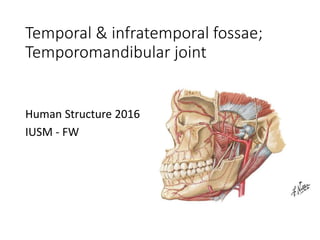
- 1. Temporal & infratemporal fossae; Temporomandibular joint Human Structure 2016 IUSM - FW
- 2. Osteology – Lateral skull • Temporal lines • Greater wing of sphenoid • Squamous part of temporal bone • Zygomatic arch • Infratemporal crest Infratemporal crest
- 3. Osteology – Base of skull • Greater wing of sphenoid • Pterygoid process • Medial plate • Lateral plate • Mandibular fossa • Foramen ovale • Foramen spinosum
- 4. Osteology – Mandible • Body • Mental foramen • Ramus • Angle • Coronoid process • Condylar process • Head • Neck • Lingula • Mandibular foramen • Mylohyoid groove
- 5. Temporal fossa • Located on the lateral side of skull • Posterior to orbit • Deep to zygomatic arch • Superior to infratemporal crest • Contents: • Temporalis m. enclosed within temporalis fascia
- 6. Infratemporal fossa • Located inferior to infratemporal crest, posterior to maxilla, deep to ramus of mandible • Contents: • Inferior part of temporalis m. • Medial & lateral pterygoid muscles • Maxillary artery & pterygoid venous plexus • Branches of mandibular n. (CN V3) • Otic ganglion • Chorda tympani
- 7. Temporomandibular joint • Modified hinge joint • Elevation and depression • Also allows protrusion and retraction; pivoting • Mandibular fossa of temporal bone • Head of mandible • Articular disc
- 8. Temporomandibular joint • Joint capsule • Lateral ligament of TMJ – strengthens capsule laterally • Extrinsic ligaments: • Stylomandibular ligament • Sphenomandibular ligament • Passive support of mandible • Check ligaments Sphenomandibular ligament Medial view
- 9. Muscles of mastication • Temporalis m. • Elevates & retracts mandible • Masseter m. • Elevates mandible • Assists in protrusion
- 10. Muscles of mastication • Lateral pterygoid m. • Protrudes & depresses mandible • Medial pterygoid m. • Elevates and protrudes mandible • Acting unilaterally both produce side-to-side movements of mandible Lateral pterygoid Medial pterygoid
- 11. Movements of mandible • Elevation • Closes mandible • Temporalis, masseter, medial pterygoid • Depression • Opens mandible • Gravity; suprahyoid muscles; lateral pterygoid • Protrusion • Lateral pterygoid • Retraction • Temporalis; suprahyoid muscles
- 12. Movements of mandible • Depression of the mandible (i.e. opening the mouth) • Slight depression • Occurs between head of mandible & articular disc • Full depression • Articular disc and head of mandible to move anteriorly • Occurs between articular disc & mandibular fossa Slight depression Full depression
- 13. Dislocation of TMJ • Head of mandible passes anterior to articular tubercle • Excessive contraction of lateral pterygoids (yawning, taking large bite, etc.) • Mandible remains depressed; unable to close mouth • Reduced by applying pressure to mandibular molars in downward & posterior direction
- 14. Maxillary artery • Arises from ECA posterior neck of mandible • Divided into 3 parts relative to lateral pterygoid m.
- 15. Branches of maxillary artery
- 17. Mandibular nerve (CN V3) • Traverses foramen ovale to enter infratemporal fossa
- 18. Mandibular nerve (CN V3)
- 19. Parasympathetics in infratemporal fossa
Editor's Notes
- The temporal fossa is located on the lateral side of skull, posterior to orbit. Its floor is formed by the squamous part of the temporal bone and the greater wing of the sphenoid, as well as small parts of the parietal and frontal bones. It is bounded superiorly and posteriorly by the temporal lines of the skull, and laterally by the zygomatic arch. It extends inferiorly to the infratemporal crest, which is a transverse ridge along the inferior aspect of the greater wing of the sphonoid and temporal bones. The temporal fossa is primarily occupied by the temporalis muscle, which arises from the floor of the temporal fossa and passes deep to the zygomatic arch to insert on the medial aspect of the coronoid process and ramus of mandible.
- The TMJ is a modified hinge joint that primarily allows elevation & depression of the mandible. In addition it allows gliding movements (protrusion & retraction), as well as slight rotational (pivoting) movements. The TMJ is formed by the mandibular fossa of the temporal bone and the head (condyle) of the mandible. An articular disc is located between the bones and separates the joint into superior and inferior articular cavities. The articular disc is made of fibrocartilage and is attached at its periphery to the internal aspect of the joint capsule. The hinge movements (elevation and depression) occur in the inferior compartment of the joint capsule, between the head of the mandible and the articular disc.
- The lateral ligament of the TMJ is a thickening of the lateral aspect of the joint capsule and helps to prevent posterior dislocation of the TMJ. 2 extrinsic ligaments also connect the mandible to the cranium: the Stylomandilar ligament attaches from the styoid process to the angle of the mandible. It is a thickening of the fibrous capsule of the parotid gland, and does not contribute significantly to the strength of the joint. The sphenomandibular ligament extends from the spine of the sphenoid bone to the lingula of the mandible. Recall that the sphenomandicular ligament is derived from Meckel’s cartilage from the 1st pharyngeal arch. It is the primary passive support for the mandible and also serves as a check ligament.
- The lateral pterygoid muscle has 2 heads: the superior head arises from the inferior surface of the greater wing of sphenoid bone & the inferior head arises from the lateral surface of lateral pterygoid plate. The superior head inserts on the joint capsule and articular disc while the inferior head inserts on the neck of the condylar process. Head of mandible & articular disc
- Divided into 3 parts relative to lateral pterygoid m. Mandibular part – Posterior to lateral pterygoid m. Pterygoid part – adjacent to lateral pterygoid m. (superficial or deep) Pterygopalatine part – Anteromedial to lateral pterygoid m.
- Not required to know all branches of maxillary a. Just larger branches that we discuss in lecture and identify in lab. Mandibular part: Middle meningeal a. ascends from maxillary a. and passes through foramen spinosum to enter cranial cavity. Supplies dura. Passes deep to pterion where it is susceptible to injury resulting in epidural hematoma. Inferior alveolar a. passes inferiorly to enter mandibular foramen. Travels through mandible within mandibular canal. Pterygoid part: branches primarily supply muscles of mastication & buccinators m.: Deep temporal arteries pass deep to temporalis m. Masseteric arteries pass through mandibular notch to supply masseter m. Pterygoid branches supply pterygoid muscles Buccal artery supplies the buccinator muscle, skin of cheek and oral mucosa. Pterygopalatine part enters the pterygopalatine fossa where it divides into 4 branches, which will be discussed in greater detail when we reach the pterygopalatine fossa. Posterior superior alveolar artery Infraorbital artery Descending palatine artery Sphenopalatine artery
- The pterygoid plexus of veins is located within the infratemporal fossa between temporalis and pterygoid muscles. It drains primarily to maxillary vein, but also anastomoses with: Facial vein superficially Cavernous sinus (within skull) Retromandibular vein Union of maxillary vein and superficial temporal vein
- Auriculotemporal n. Originates as 2 roots that encircles middle meningeal artery before joining together. and passes deep to neck of mandible. It then ascends through the parotid gland between the TMJ and ear to supply sensory innervation to the lateral aspect of the head. Also carries postganglionic parasympathetic fibers from the otic ganglion to parotid gland. Inferior alveolar n. Large branch that descends toward the mandibular foramen, which it enters. It travels through the mandible within the mandibular canal and emerges as the mental nerve through the mental foramen. It supplies sensory innervation to the mandibular teeth and gums and the skin of the lower lip, jaw, and chin. Prior to entering the mandibular foramen it gives off Nerve to mylohyoid, which travels in they mylohyoid groove on the deep surface of the mandible and supplies motor innervation to mylohyoid m. and anterior belly of digastric m. Lingual n. Large nerve that descends through infratemporal fossa anterior to inferior alveolar n. Supplies sensory innervation to anterior 2/3 of tongue. Joined by chorda tympani (branch of facial n.) which carries taste sensation to anterior 2/3 of tongue, and preganglionic parasympathetic fibers to the submandibular ganglion in the floor of the mouth.
- Deep temporal nerves pass superiorly to supply the temporalis m. Buccal n. Passes anteriorly along the anterior border of the temporalis m. with the buccal a. (from maxillary a.) and supplies sensory innervation to the skin and oral mucosa of the cheek. Note that the buccal n. does NOT supply motor innervation to buccinator m. Branches to other muscles of mastication
- Chorda tympani is a branch of facial n. (CN VII) that arises within the facial canal and enters the infratemporal fossa via the petrotympanic fissure and joins the lingual n. It provides preganglionic parasympathetic fibers to the submandibular ganglion, located in the floor of the mouth, which supplies the submandibular & sublingual glands. These will be discussed further in the oral cavity. It also provides taste senstation to the anterior 2/3 of tongue This image also indicates the otic ganglion, which is located just inferior to foramen ovale on the medial aspect of the mandibular n. Preganglionic fibers arise from the glossopharyngeal n. (CN IX) and enter the otic ganglion via the lesser petrosal n. Postganglionic fibers are distributed to the parotid gland via the auriculotemporal n.