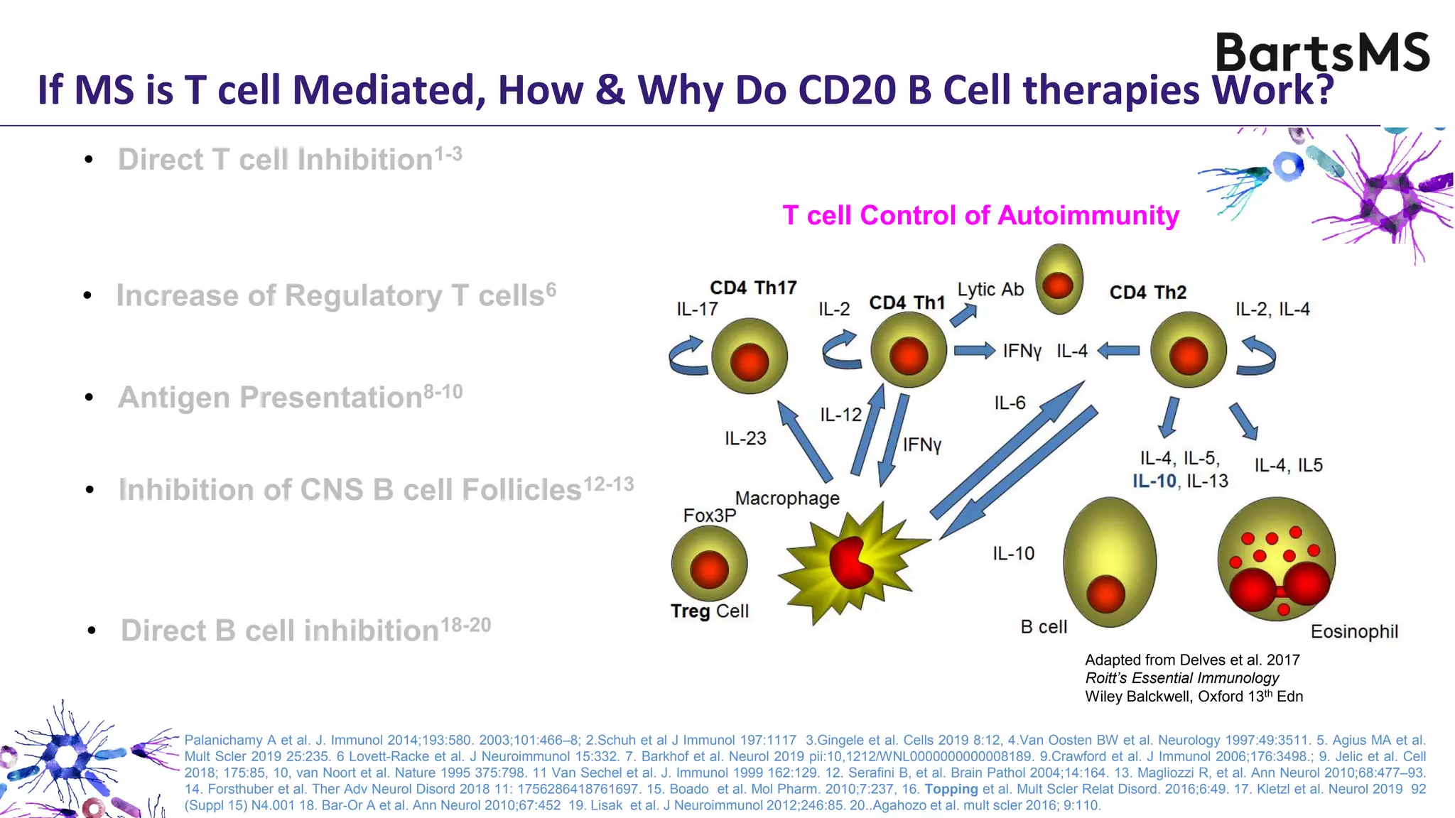
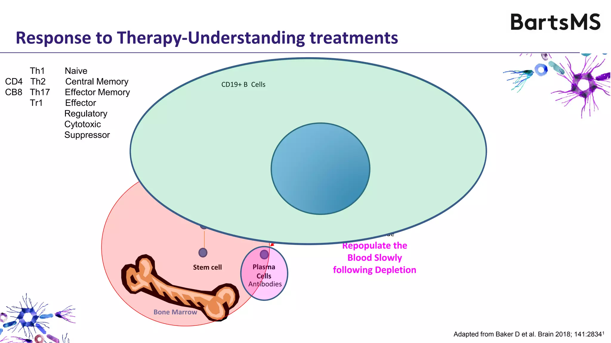
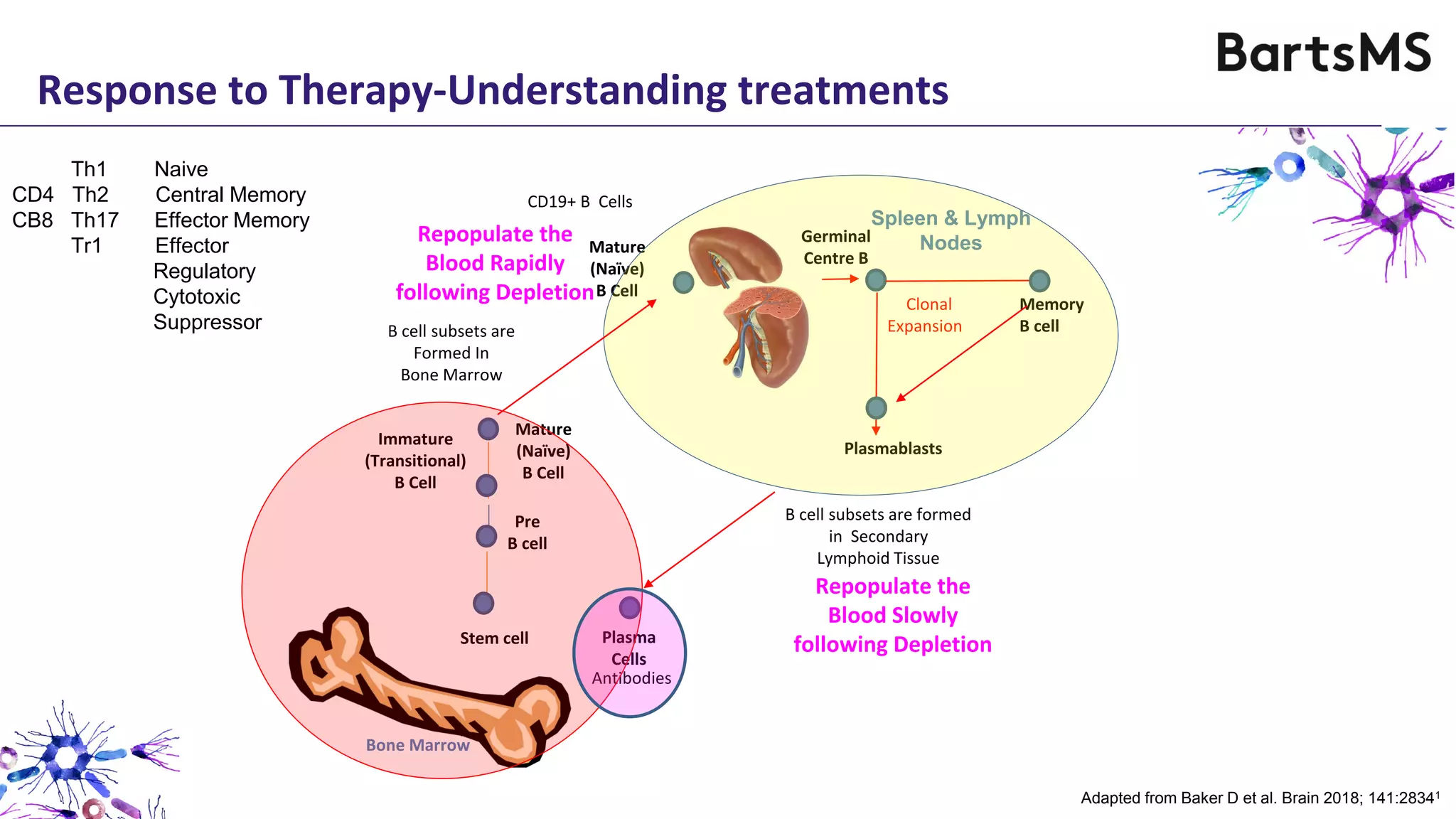
Professor David Baker
(david.baker@qmul.ac.uk)
ECTRIMS 2019
Stockholm
Cell TherapyB Therapy
The Rationale for
Hot Topic: B-cells in the pathogenesis of MS
Slides available www.ms-res.org
• The content and slides were all designed and made by David Baker
Conflict of Interest Statement
• This meeting segment was sponsored by ECTRIMS
• Although considered to be irrelevant, D. Baker has received compensation
in the past 3 years from: Canbex Therapeutics, Japan Tobacco, Merck,
• Contributor: BartsMS blog (www.ms-res.org)
Roche (Manufacturer of Ocrelizumab).
but are used here to help identify mechanisms of disease activity
• Compounds in red are not (currently) indicated for multiple sclerosis,
Slides available www.ms-res.org
Therapy
ECTRIMS 2019
Stockholm
CellB
Professor David Baker
(david.baker@qmul.ac.uk)
hy?w
Hot Topic: B-cells in the pathogenesis of MS
Slides available www.ms-res.org
iology
ECTRIMS 2019
Stockholm
B
r
s
k
hy?w
Hot Topic: B-cells in the pathogenesis of MS
Slides available www.ms-res.org
If MS is T cell Mediated, How & Why Do CD20 B Cell therapies Work?
• Direct T cell Inhibition1-3
• Increase of Regulatory T cells6
• Direct B cell inhibition18-20
Palanichamy A et al. J. Immunol 2014;193:580. 2003;101:466–8; 2.Schuh et al J Immunol 197:1117 3.Gingele et al. Cells 2019 8:12, 4.Van Oosten BW et al. Neurology 1997:49:3511. 5. Agius MA et al.
Mult Scler 2019 25:235. 6 Lovett-Racke et al. J Neuroimmunol 15:332. 7. Barkhof et al. Neurol 2019 pii:10,1212/WNL0000000000008189. 9.Crawford et al. J Immunol 2006;176:3498.; 9. Jelic et al. Cell
2018; 175:85, 10, van Noort et al. Nature 1995 375:798. 11 Van Sechel et al. J. Immunol 1999 162:129. 12. Serafini B, et al. Brain Pathol 2004;14:164. 13. Magliozzi R, et al. Ann Neurol 2010;68:477–93.
14. Forsthuber et al. Ther Adv Neurol Disord 2018 11: 1756286418761697. 15. Boado et al. Mol Pharm. 2010;7:237, 16. Topping et al. Mult Scler Relat Disord. 2016;6:49. 17. Kletzl et al. Neurol 2019 92
(Suppl 15) N4.001 18. Bar-Or A et al. Ann Neurol 2010;67:452 19. Lisak et al. J Neuroimmunol 2012;246:85. 20..Agahozo et al. mult scler 2016; 9:110.
• Antigen Presentation8-10
• Inhibition of CNS B cell Follicles12-13
Adapted from Delves et al. 2017
Roitt’s Essential Immunology
Wiley Balckwell, Oxford 13th Edn
T cell Control of Autoimmunity
If MS is T cell Mediated, How & Why Do B Cell therapies Work?
Block Pathogenic CD20+ T Cells1,2,3
Minor population of T cells Population Important3
CD3
CD19
CD20
Tissue Expression in Cells (BioGPS www.biogps.org)
CD19 monoclonal antibody (inebilizumab)
can inhibit lesion formation5
(Not indicated for multiple sclerosis)
CD4 T cell (~70%) depletion = Perceived to have FAILED4
CD3+ T cells express no or limited CD19 antigen2
18.4% of CD20+ cells are CD3+ T cells3
1.8 ± 0.3% of CD4+ cells are CD20+3
6.9 ± 1.0% of CD8+ cells are CD20+3
• Increase of Regulatory T cells6
• Direct T cell Inhibition1-3
• Antigen Presentation8-10
Palanichamy A et al. J. Immunol 2014;193:580. 2003;101:466–8; 2.Schuh et al J Immunol 197:1117 3.Gingele et al. Cells 2019 8:12, 4.Van Oosten BW et al. Neurology 1997:49:3511. 5. Agius MA et al.
Mult Scler 2019 25:235. 6 Lovett-Racke et al. J Neuroimmunol 15:332. 7. Barkhof et al. Neurol 2019 pii:10,1212/WNL0000000000008189. 9.Crawford et al. J Immunol 2006;176:3498.; 9. Jelic et al. Cell
2018; 175:85, 10, van Noort et al. Nature 1995 375:798. 11 Van Sechel et al. J. Immunol 1999 162:129. 12. Serafini B, et al. Brain Pathol 2004;14:164. 13. Magliozzi R, et al. Ann Neurol 2010;68:477–93.
14. Forsthuber et al. Ther Adv Neurol Disord 2018 11: 1756286418761697. 15. Boado et al. Mol Pharm. 2010;7:237, 16. Topping et al. Mult Scler Relat Disord. 2016;6:49. 17. Kletzl et al. Neurol 2019 92
(Suppl 15) N4.001 18. Bar-Or A et al. Ann Neurol 2010;67:452 19. Lisak et al. J Neuroimmunol 2012;246:85. 20..Agahozo et al. mult scler 2016; 9:110.
• Direct B cell inhibition18-20
• Inhibition of CNS B cell Follicles12-13
If MS is T cell Mediated, How & Why Do B Cell therapies Work?
• Increase of Regulatory T cells6
%CD3+,CD4+,CD25hi,FoxP3+
Increase in T regulatory cells6
Study Time Point (weeks)
0 1 2 3 4 4 8 12 16 20 24
0.0
0.2
0.4
0.6
0.8
1.0
1.2
P<0.001
**
• Antigen Presentation8-10
Block Pathogenic CD20+ T Cells1,2,3
Ocrelizumab controls MRI lesions within 4 weeks7
Ocrelizumab controls clinical disease within 8 weeks7
• Direct T cell Inhibition1-3
Palanichamy A et al. J. Immunol 2014;193:580. 2003;101:466–8; 2.Schuh et al J Immunol 197:1117 3.Gingele et al. Cells 2019 8:12, 4.Van Oosten BW et al. Neurology 1997:49:3511. 5. Agius MA et al.
Mult Scler 2019 25:235. 6 Lovett-Racke et al. J Neuroimmunol 15:332. 7. Barkhof et al. Neurol 2019 pii:10,1212/WNL0000000000008189. 9.Crawford et al. J Immunol 2006;176:3498.; 9. Jelic et al. Cell
2018; 175:85, 10, van Noort et al. Nature 1995 375:798. 11 Van Sechel et al. J. Immunol 1999 162:129. 12. Serafini B, et al. Brain Pathol 2004;14:164. 13. Magliozzi R, et al. Ann Neurol 2010;68:477–93.
14. Forsthuber et al. Ther Adv Neurol Disord 2018 11: 1756286418761697. 15. Boado et al. Mol Pharm. 2010;7:237, 16. Topping et al. Mult Scler Relat Disord. 2016;6:49. 17. Kletzl et al. Neurol 2019 92
(Suppl 15) N4.001 18. Bar-Or A et al. Ann Neurol 2010;67:452 19. Lisak et al. J Neuroimmunol 2012;246:85. 20..Agahozo et al. mult scler 2016; 9:110.
• Direct B cell inhibition18-20
• Inhibition of CNS B cell Follicles12-13
If MS is T cell Mediated, How & Why Do B Cell therapies Work?
B cells induce Autoproliferation of T cells in multiple sclerosis9
P=0.021
1.5
1.0
0.5
0.0
CSFEdimofCD4+(%)
CD4+ (CFSE) + + +
CD19+ (CD27-) - + -
CD19+ (CD27+) - - +
Healthy
Donor
Remission
MS
P=0.036
4.0
0.0
CSFEdim/CSFEhiCD8+memorycell
HD REM
3.0
2.0
1.0
P=0.019
0.0
CSFEdim/CSFEhiCD4+memorycell
HD REM
2.0
1.0
Healthy
Donor
Remission
MS
CD8+, CCR7-, CD45RA-CD4+, CCR7-, CD45RA-
Effector Memory T cell Effector Memory T cell
CD19+, CD27+
Memory B cell
• Increase of Regulatory T cells6
Block Pathogenic CD20+ T Cells1,2,3
• Direct T cell Inhibition1-3
• Antigen Presentation8-10
Palanichamy A et al. J. Immunol 2014;193:580. 2003;101:466–8; 2.Schuh et al J Immunol 197:1117 3.Gingele et al. Cells 2019 8:12, 4.Van Oosten BW et al. Neurology 1997:49:3511. 5. Agius MA et al.
Mult Scler 2019 25:235. 6 Lovett-Racke et al. J Neuroimmunol 15:332. 7. Barkhof et al. Neurol 2019 pii:10,1212/WNL0000000000008189. 9.Crawford et al. J Immunol 2006;176:3498.; 9. Jelic et al. Cell
2018; 175:85, 10, van Noort et al. Nature 1995 375:798. 11 Van Sechel et al. J. Immunol 1999 162:129. 12. Serafini B, et al. Brain Pathol 2004;14:164. 13. Magliozzi R, et al. Ann Neurol 2010;68:477–93.
14. Forsthuber et al. Ther Adv Neurol Disord 2018 11: 1756286418761697. 15. Boado et al. Mol Pharm. 2010;7:237, 16. Topping et al. Mult Scler Relat Disord. 2016;6:49. 17. Kletzl et al. Neurol 2019 92
(Suppl 15) N4.001 18. Bar-Or A et al. Ann Neurol 2010;67:452 19. Lisak et al. J Neuroimmunol 2012;246:85. 20..Agahozo et al. mult scler 2016; 9:110.
• Direct B cell inhibition18-20
• Inhibition of CNS B cell Follicles12-13
If MS is T cell Mediated, How & Why Do B Cell therapies Work?
Virally infected B cells present αB crystallin (HSPB5),
the dominant immunogenic antigen present in the inflammed MS brain10,11
HSPB5
HSPB5
Actin
PBMC
Protein
Message
EBV-BCell
PBMC
EBV-BCell
Donor 1
14
0
2
4
6
8
10
12
0
2
4
6
8
10
12
ProliferativeResponse(cpmx10-3)
0 5 25 0 5 25
PBMC EBV-B cell
EBV-B cell
PBMC
HSPB5 antigen (μg/ml)
Donor S5L5
Donor S5L5
Donor 2
• Increase of Regulatory T cells6
Block Pathogenic CD20+ T Cells1,2,3
• Direct T cell Inhibition1-3
• Antigen Presentation8-10
Palanichamy A et al. J. Immunol 2014;193:580. 2003;101:466–8; 2.Schuh et al J Immunol 197:1117 3.Gingele et al. Cells 2019 8:12, 4.Van Oosten BW et al. Neurology 1997:49:3511. 5. Agius MA et al.
Mult Scler 2019 25:235. 6 Lovett-Racke et al. J Neuroimmunol 15:332. 7. Barkhof et al. Neurol 2019 pii:10,1212/WNL0000000000008189. 9.Crawford et al. J Immunol 2006;176:3498.; 9. Jelic et al. Cell
2018; 175:85, 10, van Noort et al. Nature 1995 375:798. 11 Van Sechel et al. J. Immunol 1999 162:129. 12. Serafini B, et al. Brain Pathol 2004;14:164. 13. Magliozzi R, et al. Ann Neurol 2010;68:477–93.
14. Forsthuber et al. Ther Adv Neurol Disord 2018 11: 1756286418761697. 15. Boado et al. Mol Pharm. 2010;7:237, 16. Topping et al. Mult Scler Relat Disord. 2016;6:49. 17. Kletzl et al. Neurol 2019 92
(Suppl 15) N4.001 18. Bar-Or A et al. Ann Neurol 2010;67:452 19. Lisak et al. J Neuroimmunol 2012;246:85. 20..Agahozo et al. mult scler 2016; 9:110.
• Direct B cell inhibition18-20
• Inhibition of CNS B cell Follicles12-13
If MS is T cell Mediated, How & Why Do B Cell therapies Work?
Low CNS penetration of Immunoglobulin16
• Increase of Regulatory T cells6
Block Pathogenic CD20+ T Cells1,2,3
Lack of CD20 expression by plasmablasts and plasma cells15
CD20 CNS Follicle13
Disability progression related to ocrelizumab exposure levels18
• Direct T cell Inhibition1-3
• Antigen Presentation8-10
Relative lack of effect on oligoclonal bands and CNS B cells17
Palanichamy A et al. J. Immunol 2014;193:580. 2003;101:466–8; 2.Schuh et al J Immunol 197:1117 3.Gingele et al. Cells 2019 8:12, 4.Van Oosten BW et al. Neurology 1997:49:3511. 5. Agius MA et al.
Mult Scler 2019 25:235. 6 Lovett-Racke et al. J Neuroimmunol 15:332. 7. Barkhof et al. Neurol 2019 pii:10,1212/WNL0000000000008189. 9.Crawford et al. J Immunol 2006;176:3498.; 9. Jelic et al. Cell
2018; 175:85, 10, van Noort et al. Nature 1995 375:798. 11 Van Sechel et al. J. Immunol 1999 162:129. 12. Serafini B, et al. Brain Pathol 2004;14:164. 13. Magliozzi R, et al. Ann Neurol 2010;68:477–93.
14. Forsthuber et al. Ther Adv Neurol Disord 2018 11: 1756286418761697. 15. Boado et al. Mol Pharm. 2010;7:237, 16. Topping et al. Mult Scler Relat Disord. 2016;6:49. 17. Kletzl et al. Neurol 2019 92
(Suppl 15) N4.001 18. Bar-Or A et al. Ann Neurol 2010;67:452 19. Lisak et al. J Neuroimmunol 2012;246:85. 20..Agahozo et al. mult scler 2016; 9:110.
• Direct B cell inhibition18-20
• Inhibition of CNS B cell Follicles12-13
If MS is T cell Mediated, How & Why Do B Cell therapies Work?
• Increase of Regulatory T cells6
Block Pathogenic CD20+ T Cells1,2,3
Blockade of Cytokines19
Blockade of Neurotoxic and Oligodendrocyte toxic molecules20
Blockade of Pathogenic Antibody production21
Increased proinflammatory
cytokines
n=18 n=18
900
0
300
600
1200
Lymphotoxininduction(pg/ml)
p=0.025
HD
controls
Patients
with MS
Decreased anti-
inflammatory cytokines
n=18n=18
600
400
200
0
IL-10induction(pg/ml)
p=0.009
HD
controls
Patients
with MS
Cytokine production by B cells I multiple sclerosis19
• Direct T cell Inhibition1-3
• Antigen Presentation8-10
Palanichamy A et al. J. Immunol 2014;193:580. 2003;101:466–8; 2.Schuh et al J Immunol 197:1117 3.Gingele et al. Cells 2019 8:12, 4.Van Oosten BW et al. Neurology 1997:49:3511. 5. Agius MA et al.
Mult Scler 2019 25:235. 6 Lovett-Racke et al. J Neuroimmunol 15:332. 7. Barkhof et al. Neurol 2019 pii:10,1212/WNL0000000000008189. 9.Crawford et al. J Immunol 2006;176:3498.; 9. Jelic et al. Cell
2018; 175:85, 10, van Noort et al. Nature 1995 375:798. 11 Van Sechel et al. J. Immunol 1999 162:129. 12. Serafini B, et al. Brain Pathol 2004;14:164. 13. Magliozzi R, et al. Ann Neurol 2010;68:477–93.
14. Forsthuber et al. Ther Adv Neurol Disord 2018 11: 1756286418761697. 15. Boado et al. Mol Pharm. 2010;7:237, 16. Topping et al. Mult Scler Relat Disord. 2016;6:49. 17. Kletzl et al. Neurol 2019 92
(Suppl 15) N4.001 18. Bar-Or A et al. Ann Neurol 2010;67:452 19. Lisak et al. J Neuroimmunol 2012;246:85. 20..Agahozo et al. mult scler 2016; 9:110.
• Direct B cell inhibition18-20
• Inhibition of CNS B cell Follicles12-13
Autoantibodies in Multiple Sclerosis
• PATHOLOGY
Antibodies within Lesions with activated Complement1,2
• AUTOANTIBODY
Binding to Central Nervous System Targets3
Present in 28/108 pwMS who reacted to 30 different targets.
Autoantibodies often bind to Intracellular Targets. These may be secondary to Tissue Destruction.4
• PATHOGENICITY
Pathogenic effects of antibodies in vitro or when administered to animals5,6,7
• THERAPY
Benefit from Plasma Exchange in Subset of PwMS8,9
____________________________________________________________________________________
Lesion Type Feature Success of Plasma Exchange
Pattern I Mononuclear Cells 0/3 (0%)8 5/16 (31%)9
Pattern II Mononuclear cells + Antibody/Complement 10/10 (100%) 22/40 (55%)
Pattern III Hypoxia & Oligodendrocyte Damage 0/6 (0%) 0/13 (0%)
_______________________________________________________________________________
ComplémentDéposition2
1. Lucchinetti et al. Ann Neurol 2000;55:458; 2.Breij et al. Ann Neurol 2008;63:16. 3. Prineas & Paratt Mult scler 2018;24:610; 4. Willis et al. Front Immunol. 2015;6:600
5. Linder et al. Brain 2018. 6. Agahozo et al. mult scler 2016; 9:110. 7 Spadaro et al. Ann Neurol 2018;84:315. 8 Keegan et al. Lancet Neurol 2005;366:579, 9. Stock et al.
2018. 75:428.
Memory
B cell
Mature
(Naïve)
B Cell
Immature
(Transitional)
B Cell
Plasmablasts
Germinal
Centre B
Clonal
Expansion
Spleen & Lymph
Nodes
Bone Marrow
Plasma
Cells
Pre
B cell
Antibodies
Mature
(Naïve)
B Cell
Stem cell
B cell subsets are
Formed In
Bone Marrow
B cell subsets are formed
in Secondary
Lymphoid Tissue
Repopulate the
Blood Rapidly
following Depletion
Repopulate the
Blood Slowly
following Depletion
CD19+ B Cells
Adapted from Baker D et al. Brain 2018; 141:28341
Response to Therapy-Understanding treatments
Th1 Naive
CD4 Th2 Central Memory
CB8 Th17 Effector Memory
Tr1 Effector
Regulatory
Cytotoxic
Suppressor
Treatment B Memory Cell
in Blood
Availability to Enter
the CNS
Relapse Rate Reference
Atacicept
Tabalumab
Glatiramer acetate
IFN β
Teriflunomide
Daclizumab*
Dimethyl fumarate
Fingolimod
Mitoxantrone
Natalizumab
rituximab
Ocrelizumab
Cladribine
Alemtuzumab
HSCT
Increased
Increased
Reduced
Reduced
Reduced
Reduced*
Reduced
Reduced
Reduced
Increased
Reduced
Reduced
Reduced
Reduced
Reduced
Increased
Increased
Reduced
Reduced
Reduced
Reduced
Reduced
Reduced
Reduced
Reduced
Reduced
Reduced
Reduced
Reduced
Reduced
Increased/No Effect#
No Effect
Reduced
Reduced
Reduced
Reduced
Reduced
Reduced
Reduced
Reduced
Reduced
Reduced
Reduced
Reduced
Reduced
Sergott RC et al. Neurol Sci 2015; 15:175
Silk & Nantz Neurol 2018 90 (Suppl 15) P3.397 (NCT00882999)
Ireland SJ et al. JAMA Neurol 2014;71:1421
Rizzo F et al. J. Immunol Cell Biol 2016; 94:886
Gandoglia I et al. Neurol Neuroimmunol and Neuroinflamm 2017; 4:e403
Gold et al. BMC Neurol 2016;16:117
Lundy SK et al. Neurol Neuroimmunol and Neuroinflamm 2016; 3:e211Hh
Grutzke B et al Ann Clin Transl Neurol 215; 2: 119
Duddy M et al. J. Immunol 2007; 178;6092
Planas R et al. Eur J Immunol 2012;42;790
Palanichamy A et al. J. Immunol 2014;193:580
Fernandez Velasco et al. Mult scler 25 (S2) P686 ECTRIMS 2019
Ceronie B et al. J Neurol 2018;265:1199, Ruggieri et al. EPO1241 EAN 2019
Thompson et al. J Clin Immunol 2010;30;99
Storek J et al. Biol Blood Marrow Transplant 2008; 14:1379
Adapted from Baker D et al. EBioMed 2017 16:41.
DEPLETION OF MEMORY B CELLS BY CURRENT MS THERAPEUTICS
* Data not from multiple sclerosis.
Response to Therapy-Understanding Treatments
*Licensed in the US, Germany, Austria and France
^Licensed in Europe, # Unlicensed in Europe (not EMA approved)
# primary imaging endpoint showed no difference
INDICATED FOR MS
NOT INDICATED FOR MS
Lower
Efficacy
Higher
Efficacy
No
Efficacy
Hierarchy
Memory B cells: Pathology
Peripheral Memory Cells Populate relatively slowly1
B Cells accumulating in the CSF during active MS
are CD27+ memory B cells and plasmablasts3
________________________________________
CD19+, CD27+ Memory B cells
Blood CSF
MS Type Remission Relapse Remission
________________________________________
Paediatric MS2 30.1% 20.9% 66.4%
Adult MS3 32.2% 26.7% 75.9%
________________________________________
Memory B cells are more common in Childhood MS2
Memory B Cells are Reduced from the Circulation
during Relapse2,3
Memory B cells
Population in Western Societies
is relatively Slow
Duchamp M et al. Immun Inflamm 2014: 2:1311
Childhood
(1-13year)
PercentageofMemoryBcells+SD
0
10
20
30
40
50
60
Healthy
multiple sclerosis
Adolescent
(14-17year)
Adult
(25-55 year)
Adult
Onset
Paediatric
Onset
Schwarz A et al. 2017. Neurol Neuroimmunol Neuroinflamm 4:e3092
Memory B cells appear in paediatric MS
more rapidly than during aging
Memory B cells are the Major B cell Subset
Entering the CNS During MS
Image from Eggers EL et al. JCI Insight 2017;
2:pii 927243
1. Duchamp M et al. Immun Inflamm Disord 2014;2:131–140; 2. Schwarz A et al. 2017. Neurol Neuroimmunol Neuroinflamm 4:e309; 3. Eggers EL et al.
JCI Insight 2017;2:pii 92724 4. Serafini B et al. J Neuropathol Exp Neurol. 2010; 69:677;
B Cells accumulating in the CNS parenchyma are
CD27+ memory B cells and plasmablasts/Plasma
cells4
Courtesy of Sandra Amor, NLCD20 B Cells
Memory B Cells: Aetiology
B Cell Activating Factor (BAFF = TNFSF13B )
Genetic Association in Sardinian MS1
High BAFF levels Associated with Enhanced
Memory B cell Levels
MS Susceptibility Genes2 with presumed T cell function are present in or acts on B cells3
HLA-DRB1*15012: Expressed by B cells
IL2RA (CD25) Expressed by memory B cells
IL7RA (CD127) Expressed by pre-B cells
TNFRSF1A Expressed by pre B cells
STAT4 Expressed by pre B cells
IL12A Expressed by Immature B cells
BCL10 Expressed by Immature B cells
CXCR5 Expressed by Memory B cells
CD40 Expressed by Memory B cells
Monocytes
Increased
Immunoglobulin
Decreased
Monocytes
Increased Memory B cells
Increased
Soluble BAFF
Y Y Y
TNFS13B mRNA
Gene expression BioGPS: (www.biogps.org)
using Primary Cell Atlas.
Adapted from concepts in Baker D et al. Ebiomedicine 2017; 16:41–50; Baker D et al. Brain 2018;141:2834–7; 1. Steri M et al. N Engl Med 2017
376;1615–26; 2.Baranzini SE, Oksenberg JR. Trends Genet. 2017; 33:960–70. 3. Madireddy et al. Nat Commun 2019:10:2236
Genome studies indicate3
233 significant gene associations
416 additional possible associations
Cell-related Gene Networks connectivity3
30% B cell exclusive
22% T cell exclusive
6% monocyte exclusive
Differentiation for Antibody Secretion
Cytokine
Secretion
B cell
CD22
MHC
Y
EBV-Epstein Barr Virus5
EBV infects all People with
Multiple sclerosis1
EBV proteins (EBNA2) activate
autoimmune risk genes2
EBV proteins (EBNA3) Induce memory
B cells3
EBV produces viral interleukin 10-like
molecule4
APC Antigen presenting cell
EBV Epstein Barr Virus
LMP = Latent Membrane Protein
MHC Major histocompatibility complex
EBNA Epstein Barr Nuclear Antigen
NAWM Normal appearing White Matter
Adapted from Baker D et al. Brain 2018; 141:2834 ; 1. Pakpoor et al. Mult Scler 2013;19:162 ; 2. Harley JB et al. Nat Genet 2018;50:699 ; 3. Styles CT et al. PLoS Biol 2017;15:e2001992;
4. Moore KW et al. Springer Semin Immunopathol 1991;13:157; 5. Burns DM et al. Blood 2015;126:2665; 6. Van Nierop GP et al. Acta Neuropath 2017;134:383; 7. Jelcic I et al. Cell
2018;175:85 ; 8 Choi et al. PNAS 2018 115: E6868. 9. Uchida et al. Science 1999;286:300; 10. Portis T et al. Oncogene 2004; 23:8619
6
Memory B Cells: Aetiology
Viral Load associated with HLA-DRB1*15
and lower viral load with with HLA-A*02
Agostini et al. J Transl Med (2018 16:80)
Haematopoeitic Stem Cell Therapy5
Differentiation for Antibody Secretion
Cytokine
Secretion
B cell
CD22
MHC
Y
APC Antigen presenting cell
EBV Epstein Barr Virus
LMP = Latent Membrane Protein
MHC Major histocompatibility complex
EBNA Epstein Barr Nuclear Antigen
NAWM Normal appearing White Matter
Adapted from Baker D et al. Brain 2018; 141:2834 ; 1. Pakpoor et al. Mult Scler 2013;19:162 ; 2. Harley JB et al. Nat Genet 2018;50:699 ; 3. Styles CT et al. PLoS Biol 2017;15:e2001992;
4. Moore KW et al. Springer Semin Immunopathol 1991;13:157; 5. Burns DM et al. Blood 2015;126:2665; 6. Van Nierop GP et al. Acta Neuropath 2017;134:383; 7. Jelcic I et al. Cell
2018;175:85 ; 8 Choi et al. PNAS 2018 115: E6868. 9. Uchida et al. Science 1999;286:300; 10. Portis T et al. Oncogene 2004; 23:8619
6
Memory B Cells: Aetiology
Memory
APC
Function7
T
cell
Differentiation for Antibody Secretion
Cytokine
Secretion
APC
Function7
B cell
CD22
MHC
Y
EBV-Epstein Barr Virus5
Memory
LMP210
T
cell
X
APC Antigen presenting cell
EBV Epstein Barr Virus
LMP = Latent Membrane Protein
MHC Major histocompatibility complex
EBNA Epstein Barr Nuclear Antigen
NAWM Normal appearing White Matter
Th
cell
cytokines
Adapted from Baker D et al. Brain 2018; 141:2834 ; 1. Pakpoor et al. Mult Scler 2013;19:162 ; 2. Harley JB et al. Nat Genet 2018;50:699 ; 3. Styles CT et al. PLoS Biol 2017;15:e2001992;
4. Moore KW et al. Springer Semin Immunopathol 1991;13:157; 5. Burns DM et al. Blood 2015;126:2665; 6. Van Nierop GP et al. Acta Neuropath 2017;134:383; 7. Jelcic I et al. Cell
2018;175:85 ; 8 Choi et al. PNAS 2018 115: E6868. 9. Uchida et al. Science 1999;286:300; 10. Portis T et al. Oncogene 2004; 23:8619
Memory B Cells: Aetiology
T-Cell co-stimulation Molecules8
Differentiation for Antibody Secretion
Cytokine
Secretion
APC
Function6
B cell
CD22
MHC
Y
EBV-Epstein Barr Virus5
Memory
EBV
CD8
Lesion
Lesion
LMP29
T
cell
X
APC Antigen presenting cell
EBV Epstein Barr Virus
LMP = Latent Membrane Protein
MHC Major histocompatibility complex
EBNA Epstein Barr Nuclear Antigen
NAWM Normal appearing White Matter
Th
cell
cytokines
Adapted from Baker D et al. Brain 2018; 141:2834 ; 1. Pakpoor et al. Mult Scler 2013;19:162 ; 2. Harley JB et al. Nat Genet 2018;50:699 ; 3. Styles CT et al. PLoS Biol 2017;15:e2001992;
4. Moore KW et al. Springer Semin Immunopathol 1991;13:157; 5. Van Nierop GP et al. Acta Neuropath 2017;134:383; 6. Jelcic I et al. Cell 2018;175:85 ; 7 Choi et al. PNAS 2018 115:
E6868. 8. Uchida et al. Science 1999;286:300; 9. Portis T et al. Oncogene 2004; 23:8619
5
Memory B Cells: Aetiology
CD137/4-1BBL/TNFSF9 (CD8)
CD252/OX40L/TNFSF4 (CD4 & CD8)
CD70/CD27L/TNFSF7 (CD4 & CD8)
T-Cell co-stimulation Molecules7
Memory B Cells: Biology
• Animals do not get infected with Epstein Barr Virus and have distinct B cell biology1
• Multiple sclerosis is a uniquely human disease, that has become recently more common in affluent societies2,3
• Viral infection allows escape of immune-tolerance by creating memory B cells5,6.
Response to infection will be quicker, as primary immune responses do not need to be generated7.
This process would be augmented by T cell help but class-switched B cells can occur in absence of T cells8
Viral infection may be beneficial to the health of the human population
• The virus infects most of the human population2,4
• Consequences of viral infection4,8,9,10,:
• Mononucleosis/Glandular fever in adolescence8 (produce to affluence and later infection) and distinct memory cell repertoire.
• Increased risk of B cell cancers9 (Hodgkin's/Burkitt’s Lymphoma)
• Increased risk of autoimmunity4 (Increasing incidence of autoimmunity)10.
• Historically age of child birth and death is earlier11,12 than the current age of adult autoimmunities 2,3,11,12
1.Moghaddam et al. Science 1997 276:2030, 2. Dobson & Giovannoni 2016;7:67551, 3. GBD 2016. Lancet Neurol 2019 18:269. 4. Ascherio & Munger. Curr top microbial Immunol
2015; 390:365, 5. Burns DM et al. Blood 2015;126:2665. 6. Kuosaki et al. Nat Rev Immunol 2015;15:149. 7. Kotov & Jenkins. J Immunol 2019;202:401, 8. Barros et al. Front Immunol
2019;10:146. 9. Nagpal et al. Oncotarget 2016:7:67551, 10. Lerner et al. Int J Celiac Dis. 2015; 3:151. 11. Roser. https://porworldindata.org. Accessed March 2019). 12. Lawrence. Am
J physical Anthropol 1969:30:427.
Perivascular Lesion
driving Relapsing MS
Blood Vessel
Blood Brain Barrier
Dysfunction in MS
Immunglobulins
Enter CNS from
the Periphery
Y
Y
YY
Y
Y
Y
Y
Antibody-Dependent
Damage
Y
Microglia
Y
Y
Y
Y
Y
CNS-specific Ig
Specificity
Irrelevant Ig
Mitochondrial Energy DeficitsDemyelination
Nerve
Oligodendrocyte
Myelin Formation
Y
Y
Y
Fc Receptor
Binding
Advanced
MS
Adapted from:
1. Baker D et al. EBioMedicine 2017; 16:41–50; 2. Baker D et al. Brain 2018;141:2834–7.
Y
Y
Y
Activated
Microglia
Pro-Inflammation
Anti—Inflammation
Glial Cell Activation
Cytokines & Toxins
Cytokines & Toxins
Activated
Astrocyte
Fc Receptor
Binding
Microglia
Ectopic
B cell Follicle
B cell Growth
& Survival
(IL-6, IL-10, TNF)
Y
YY
Y Y
Oligoclonal
Immunoglobulin
Formation of Immune Niche in CNS
Pathogenesis of MS1,2
Slides available www.ms-res.org
Therapy?
ECTRIMS 2019
Stockholm
CellB
Thank you for Listening
NotWhy
Hot Topic: B-cells in the pathogenesis of MS
Slides available www.ms-res.org

Baker slides ectrims hot topic

  • 1.
    Professor David Baker (david.baker@qmul.ac.uk) ECTRIMS2019 Stockholm Cell TherapyB Therapy The Rationale for Hot Topic: B-cells in the pathogenesis of MS Slides available www.ms-res.org
  • 2.
    • The contentand slides were all designed and made by David Baker Conflict of Interest Statement • This meeting segment was sponsored by ECTRIMS • Although considered to be irrelevant, D. Baker has received compensation in the past 3 years from: Canbex Therapeutics, Japan Tobacco, Merck, • Contributor: BartsMS blog (www.ms-res.org) Roche (Manufacturer of Ocrelizumab). but are used here to help identify mechanisms of disease activity • Compounds in red are not (currently) indicated for multiple sclerosis, Slides available www.ms-res.org
  • 3.
    Therapy ECTRIMS 2019 Stockholm CellB Professor DavidBaker (david.baker@qmul.ac.uk) hy?w Hot Topic: B-cells in the pathogenesis of MS Slides available www.ms-res.org
  • 4.
    iology ECTRIMS 2019 Stockholm B r s k hy?w Hot Topic:B-cells in the pathogenesis of MS Slides available www.ms-res.org
  • 5.
    If MS isT cell Mediated, How & Why Do CD20 B Cell therapies Work? • Direct T cell Inhibition1-3 • Increase of Regulatory T cells6 • Direct B cell inhibition18-20 Palanichamy A et al. J. Immunol 2014;193:580. 2003;101:466–8; 2.Schuh et al J Immunol 197:1117 3.Gingele et al. Cells 2019 8:12, 4.Van Oosten BW et al. Neurology 1997:49:3511. 5. Agius MA et al. Mult Scler 2019 25:235. 6 Lovett-Racke et al. J Neuroimmunol 15:332. 7. Barkhof et al. Neurol 2019 pii:10,1212/WNL0000000000008189. 9.Crawford et al. J Immunol 2006;176:3498.; 9. Jelic et al. Cell 2018; 175:85, 10, van Noort et al. Nature 1995 375:798. 11 Van Sechel et al. J. Immunol 1999 162:129. 12. Serafini B, et al. Brain Pathol 2004;14:164. 13. Magliozzi R, et al. Ann Neurol 2010;68:477–93. 14. Forsthuber et al. Ther Adv Neurol Disord 2018 11: 1756286418761697. 15. Boado et al. Mol Pharm. 2010;7:237, 16. Topping et al. Mult Scler Relat Disord. 2016;6:49. 17. Kletzl et al. Neurol 2019 92 (Suppl 15) N4.001 18. Bar-Or A et al. Ann Neurol 2010;67:452 19. Lisak et al. J Neuroimmunol 2012;246:85. 20..Agahozo et al. mult scler 2016; 9:110. • Antigen Presentation8-10 • Inhibition of CNS B cell Follicles12-13 Adapted from Delves et al. 2017 Roitt’s Essential Immunology Wiley Balckwell, Oxford 13th Edn T cell Control of Autoimmunity
  • 6.
    If MS isT cell Mediated, How & Why Do B Cell therapies Work? Block Pathogenic CD20+ T Cells1,2,3 Minor population of T cells Population Important3 CD3 CD19 CD20 Tissue Expression in Cells (BioGPS www.biogps.org) CD19 monoclonal antibody (inebilizumab) can inhibit lesion formation5 (Not indicated for multiple sclerosis) CD4 T cell (~70%) depletion = Perceived to have FAILED4 CD3+ T cells express no or limited CD19 antigen2 18.4% of CD20+ cells are CD3+ T cells3 1.8 ± 0.3% of CD4+ cells are CD20+3 6.9 ± 1.0% of CD8+ cells are CD20+3 • Increase of Regulatory T cells6 • Direct T cell Inhibition1-3 • Antigen Presentation8-10 Palanichamy A et al. J. Immunol 2014;193:580. 2003;101:466–8; 2.Schuh et al J Immunol 197:1117 3.Gingele et al. Cells 2019 8:12, 4.Van Oosten BW et al. Neurology 1997:49:3511. 5. Agius MA et al. Mult Scler 2019 25:235. 6 Lovett-Racke et al. J Neuroimmunol 15:332. 7. Barkhof et al. Neurol 2019 pii:10,1212/WNL0000000000008189. 9.Crawford et al. J Immunol 2006;176:3498.; 9. Jelic et al. Cell 2018; 175:85, 10, van Noort et al. Nature 1995 375:798. 11 Van Sechel et al. J. Immunol 1999 162:129. 12. Serafini B, et al. Brain Pathol 2004;14:164. 13. Magliozzi R, et al. Ann Neurol 2010;68:477–93. 14. Forsthuber et al. Ther Adv Neurol Disord 2018 11: 1756286418761697. 15. Boado et al. Mol Pharm. 2010;7:237, 16. Topping et al. Mult Scler Relat Disord. 2016;6:49. 17. Kletzl et al. Neurol 2019 92 (Suppl 15) N4.001 18. Bar-Or A et al. Ann Neurol 2010;67:452 19. Lisak et al. J Neuroimmunol 2012;246:85. 20..Agahozo et al. mult scler 2016; 9:110. • Direct B cell inhibition18-20 • Inhibition of CNS B cell Follicles12-13
  • 7.
    If MS isT cell Mediated, How & Why Do B Cell therapies Work? • Increase of Regulatory T cells6 %CD3+,CD4+,CD25hi,FoxP3+ Increase in T regulatory cells6 Study Time Point (weeks) 0 1 2 3 4 4 8 12 16 20 24 0.0 0.2 0.4 0.6 0.8 1.0 1.2 P<0.001 ** • Antigen Presentation8-10 Block Pathogenic CD20+ T Cells1,2,3 Ocrelizumab controls MRI lesions within 4 weeks7 Ocrelizumab controls clinical disease within 8 weeks7 • Direct T cell Inhibition1-3 Palanichamy A et al. J. Immunol 2014;193:580. 2003;101:466–8; 2.Schuh et al J Immunol 197:1117 3.Gingele et al. Cells 2019 8:12, 4.Van Oosten BW et al. Neurology 1997:49:3511. 5. Agius MA et al. Mult Scler 2019 25:235. 6 Lovett-Racke et al. J Neuroimmunol 15:332. 7. Barkhof et al. Neurol 2019 pii:10,1212/WNL0000000000008189. 9.Crawford et al. J Immunol 2006;176:3498.; 9. Jelic et al. Cell 2018; 175:85, 10, van Noort et al. Nature 1995 375:798. 11 Van Sechel et al. J. Immunol 1999 162:129. 12. Serafini B, et al. Brain Pathol 2004;14:164. 13. Magliozzi R, et al. Ann Neurol 2010;68:477–93. 14. Forsthuber et al. Ther Adv Neurol Disord 2018 11: 1756286418761697. 15. Boado et al. Mol Pharm. 2010;7:237, 16. Topping et al. Mult Scler Relat Disord. 2016;6:49. 17. Kletzl et al. Neurol 2019 92 (Suppl 15) N4.001 18. Bar-Or A et al. Ann Neurol 2010;67:452 19. Lisak et al. J Neuroimmunol 2012;246:85. 20..Agahozo et al. mult scler 2016; 9:110. • Direct B cell inhibition18-20 • Inhibition of CNS B cell Follicles12-13
  • 8.
    If MS isT cell Mediated, How & Why Do B Cell therapies Work? B cells induce Autoproliferation of T cells in multiple sclerosis9 P=0.021 1.5 1.0 0.5 0.0 CSFEdimofCD4+(%) CD4+ (CFSE) + + + CD19+ (CD27-) - + - CD19+ (CD27+) - - + Healthy Donor Remission MS P=0.036 4.0 0.0 CSFEdim/CSFEhiCD8+memorycell HD REM 3.0 2.0 1.0 P=0.019 0.0 CSFEdim/CSFEhiCD4+memorycell HD REM 2.0 1.0 Healthy Donor Remission MS CD8+, CCR7-, CD45RA-CD4+, CCR7-, CD45RA- Effector Memory T cell Effector Memory T cell CD19+, CD27+ Memory B cell • Increase of Regulatory T cells6 Block Pathogenic CD20+ T Cells1,2,3 • Direct T cell Inhibition1-3 • Antigen Presentation8-10 Palanichamy A et al. J. Immunol 2014;193:580. 2003;101:466–8; 2.Schuh et al J Immunol 197:1117 3.Gingele et al. Cells 2019 8:12, 4.Van Oosten BW et al. Neurology 1997:49:3511. 5. Agius MA et al. Mult Scler 2019 25:235. 6 Lovett-Racke et al. J Neuroimmunol 15:332. 7. Barkhof et al. Neurol 2019 pii:10,1212/WNL0000000000008189. 9.Crawford et al. J Immunol 2006;176:3498.; 9. Jelic et al. Cell 2018; 175:85, 10, van Noort et al. Nature 1995 375:798. 11 Van Sechel et al. J. Immunol 1999 162:129. 12. Serafini B, et al. Brain Pathol 2004;14:164. 13. Magliozzi R, et al. Ann Neurol 2010;68:477–93. 14. Forsthuber et al. Ther Adv Neurol Disord 2018 11: 1756286418761697. 15. Boado et al. Mol Pharm. 2010;7:237, 16. Topping et al. Mult Scler Relat Disord. 2016;6:49. 17. Kletzl et al. Neurol 2019 92 (Suppl 15) N4.001 18. Bar-Or A et al. Ann Neurol 2010;67:452 19. Lisak et al. J Neuroimmunol 2012;246:85. 20..Agahozo et al. mult scler 2016; 9:110. • Direct B cell inhibition18-20 • Inhibition of CNS B cell Follicles12-13
  • 9.
    If MS isT cell Mediated, How & Why Do B Cell therapies Work? Virally infected B cells present αB crystallin (HSPB5), the dominant immunogenic antigen present in the inflammed MS brain10,11 HSPB5 HSPB5 Actin PBMC Protein Message EBV-BCell PBMC EBV-BCell Donor 1 14 0 2 4 6 8 10 12 0 2 4 6 8 10 12 ProliferativeResponse(cpmx10-3) 0 5 25 0 5 25 PBMC EBV-B cell EBV-B cell PBMC HSPB5 antigen (μg/ml) Donor S5L5 Donor S5L5 Donor 2 • Increase of Regulatory T cells6 Block Pathogenic CD20+ T Cells1,2,3 • Direct T cell Inhibition1-3 • Antigen Presentation8-10 Palanichamy A et al. J. Immunol 2014;193:580. 2003;101:466–8; 2.Schuh et al J Immunol 197:1117 3.Gingele et al. Cells 2019 8:12, 4.Van Oosten BW et al. Neurology 1997:49:3511. 5. Agius MA et al. Mult Scler 2019 25:235. 6 Lovett-Racke et al. J Neuroimmunol 15:332. 7. Barkhof et al. Neurol 2019 pii:10,1212/WNL0000000000008189. 9.Crawford et al. J Immunol 2006;176:3498.; 9. Jelic et al. Cell 2018; 175:85, 10, van Noort et al. Nature 1995 375:798. 11 Van Sechel et al. J. Immunol 1999 162:129. 12. Serafini B, et al. Brain Pathol 2004;14:164. 13. Magliozzi R, et al. Ann Neurol 2010;68:477–93. 14. Forsthuber et al. Ther Adv Neurol Disord 2018 11: 1756286418761697. 15. Boado et al. Mol Pharm. 2010;7:237, 16. Topping et al. Mult Scler Relat Disord. 2016;6:49. 17. Kletzl et al. Neurol 2019 92 (Suppl 15) N4.001 18. Bar-Or A et al. Ann Neurol 2010;67:452 19. Lisak et al. J Neuroimmunol 2012;246:85. 20..Agahozo et al. mult scler 2016; 9:110. • Direct B cell inhibition18-20 • Inhibition of CNS B cell Follicles12-13
  • 10.
    If MS isT cell Mediated, How & Why Do B Cell therapies Work? Low CNS penetration of Immunoglobulin16 • Increase of Regulatory T cells6 Block Pathogenic CD20+ T Cells1,2,3 Lack of CD20 expression by plasmablasts and plasma cells15 CD20 CNS Follicle13 Disability progression related to ocrelizumab exposure levels18 • Direct T cell Inhibition1-3 • Antigen Presentation8-10 Relative lack of effect on oligoclonal bands and CNS B cells17 Palanichamy A et al. J. Immunol 2014;193:580. 2003;101:466–8; 2.Schuh et al J Immunol 197:1117 3.Gingele et al. Cells 2019 8:12, 4.Van Oosten BW et al. Neurology 1997:49:3511. 5. Agius MA et al. Mult Scler 2019 25:235. 6 Lovett-Racke et al. J Neuroimmunol 15:332. 7. Barkhof et al. Neurol 2019 pii:10,1212/WNL0000000000008189. 9.Crawford et al. J Immunol 2006;176:3498.; 9. Jelic et al. Cell 2018; 175:85, 10, van Noort et al. Nature 1995 375:798. 11 Van Sechel et al. J. Immunol 1999 162:129. 12. Serafini B, et al. Brain Pathol 2004;14:164. 13. Magliozzi R, et al. Ann Neurol 2010;68:477–93. 14. Forsthuber et al. Ther Adv Neurol Disord 2018 11: 1756286418761697. 15. Boado et al. Mol Pharm. 2010;7:237, 16. Topping et al. Mult Scler Relat Disord. 2016;6:49. 17. Kletzl et al. Neurol 2019 92 (Suppl 15) N4.001 18. Bar-Or A et al. Ann Neurol 2010;67:452 19. Lisak et al. J Neuroimmunol 2012;246:85. 20..Agahozo et al. mult scler 2016; 9:110. • Direct B cell inhibition18-20 • Inhibition of CNS B cell Follicles12-13
  • 11.
    If MS isT cell Mediated, How & Why Do B Cell therapies Work? • Increase of Regulatory T cells6 Block Pathogenic CD20+ T Cells1,2,3 Blockade of Cytokines19 Blockade of Neurotoxic and Oligodendrocyte toxic molecules20 Blockade of Pathogenic Antibody production21 Increased proinflammatory cytokines n=18 n=18 900 0 300 600 1200 Lymphotoxininduction(pg/ml) p=0.025 HD controls Patients with MS Decreased anti- inflammatory cytokines n=18n=18 600 400 200 0 IL-10induction(pg/ml) p=0.009 HD controls Patients with MS Cytokine production by B cells I multiple sclerosis19 • Direct T cell Inhibition1-3 • Antigen Presentation8-10 Palanichamy A et al. J. Immunol 2014;193:580. 2003;101:466–8; 2.Schuh et al J Immunol 197:1117 3.Gingele et al. Cells 2019 8:12, 4.Van Oosten BW et al. Neurology 1997:49:3511. 5. Agius MA et al. Mult Scler 2019 25:235. 6 Lovett-Racke et al. J Neuroimmunol 15:332. 7. Barkhof et al. Neurol 2019 pii:10,1212/WNL0000000000008189. 9.Crawford et al. J Immunol 2006;176:3498.; 9. Jelic et al. Cell 2018; 175:85, 10, van Noort et al. Nature 1995 375:798. 11 Van Sechel et al. J. Immunol 1999 162:129. 12. Serafini B, et al. Brain Pathol 2004;14:164. 13. Magliozzi R, et al. Ann Neurol 2010;68:477–93. 14. Forsthuber et al. Ther Adv Neurol Disord 2018 11: 1756286418761697. 15. Boado et al. Mol Pharm. 2010;7:237, 16. Topping et al. Mult Scler Relat Disord. 2016;6:49. 17. Kletzl et al. Neurol 2019 92 (Suppl 15) N4.001 18. Bar-Or A et al. Ann Neurol 2010;67:452 19. Lisak et al. J Neuroimmunol 2012;246:85. 20..Agahozo et al. mult scler 2016; 9:110. • Direct B cell inhibition18-20 • Inhibition of CNS B cell Follicles12-13
  • 12.
    Autoantibodies in MultipleSclerosis • PATHOLOGY Antibodies within Lesions with activated Complement1,2 • AUTOANTIBODY Binding to Central Nervous System Targets3 Present in 28/108 pwMS who reacted to 30 different targets. Autoantibodies often bind to Intracellular Targets. These may be secondary to Tissue Destruction.4 • PATHOGENICITY Pathogenic effects of antibodies in vitro or when administered to animals5,6,7 • THERAPY Benefit from Plasma Exchange in Subset of PwMS8,9 ____________________________________________________________________________________ Lesion Type Feature Success of Plasma Exchange Pattern I Mononuclear Cells 0/3 (0%)8 5/16 (31%)9 Pattern II Mononuclear cells + Antibody/Complement 10/10 (100%) 22/40 (55%) Pattern III Hypoxia & Oligodendrocyte Damage 0/6 (0%) 0/13 (0%) _______________________________________________________________________________ ComplémentDéposition2 1. Lucchinetti et al. Ann Neurol 2000;55:458; 2.Breij et al. Ann Neurol 2008;63:16. 3. Prineas & Paratt Mult scler 2018;24:610; 4. Willis et al. Front Immunol. 2015;6:600 5. Linder et al. Brain 2018. 6. Agahozo et al. mult scler 2016; 9:110. 7 Spadaro et al. Ann Neurol 2018;84:315. 8 Keegan et al. Lancet Neurol 2005;366:579, 9. Stock et al. 2018. 75:428.
  • 13.
    Memory B cell Mature (Naïve) B Cell Immature (Transitional) BCell Plasmablasts Germinal Centre B Clonal Expansion Spleen & Lymph Nodes Bone Marrow Plasma Cells Pre B cell Antibodies Mature (Naïve) B Cell Stem cell B cell subsets are Formed In Bone Marrow B cell subsets are formed in Secondary Lymphoid Tissue Repopulate the Blood Rapidly following Depletion Repopulate the Blood Slowly following Depletion CD19+ B Cells Adapted from Baker D et al. Brain 2018; 141:28341 Response to Therapy-Understanding treatments Th1 Naive CD4 Th2 Central Memory CB8 Th17 Effector Memory Tr1 Effector Regulatory Cytotoxic Suppressor
  • 14.
    Memory B cell Mature (Naïve) B Cell Immature (Transitional) BCell Plasmablasts Germinal Centre B Clonal Expansion Spleen & Lymph Nodes Bone Marrow Plasma Cells Pre B cell Antibodies Mature (Naïve) B Cell Stem cell B cell subsets are Formed In Bone Marrow B cell subsets are formed in Secondary Lymphoid Tissue Repopulate the Blood Rapidly following Depletion Repopulate the Blood Slowly following Depletion CD19+ B Cells Adapted from Baker D et al. Brain 2018; 141:28341 Response to Therapy-Understanding treatments Th1 Naive CD4 Th2 Central Memory CB8 Th17 Effector Memory Tr1 Effector Regulatory Cytotoxic Suppressor
  • 15.
    Treatment B MemoryCell in Blood Availability to Enter the CNS Relapse Rate Reference Atacicept Tabalumab Glatiramer acetate IFN β Teriflunomide Daclizumab* Dimethyl fumarate Fingolimod Mitoxantrone Natalizumab rituximab Ocrelizumab Cladribine Alemtuzumab HSCT Increased Increased Reduced Reduced Reduced Reduced* Reduced Reduced Reduced Increased Reduced Reduced Reduced Reduced Reduced Increased Increased Reduced Reduced Reduced Reduced Reduced Reduced Reduced Reduced Reduced Reduced Reduced Reduced Reduced Increased/No Effect# No Effect Reduced Reduced Reduced Reduced Reduced Reduced Reduced Reduced Reduced Reduced Reduced Reduced Reduced Sergott RC et al. Neurol Sci 2015; 15:175 Silk & Nantz Neurol 2018 90 (Suppl 15) P3.397 (NCT00882999) Ireland SJ et al. JAMA Neurol 2014;71:1421 Rizzo F et al. J. Immunol Cell Biol 2016; 94:886 Gandoglia I et al. Neurol Neuroimmunol and Neuroinflamm 2017; 4:e403 Gold et al. BMC Neurol 2016;16:117 Lundy SK et al. Neurol Neuroimmunol and Neuroinflamm 2016; 3:e211Hh Grutzke B et al Ann Clin Transl Neurol 215; 2: 119 Duddy M et al. J. Immunol 2007; 178;6092 Planas R et al. Eur J Immunol 2012;42;790 Palanichamy A et al. J. Immunol 2014;193:580 Fernandez Velasco et al. Mult scler 25 (S2) P686 ECTRIMS 2019 Ceronie B et al. J Neurol 2018;265:1199, Ruggieri et al. EPO1241 EAN 2019 Thompson et al. J Clin Immunol 2010;30;99 Storek J et al. Biol Blood Marrow Transplant 2008; 14:1379 Adapted from Baker D et al. EBioMed 2017 16:41. DEPLETION OF MEMORY B CELLS BY CURRENT MS THERAPEUTICS * Data not from multiple sclerosis. Response to Therapy-Understanding Treatments *Licensed in the US, Germany, Austria and France ^Licensed in Europe, # Unlicensed in Europe (not EMA approved) # primary imaging endpoint showed no difference INDICATED FOR MS NOT INDICATED FOR MS Lower Efficacy Higher Efficacy No Efficacy Hierarchy
  • 16.
    Memory B cells:Pathology Peripheral Memory Cells Populate relatively slowly1 B Cells accumulating in the CSF during active MS are CD27+ memory B cells and plasmablasts3 ________________________________________ CD19+, CD27+ Memory B cells Blood CSF MS Type Remission Relapse Remission ________________________________________ Paediatric MS2 30.1% 20.9% 66.4% Adult MS3 32.2% 26.7% 75.9% ________________________________________ Memory B cells are more common in Childhood MS2 Memory B Cells are Reduced from the Circulation during Relapse2,3 Memory B cells Population in Western Societies is relatively Slow Duchamp M et al. Immun Inflamm 2014: 2:1311 Childhood (1-13year) PercentageofMemoryBcells+SD 0 10 20 30 40 50 60 Healthy multiple sclerosis Adolescent (14-17year) Adult (25-55 year) Adult Onset Paediatric Onset Schwarz A et al. 2017. Neurol Neuroimmunol Neuroinflamm 4:e3092 Memory B cells appear in paediatric MS more rapidly than during aging Memory B cells are the Major B cell Subset Entering the CNS During MS Image from Eggers EL et al. JCI Insight 2017; 2:pii 927243 1. Duchamp M et al. Immun Inflamm Disord 2014;2:131–140; 2. Schwarz A et al. 2017. Neurol Neuroimmunol Neuroinflamm 4:e309; 3. Eggers EL et al. JCI Insight 2017;2:pii 92724 4. Serafini B et al. J Neuropathol Exp Neurol. 2010; 69:677; B Cells accumulating in the CNS parenchyma are CD27+ memory B cells and plasmablasts/Plasma cells4 Courtesy of Sandra Amor, NLCD20 B Cells
  • 17.
    Memory B Cells:Aetiology B Cell Activating Factor (BAFF = TNFSF13B ) Genetic Association in Sardinian MS1 High BAFF levels Associated with Enhanced Memory B cell Levels MS Susceptibility Genes2 with presumed T cell function are present in or acts on B cells3 HLA-DRB1*15012: Expressed by B cells IL2RA (CD25) Expressed by memory B cells IL7RA (CD127) Expressed by pre-B cells TNFRSF1A Expressed by pre B cells STAT4 Expressed by pre B cells IL12A Expressed by Immature B cells BCL10 Expressed by Immature B cells CXCR5 Expressed by Memory B cells CD40 Expressed by Memory B cells Monocytes Increased Immunoglobulin Decreased Monocytes Increased Memory B cells Increased Soluble BAFF Y Y Y TNFS13B mRNA Gene expression BioGPS: (www.biogps.org) using Primary Cell Atlas. Adapted from concepts in Baker D et al. Ebiomedicine 2017; 16:41–50; Baker D et al. Brain 2018;141:2834–7; 1. Steri M et al. N Engl Med 2017 376;1615–26; 2.Baranzini SE, Oksenberg JR. Trends Genet. 2017; 33:960–70. 3. Madireddy et al. Nat Commun 2019:10:2236 Genome studies indicate3 233 significant gene associations 416 additional possible associations Cell-related Gene Networks connectivity3 30% B cell exclusive 22% T cell exclusive 6% monocyte exclusive
  • 18.
    Differentiation for AntibodySecretion Cytokine Secretion B cell CD22 MHC Y EBV-Epstein Barr Virus5 EBV infects all People with Multiple sclerosis1 EBV proteins (EBNA2) activate autoimmune risk genes2 EBV proteins (EBNA3) Induce memory B cells3 EBV produces viral interleukin 10-like molecule4 APC Antigen presenting cell EBV Epstein Barr Virus LMP = Latent Membrane Protein MHC Major histocompatibility complex EBNA Epstein Barr Nuclear Antigen NAWM Normal appearing White Matter Adapted from Baker D et al. Brain 2018; 141:2834 ; 1. Pakpoor et al. Mult Scler 2013;19:162 ; 2. Harley JB et al. Nat Genet 2018;50:699 ; 3. Styles CT et al. PLoS Biol 2017;15:e2001992; 4. Moore KW et al. Springer Semin Immunopathol 1991;13:157; 5. Burns DM et al. Blood 2015;126:2665; 6. Van Nierop GP et al. Acta Neuropath 2017;134:383; 7. Jelcic I et al. Cell 2018;175:85 ; 8 Choi et al. PNAS 2018 115: E6868. 9. Uchida et al. Science 1999;286:300; 10. Portis T et al. Oncogene 2004; 23:8619 6 Memory B Cells: Aetiology Viral Load associated with HLA-DRB1*15 and lower viral load with with HLA-A*02 Agostini et al. J Transl Med (2018 16:80)
  • 19.
    Haematopoeitic Stem CellTherapy5 Differentiation for Antibody Secretion Cytokine Secretion B cell CD22 MHC Y APC Antigen presenting cell EBV Epstein Barr Virus LMP = Latent Membrane Protein MHC Major histocompatibility complex EBNA Epstein Barr Nuclear Antigen NAWM Normal appearing White Matter Adapted from Baker D et al. Brain 2018; 141:2834 ; 1. Pakpoor et al. Mult Scler 2013;19:162 ; 2. Harley JB et al. Nat Genet 2018;50:699 ; 3. Styles CT et al. PLoS Biol 2017;15:e2001992; 4. Moore KW et al. Springer Semin Immunopathol 1991;13:157; 5. Burns DM et al. Blood 2015;126:2665; 6. Van Nierop GP et al. Acta Neuropath 2017;134:383; 7. Jelcic I et al. Cell 2018;175:85 ; 8 Choi et al. PNAS 2018 115: E6868. 9. Uchida et al. Science 1999;286:300; 10. Portis T et al. Oncogene 2004; 23:8619 6 Memory B Cells: Aetiology Memory APC Function7 T cell
  • 20.
    Differentiation for AntibodySecretion Cytokine Secretion APC Function7 B cell CD22 MHC Y EBV-Epstein Barr Virus5 Memory LMP210 T cell X APC Antigen presenting cell EBV Epstein Barr Virus LMP = Latent Membrane Protein MHC Major histocompatibility complex EBNA Epstein Barr Nuclear Antigen NAWM Normal appearing White Matter Th cell cytokines Adapted from Baker D et al. Brain 2018; 141:2834 ; 1. Pakpoor et al. Mult Scler 2013;19:162 ; 2. Harley JB et al. Nat Genet 2018;50:699 ; 3. Styles CT et al. PLoS Biol 2017;15:e2001992; 4. Moore KW et al. Springer Semin Immunopathol 1991;13:157; 5. Burns DM et al. Blood 2015;126:2665; 6. Van Nierop GP et al. Acta Neuropath 2017;134:383; 7. Jelcic I et al. Cell 2018;175:85 ; 8 Choi et al. PNAS 2018 115: E6868. 9. Uchida et al. Science 1999;286:300; 10. Portis T et al. Oncogene 2004; 23:8619 Memory B Cells: Aetiology T-Cell co-stimulation Molecules8
  • 21.
    Differentiation for AntibodySecretion Cytokine Secretion APC Function6 B cell CD22 MHC Y EBV-Epstein Barr Virus5 Memory EBV CD8 Lesion Lesion LMP29 T cell X APC Antigen presenting cell EBV Epstein Barr Virus LMP = Latent Membrane Protein MHC Major histocompatibility complex EBNA Epstein Barr Nuclear Antigen NAWM Normal appearing White Matter Th cell cytokines Adapted from Baker D et al. Brain 2018; 141:2834 ; 1. Pakpoor et al. Mult Scler 2013;19:162 ; 2. Harley JB et al. Nat Genet 2018;50:699 ; 3. Styles CT et al. PLoS Biol 2017;15:e2001992; 4. Moore KW et al. Springer Semin Immunopathol 1991;13:157; 5. Van Nierop GP et al. Acta Neuropath 2017;134:383; 6. Jelcic I et al. Cell 2018;175:85 ; 7 Choi et al. PNAS 2018 115: E6868. 8. Uchida et al. Science 1999;286:300; 9. Portis T et al. Oncogene 2004; 23:8619 5 Memory B Cells: Aetiology CD137/4-1BBL/TNFSF9 (CD8) CD252/OX40L/TNFSF4 (CD4 & CD8) CD70/CD27L/TNFSF7 (CD4 & CD8) T-Cell co-stimulation Molecules7
  • 22.
    Memory B Cells:Biology • Animals do not get infected with Epstein Barr Virus and have distinct B cell biology1 • Multiple sclerosis is a uniquely human disease, that has become recently more common in affluent societies2,3 • Viral infection allows escape of immune-tolerance by creating memory B cells5,6. Response to infection will be quicker, as primary immune responses do not need to be generated7. This process would be augmented by T cell help but class-switched B cells can occur in absence of T cells8 Viral infection may be beneficial to the health of the human population • The virus infects most of the human population2,4 • Consequences of viral infection4,8,9,10,: • Mononucleosis/Glandular fever in adolescence8 (produce to affluence and later infection) and distinct memory cell repertoire. • Increased risk of B cell cancers9 (Hodgkin's/Burkitt’s Lymphoma) • Increased risk of autoimmunity4 (Increasing incidence of autoimmunity)10. • Historically age of child birth and death is earlier11,12 than the current age of adult autoimmunities 2,3,11,12 1.Moghaddam et al. Science 1997 276:2030, 2. Dobson & Giovannoni 2016;7:67551, 3. GBD 2016. Lancet Neurol 2019 18:269. 4. Ascherio & Munger. Curr top microbial Immunol 2015; 390:365, 5. Burns DM et al. Blood 2015;126:2665. 6. Kuosaki et al. Nat Rev Immunol 2015;15:149. 7. Kotov & Jenkins. J Immunol 2019;202:401, 8. Barros et al. Front Immunol 2019;10:146. 9. Nagpal et al. Oncotarget 2016:7:67551, 10. Lerner et al. Int J Celiac Dis. 2015; 3:151. 11. Roser. https://porworldindata.org. Accessed March 2019). 12. Lawrence. Am J physical Anthropol 1969:30:427.
  • 23.
    Perivascular Lesion driving RelapsingMS Blood Vessel Blood Brain Barrier Dysfunction in MS Immunglobulins Enter CNS from the Periphery Y Y YY Y Y Y Y Antibody-Dependent Damage Y Microglia Y Y Y Y Y CNS-specific Ig Specificity Irrelevant Ig Mitochondrial Energy DeficitsDemyelination Nerve Oligodendrocyte Myelin Formation Y Y Y Fc Receptor Binding Advanced MS Adapted from: 1. Baker D et al. EBioMedicine 2017; 16:41–50; 2. Baker D et al. Brain 2018;141:2834–7. Y Y Y Activated Microglia Pro-Inflammation Anti—Inflammation Glial Cell Activation Cytokines & Toxins Cytokines & Toxins Activated Astrocyte Fc Receptor Binding Microglia Ectopic B cell Follicle B cell Growth & Survival (IL-6, IL-10, TNF) Y YY Y Y Oligoclonal Immunoglobulin Formation of Immune Niche in CNS Pathogenesis of MS1,2 Slides available www.ms-res.org
  • 24.
    Therapy? ECTRIMS 2019 Stockholm CellB Thank youfor Listening NotWhy Hot Topic: B-cells in the pathogenesis of MS Slides available www.ms-res.org