
Recommended
More Related Content
What's hot
What's hot (20)
Similar to I BDS 2019-20 Anterior median region of neck.pptx
Similar to I BDS 2019-20 Anterior median region of neck.pptx (20)
More from BJYOTHSNA1
More from BJYOTHSNA1 (16)
Recently uploaded
Recently uploaded (20)
I BDS 2019-20 Anterior median region of neck.pptx
- 1. ANTERIOR MEDIAN REGION OF NECK
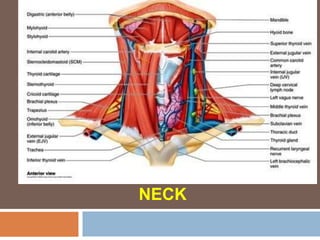
- 2. Anterior Region of Neck Triangular area on front of the neck b/n two SCM muscles
- 3. Anterior Region of Neck This area includes both (right & left) anterior triangles & Suprahyoid & Infrahyoid areas
- 4. THE NECK – EXTENT (Above & Below)
- 5. THE NECK – EXTENT (Posterior) Ligamentum nuchae – connects spinous process of all cervical vertebrae – external occipital protuberance
- 6. Anterior Median line of Neck Essential while performing tracheostomy Also useful in clinical examination of midline swellings of the
- 7. Anterior Median line of Neck Extending - Symphysis menti to suprasternal notch Lies deep to skin, Superficial Fascia & deep fascia
- 9. Mid-line Structures of Neck 1. Symphysis menti 2. Fibrous (Mylohyoid) raphae 3. Hyoid bone 4. Thyrohyoid membrane & Median Thyrohyoid Ligament 5. Upper border of thyroid
- 11. Mid-line Structures of Neck 6. Angle of thyroid cartilage 7. Median cricothyroid lig & Crico-thyroid muscles 8. Cricoid cartilage 9. Cervical part of trachea
- 12. Mid-line Structures of Neck 11. Pyramidal lobe, Levator glanudulae thyroidae 12. Thyroidea ima artery 13. Inferior thyroid veins 14. Jugular venous arch
- 14. Symphysis menti Two halves of body of mandible united in midline
- 15. Fibrous raphae Symphysis menti to Hyoid Bone Both mylohyoid muscle meet – to form floor of mouth (diaphragma oris)
- 16. Hyoid bone – C3 vertebra Horseshoe –shaped (“U”) Body, 2 cornua 2nd & 3rd branchial arches No bony articulation – suspended from base of skull by stylohyoid
- 18. Thyrohyoid membrane Extends from upper border of thyroid cartilage to lower border & greater cornua of hyoid bone Structures pierced - Internal laryngeal
- 20. Median Thyrohyoid ligament Thickening of the thyrohyoid membrane in midline Connects the upper border of thyroid cartilage to the lower border of hyoid bone
- 21. Angle of Thyroid Cartilage – C4 & C5 Forms Laryngeal prominence – Adam’s Apple More prominent in (adult Males)
- 23. Median Crico-thyroid ligament & Cricothyroid muscle Midline thickening of cricothyroid membrane extending b/n upper border of cricoid cartilage to lower border of thyroid cartilage Cricothyroid Muscle - Tensors of vocal folds
- 24. Cricoid Cartilage – C6 Unpaired cartilage of larynx Most important surface landmark on the front of the neck
- 25. Level of C6 forms a Critical plane of Neck Junction of Larynx & Trachea ; Pharynx & Esophagus Anterior tubercle of transverse process of C6 vertebra (carotid tubercle) against which common carotid artery may compressed Location of ansa cervicalis & intermediate tendon of omohyoid infront of carotid sheath
- 27. Cervical part of trachea – Isthmus of thyroid gland Isthmus – lies infront of 2,3,4 tracheal rings Above the isthmus – anastomosis of both superior thyroid arteries pyramidal lobe & a fibromuscular band – levator glanudulae
- 29. Cervical part of trachea – below the isthmus Inferior thyroid veins (lie infront of 5th, 6th & 7th tracheal rings) Sometimes arteria thyroidae ima (arises from brachiocephalic trunk) – it ascends infront of trachea to the isthmus of thyroid
- 31. Jugular venous arch Above the suprasternal notch – connects the two anterior Jugular veins
- 32. Suprasternal notch Can be felt b/n the medial ends of the clavicle Lies opposite the lower border of body of T2 Ocassionally Left brachiocephalic vein & artery may lie infront of trachea in the
Editor's Notes
- Above (From before backwards) Below ()