SlideShare a Scribd company logo
1 of 40
Download to read offline
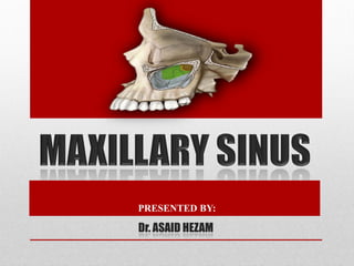
PRESENTED BY:
Dr. ASAID HEZAM
CONTENTS
• INTRODUCTION………………………………………………........…1
• DEFINITION……………………………………………………….…….2
• DEVELOPMENT……………………………………………….……….3
• ANATOMY…………………………………………………………….…4
• VASCULAR SUPPLY……………………………………….……….5
• HISTOLOGY……………………………………………….……………6
• FUNCTION……………………………………………….……………..7
• DISORDER…………………………………………….………………..8
INTRODUCTION
• Paranasal sinuses (PNS) are air containing bony spaces around
the nasal cavity.
• These spaces communicates with the nasal airway and forms
the various boundaries of nasal cavity and named for the bones
in which they locates.
• Paranasal sinuses are present in a variety animals (including
most mammals, birds, and crocodile) .
INTRODUCTION
• There are 4 pairs of paranasal sinuses (bilaterally) viz
A. Frontal air sinus
B. Ethmoidal air sinus
C. Maxillary air sinus
D. Sphenoidal air sinus
DEFINITION
• Maxillary sinus is a part of series of pneumatic cavity,
which are restricted to the skull in human; called the
paranasal sinuses.
• Maxillary sinus is defined as “ the pneumatic space that is
lodged inside the body of the maxilla and that
communicates with the environment by way of the middle
nasal meatus and the nasal vestibule ”.
DEFINITION
DEVELOPMENT
• Maxillary sinus is the first of the PNS to develop at
approximately the third month of fetal life.
• The process begins by slow development of a mucosal
pouching of the ethmoid infudibulum.
• The sinus cavity continues to develop as a slit like
invagination of the nasal epithelium into the cartilagenous
nasal capsule.
• This stage of development is called the primary
pneumatization process which continues until late in the
fourth fetal month.
• The second phase of development of the maxillary sinus is
called secondary pneumatization.
DEVELOPMENT
• This process starts at approximately the fifth mouth of fetal
life, when the shallow primordium of the maxillary sinus
begins to grow into the adjacent growing bone of maxilla.
DEVELOPMENT
• This process proceeds slowly, and by birth sinus appears as a
small ovoid groove on the side of maxillary bone close to the
orbit and measures on average 7mm in anteroposterior length,
4mm in length, with an estimated volume of 6 to 8ml.
• By the 4th or 5th month of age, the sinus can be seen
radiographically on anteroposterior views as a triangular area
medial to the infra orbital foramen.
• At 7 years of age, the rapid growth of maxillary sinus resumes
and continuous for the next 4 to 5 years, corresponding to the
eruption of the permanent teeth.
DEVELOPMENT
• The final growth spurts of maxillary sinus takes place
between 12 and 14 years of age, when it extends down to
the same level as the nasal floor.
• With completion of eruption of all the maxillary permanent
teeth, expansion of the maxillary sinus fills the growing
maxillary bone to produce adult pyramidal shape of the
sinus. It reach to maximum size around 18 years of age.
• This expansion into alveolar process places the floor of the
sinus approximately 5 to 12 mm below the floor of nose.
DEVELOPMENT
 However, in some patients, some degree of expansion or
pneumatization of the sinus continue throughout life.
 In the 4th week I.D.L- dorsal of 1st pharyngeal arch forms
the maxillary process, which extends forward and beneath
the development eye to give rise to the maxilla.
DEVELOPMENT
Horizontal shift of the palatal
shelves & fusion with one another.
Nasal septum separates the 2* oral
cavity from two nasal chamber.
Influences further expansion of the
lateral nasal wall & 3 wall begin to
fold.
3 conchae & meatuses arise
DEVELOPMENT
In its development maxillary sinus is in shape :
tubular birth
at
ovoid childhood
in
pyramidal adulthood
in
 The maxillary sinus has a horizontal pyramidal shape that
consists of a base, an apex, and 4 sides.
 The base comprises the lateral wall of the nasal cavity,
whereas apex is at the junction of the maxillary and
zygomatic bone (root of the zygomatic).
 The wall of sinus (4 sided pyramid) are related to the
surface of maxilla as follow:
I. Anterior wall: to facial surface of maxilla
II. Posterior wall: to infra-temporal surface of maxilla
III.Inferior wall: to alveolar process
IV.Superior wall: floor of orbit.
ANATOMY
ROOF OF THE ANTRUM
 Formed by floor of the orbit and is transversed by the
infraorbital is flat and slopes slightly anteriorly and laterally.
 Imp structures :
1. Infraorbital canal
2. Infraorbital foramen
3. Infraorbital nerve and vessels.
ANATOMY
FLOOR OF THE SINUS
 Curved than flat in structure.
 Formed by junction of anterior sinus wall and lateral nasal wall
 Lies 1-1.2cm below nasal floor
 Close relationship between sinus and teeth facilitate spread of
pathology.
ANATOMY
ANTERIOR WALL
 Formed by the facial surface of the maxilla.
 Extend from pyriform aperture anteriorly to alveolar process
inferiorly.
 Convexity towards sinus
 Thinnest in canine fossa
 Imp structures:
1. Infraorbital foramen
2. ASA, MSA nerves
3. Canine fossa
ANATOMY
POSTERIOR WALL
 Formed by sphenomaxillary wall.
 A thin plate of bone separate the antral cavity from the
infratemporal fossa. Mede of zygomatic and greater wing of
sphenoid bone.
 Thick laterally, thin medially.
 Important structures:
1. PSA nerve
2. Maxillary artery
3. Pterygopalatine ganglion
4. Nerve of pterygoid canal
ANATOMY
MEDIAL WALL
 Formed by lateral nasal wall.
 Below-inferior, nasal conchae
 Above-uncinate process of ethmoid, lacrimal bone
 Behind-palatine bone
 Contains double layer of mucous membrane (pars membranacea)
 Imp structures:
1. Sinus ostium
2. Hiatus semilunaris
3. Ethmoidal bulla
4. Uncinate process
5. infundibulum
ANATOMY
LATERAL WALL
 Related to zygoma and cheek.
ANATOMY
ANATOMY
OSTIUM
 Opening of the maxillary sinus is called osteum.
 It opens in middle meatus at the lower part of the hiatus
semilunaris.
 Lies above the level of nasal floor.
 The ostium lies approximately 2/3rds up the medial wall of the
sinus, making drainage of the sinus inherently difficult.
• Greater palatine arteries
• Infra-orbital artery
• Facial artery also contribute.
VASCULAR SUPPLY
VASCULAR SUPPLY
• Maxillary division of the trigeminal nerve,
• i.e.
- the posterior, middle and anterior superior alveolar nerves,
- the infraorbital nerve ,
- anterior palatine nerve .
• Pterygoid venous plexus (anterior)
• Sphenopalatin (anterior)
• Facial vein contribute to venous drainage of sinus (posterior)
 Infection from maxillary sinus may spread to involve the cavernous sinus
via draining veins facial vein and emissary vein to cause cavernous sinus
thrombosis.
VASCULAR SUPPLY
VASCULAR SUPPLY
1. Submandibular lymph nodes
2. Deep cervical lymph node
3. Retro pharyngeal lymph nodes
 Lymphatic drainage is important
because infections and malignant
tumors may spread along the lymphatic
system
 Drain into deep cervical either directly
or via submandibular nodes.
• Maxillary sinus is lined by three layers: epithelial layer, basal,
lamina and subepithelial layer with periostium.
• Epithelium is pseudo stratified, columnar and cilliated.
• As cilia beats, the mucous on epithelial surface moves from
sinus interior towards nasal cavity.
• Imparts resonance to the voice.
• Increases the surface area and lightens the skull.
• Moistens and warms inspired air.
• Filters the debris from the inspired air.
• Mucus production and storage.
• Limit extent of facial injury from trauma.
• Provides thermal insulation to important.
• Serves as accessory olfactory organs.
1. Inflammatory - maxillary sinusitis.
2. Traumatic - fractured root , sinus contusion, blow out
fracture, zygomatic complex fracture.
3. Calcification - antroliths.
4. Cyst - radicular cyst, dentigerous cyst, mucous
retention cyst.
5. Tumor - antral polyps, squammous cell carcinoma.
 It is the inflammation of the maxillary sinus mucosa.
 Types: depending upon duration
I. Acute : sudden onset, duration 4week or less.
II. Subacute : duration 4-12week.
III. Chronic : duration 12week.
 The spread of pulpal disease beyond the confines of
the dental supporting tissues into the maxillary sinus
was termed endo-antral syndrome (EAS) by selden
(1974)
 The sinus is directly involved in tooth extraction due to
the relation of surrounding structures with maxillary
sinus and can lead to an oroantral communication or
complicated by displacement of root.
 Patient complained of regurgitation of food through
the nose while eating.
Oroantral communication
Escape of fluids
Epistaxis
Escape of air
Enhanced column of air
Excruciating pain
Oroantral fistula
Pain persistent purulent unilateral
nasal discharge
Post nasal drip
Possible sequale of toxememic
condition
Popping out of antral polyp
Surgical management
Buccal flap advancement procedure
Palatal pedical flap
Ashley”s operation
Caldwell luc operation
Intra nasal antrostomy
 The use of 7-10mm long implants is a greater concern
in the maxilla than the mandible because the implant
failure rate is higher in the maxilla.
 Therefore, 13mm is the recommended minimum
occlusocervical bone dimension in the maxilla
There are two main approaches to
lift the maxillary sinus
Indirect
Direct
(caldwell luc)
indirect
direct
Crouzon syndrome: (craniofcial dyostosis) there is early synostosis
of the sutures produce hypoplasia of the maxilla and therefore
maxillary sinus together with high arch palate resulting in crowding
of teeth
As shows brachycephaly, hypertelorism and orbital proptosis
Treacher collins syndrome: (mandibulofacial dysostosis)
features may include underdeveloped or absence of
zygomatic bone, downward inclination of palpebral fissure
underdeveloped maxillary sinus and mandible malformed external
ears, high arched or cleft palate.
Binder syndrome: (maxillaonasal dysplasia)
features include hypoplasia of middle third of face. There is
maxillary sinus hypoplasia, retrognathic maxilla.
Silent sinus syndrome:
spontaneous, asymptomatic collapse of the maxillary sinus and
orbital floor associated with negative sinus pressures. It can cause
painless facial asymmetry, diplopia and enophthalmos.
Usually the diagnosis is suspected clinically, and
it can be confirmrd radiologically by characteristic
imaging features that include maxillary sinus outlet
obstruction, sinus opacification, and sinus volume
loss caused by inward retraction of the sinus wall.
REFERNCES
• Human anatomy, head & neck anatomy, atlas anatomy.
• Orban's Oral histology & embryology
• Oral anatomy, histology, and embryology. B.K.B. Berkovitz. G.R. Holland. B.J
Moxham. Fourth edition.
• Atlas of oral diseases. Goeorge Laskaris.
• Maxillary sinus and its implication Killey &Kay
• Cate A.R Ten, Oral histology: development, structure, and function.
• Textbook of oral & general anatomy & maxillofacial surgary of 3 Doctors.
• Soames & Cawson's Oral pathology.
• Seminars on maxillary sinus of some doctors.
• Online location . google ..

More Related Content

What's hot

What's hot (20)

Osteology of Maxilla
Osteology of MaxillaOsteology of Maxilla
Osteology of Maxilla
 
The maxillary nerve
The maxillary nerveThe maxillary nerve
The maxillary nerve
 
Maxillary sinus
Maxillary sinusMaxillary sinus
Maxillary sinus
 
Inferior alveolar nerve
Inferior alveolar nerveInferior alveolar nerve
Inferior alveolar nerve
 
Development of Mandible
Development of MandibleDevelopment of Mandible
Development of Mandible
 
Development of Palate and Tongue PPT
Development of Palate and Tongue PPTDevelopment of Palate and Tongue PPT
Development of Palate and Tongue PPT
 
Maxillary sinus
Maxillary sinusMaxillary sinus
Maxillary sinus
 
Cementum
CementumCementum
Cementum
 
Development & Growth of Maxilla
Development & Growth of Maxilla  Development & Growth of Maxilla
Development & Growth of Maxilla
 
Mandibular nerve dental surgery
Mandibular nerve dental surgeryMandibular nerve dental surgery
Mandibular nerve dental surgery
 
Development of tooth
Development of toothDevelopment of tooth
Development of tooth
 
Congenital defects of the Face
Congenital defects of the FaceCongenital defects of the Face
Congenital defects of the Face
 
Tooth development, eruption & applied aspects
Tooth development, eruption & applied aspectsTooth development, eruption & applied aspects
Tooth development, eruption & applied aspects
 
Tongue development, applied anatomy and prosthetic implications
Tongue development, applied anatomy and prosthetic implicationsTongue development, applied anatomy and prosthetic implications
Tongue development, applied anatomy and prosthetic implications
 
Lymphatic drainage of head & neck
Lymphatic drainage of head & neckLymphatic drainage of head & neck
Lymphatic drainage of head & neck
 
Development of tongue
Development of  tongueDevelopment of  tongue
Development of tongue
 
Saliva in dentistry
Saliva in dentistrySaliva in dentistry
Saliva in dentistry
 
soft palate
soft palatesoft palate
soft palate
 
Growth & development of maxilla and mandible
Growth & development of maxilla and mandibleGrowth & development of maxilla and mandible
Growth & development of maxilla and mandible
 
Gingiva
GingivaGingiva
Gingiva
 

Similar to Maxillary sinus.pptx

Maxilla and Mandible - its applied aspect - Copy.pptx
Maxilla and Mandible - its applied aspect - Copy.pptxMaxilla and Mandible - its applied aspect - Copy.pptx
Maxilla and Mandible - its applied aspect - Copy.pptxRiddhikaKochar1
 
Imaging of paranasal sinuses RV
Imaging of paranasal sinuses RVImaging of paranasal sinuses RV
Imaging of paranasal sinuses RVRoshan Valentine
 
Maxillary sinus
Maxillary sinusMaxillary sinus
Maxillary sinussauvik2014
 
Imaging of paranasal sinuses (including anatomy and varaints)pk1 pdf ppt
Imaging of paranasal sinuses (including anatomy and varaints)pk1 pdf  pptImaging of paranasal sinuses (including anatomy and varaints)pk1 pdf  ppt
Imaging of paranasal sinuses (including anatomy and varaints)pk1 pdf pptDr pradeep Kumar
 
Development,anatomy and applied anatomy of Maxilla
Development,anatomy and applied anatomy of MaxillaDevelopment,anatomy and applied anatomy of Maxilla
Development,anatomy and applied anatomy of MaxillaSoni Bista
 
Maxillary sinus aishu.pptx
Maxillary sinus aishu.pptxMaxillary sinus aishu.pptx
Maxillary sinus aishu.pptxshilpasm3
 
Paranasal sinus diseases and their management
Paranasal sinus diseases and their managementParanasal sinus diseases and their management
Paranasal sinus diseases and their managementDibya Falgoon Sarkar
 
Anatomy of maxilla and its development
Anatomy of maxilla and its developmentAnatomy of maxilla and its development
Anatomy of maxilla and its developmentDr.komal sharma
 
paranasasinuses PPT.pptx
paranasasinuses PPT.pptxparanasasinuses PPT.pptx
paranasasinuses PPT.pptxVickyS88
 
MAXILLARY SINUS part I / oral surgery courses  
 MAXILLARY SINUS part I / oral surgery courses   MAXILLARY SINUS part I / oral surgery courses  
MAXILLARY SINUS part I / oral surgery courses  Indian dental academy
 
Surgical anatomy of Maxillary Sinus , Lateral wall of nose and Soft Palate
Surgical anatomy of Maxillary Sinus , Lateral wall of nose and Soft Palate Surgical anatomy of Maxillary Sinus , Lateral wall of nose and Soft Palate
Surgical anatomy of Maxillary Sinus , Lateral wall of nose and Soft Palate Dr. Hani Yousuf
 
Radiographic Features of Paranasal Sinuses
Radiographic Features of Paranasal SinusesRadiographic Features of Paranasal Sinuses
Radiographic Features of Paranasal SinusesHadi Munib
 
Maxillary sinus
Maxillary sinusMaxillary sinus
Maxillary sinusVyshakh Mv
 

Similar to Maxillary sinus.pptx (20)

Maxillary sinus
Maxillary sinusMaxillary sinus
Maxillary sinus
 
paranasal air sinuses
paranasal air sinusesparanasal air sinuses
paranasal air sinuses
 
Maxxilla seminar ih
Maxxilla seminar  ihMaxxilla seminar  ih
Maxxilla seminar ih
 
Maxilla and Mandible - its applied aspect - Copy.pptx
Maxilla and Mandible - its applied aspect - Copy.pptxMaxilla and Mandible - its applied aspect - Copy.pptx
Maxilla and Mandible - its applied aspect - Copy.pptx
 
Imaging of paranasal sinuses RV
Imaging of paranasal sinuses RVImaging of paranasal sinuses RV
Imaging of paranasal sinuses RV
 
Maxillary sinus
Maxillary sinusMaxillary sinus
Maxillary sinus
 
Imaging of paranasal sinuses (including anatomy and varaints)pk1 pdf ppt
Imaging of paranasal sinuses (including anatomy and varaints)pk1 pdf  pptImaging of paranasal sinuses (including anatomy and varaints)pk1 pdf  ppt
Imaging of paranasal sinuses (including anatomy and varaints)pk1 pdf ppt
 
paranasal sinuses
paranasal sinusesparanasal sinuses
paranasal sinuses
 
Maxillary sinus new
Maxillary sinus newMaxillary sinus new
Maxillary sinus new
 
Development,anatomy and applied anatomy of Maxilla
Development,anatomy and applied anatomy of MaxillaDevelopment,anatomy and applied anatomy of Maxilla
Development,anatomy and applied anatomy of Maxilla
 
Maxillary sinus part 1
Maxillary sinus part  1Maxillary sinus part  1
Maxillary sinus part 1
 
Maxillary sinus aishu.pptx
Maxillary sinus aishu.pptxMaxillary sinus aishu.pptx
Maxillary sinus aishu.pptx
 
Paranasal sinus diseases and their management
Paranasal sinus diseases and their managementParanasal sinus diseases and their management
Paranasal sinus diseases and their management
 
Anatomy of maxilla and its development
Anatomy of maxilla and its developmentAnatomy of maxilla and its development
Anatomy of maxilla and its development
 
Maxilla and Mandible
Maxilla and MandibleMaxilla and Mandible
Maxilla and Mandible
 
paranasasinuses PPT.pptx
paranasasinuses PPT.pptxparanasasinuses PPT.pptx
paranasasinuses PPT.pptx
 
MAXILLARY SINUS part I / oral surgery courses  
 MAXILLARY SINUS part I / oral surgery courses   MAXILLARY SINUS part I / oral surgery courses  
MAXILLARY SINUS part I / oral surgery courses  
 
Surgical anatomy of Maxillary Sinus , Lateral wall of nose and Soft Palate
Surgical anatomy of Maxillary Sinus , Lateral wall of nose and Soft Palate Surgical anatomy of Maxillary Sinus , Lateral wall of nose and Soft Palate
Surgical anatomy of Maxillary Sinus , Lateral wall of nose and Soft Palate
 
Radiographic Features of Paranasal Sinuses
Radiographic Features of Paranasal SinusesRadiographic Features of Paranasal Sinuses
Radiographic Features of Paranasal Sinuses
 
Maxillary sinus
Maxillary sinusMaxillary sinus
Maxillary sinus
 

Recently uploaded

Types of Journalistic Writing Grade 8.pptx
Types of Journalistic Writing Grade 8.pptxTypes of Journalistic Writing Grade 8.pptx
Types of Journalistic Writing Grade 8.pptxEyham Joco
 
Organic Name Reactions for the students and aspirants of Chemistry12th.pptx
Organic Name Reactions  for the students and aspirants of Chemistry12th.pptxOrganic Name Reactions  for the students and aspirants of Chemistry12th.pptx
Organic Name Reactions for the students and aspirants of Chemistry12th.pptxVS Mahajan Coaching Centre
 
CELL CYCLE Division Science 8 quarter IV.pptx
CELL CYCLE Division Science 8 quarter IV.pptxCELL CYCLE Division Science 8 quarter IV.pptx
CELL CYCLE Division Science 8 quarter IV.pptxJiesonDelaCerna
 
“Oh GOSH! Reflecting on Hackteria's Collaborative Practices in a Global Do-It...
“Oh GOSH! Reflecting on Hackteria's Collaborative Practices in a Global Do-It...“Oh GOSH! Reflecting on Hackteria's Collaborative Practices in a Global Do-It...
“Oh GOSH! Reflecting on Hackteria's Collaborative Practices in a Global Do-It...Marc Dusseiller Dusjagr
 
Incoming and Outgoing Shipments in 1 STEP Using Odoo 17
Incoming and Outgoing Shipments in 1 STEP Using Odoo 17Incoming and Outgoing Shipments in 1 STEP Using Odoo 17
Incoming and Outgoing Shipments in 1 STEP Using Odoo 17Celine George
 
KSHARA STURA .pptx---KSHARA KARMA THERAPY (CAUSTIC THERAPY)————IMP.OF KSHARA ...
KSHARA STURA .pptx---KSHARA KARMA THERAPY (CAUSTIC THERAPY)————IMP.OF KSHARA ...KSHARA STURA .pptx---KSHARA KARMA THERAPY (CAUSTIC THERAPY)————IMP.OF KSHARA ...
KSHARA STURA .pptx---KSHARA KARMA THERAPY (CAUSTIC THERAPY)————IMP.OF KSHARA ...M56BOOKSTORE PRODUCT/SERVICE
 
Alper Gobel In Media Res Media Component
Alper Gobel In Media Res Media ComponentAlper Gobel In Media Res Media Component
Alper Gobel In Media Res Media ComponentInMediaRes1
 
Introduction to AI in Higher Education_draft.pptx
Introduction to AI in Higher Education_draft.pptxIntroduction to AI in Higher Education_draft.pptx
Introduction to AI in Higher Education_draft.pptxpboyjonauth
 
Solving Puzzles Benefits Everyone (English).pptx
Solving Puzzles Benefits Everyone (English).pptxSolving Puzzles Benefits Everyone (English).pptx
Solving Puzzles Benefits Everyone (English).pptxOH TEIK BIN
 
MARGINALIZATION (Different learners in Marginalized Group
MARGINALIZATION (Different learners in Marginalized GroupMARGINALIZATION (Different learners in Marginalized Group
MARGINALIZATION (Different learners in Marginalized GroupJonathanParaisoCruz
 
History Class XII Ch. 3 Kinship, Caste and Class (1).pptx
History Class XII Ch. 3 Kinship, Caste and Class (1).pptxHistory Class XII Ch. 3 Kinship, Caste and Class (1).pptx
History Class XII Ch. 3 Kinship, Caste and Class (1).pptxsocialsciencegdgrohi
 
Crayon Activity Handout For the Crayon A
Crayon Activity Handout For the Crayon ACrayon Activity Handout For the Crayon A
Crayon Activity Handout For the Crayon AUnboundStockton
 
EPANDING THE CONTENT OF AN OUTLINE using notes.pptx
EPANDING THE CONTENT OF AN OUTLINE using notes.pptxEPANDING THE CONTENT OF AN OUTLINE using notes.pptx
EPANDING THE CONTENT OF AN OUTLINE using notes.pptxRaymartEstabillo3
 
Presiding Officer Training module 2024 lok sabha elections
Presiding Officer Training module 2024 lok sabha electionsPresiding Officer Training module 2024 lok sabha elections
Presiding Officer Training module 2024 lok sabha electionsanshu789521
 
18-04-UA_REPORT_MEDIALITERAСY_INDEX-DM_23-1-final-eng.pdf
18-04-UA_REPORT_MEDIALITERAСY_INDEX-DM_23-1-final-eng.pdf18-04-UA_REPORT_MEDIALITERAСY_INDEX-DM_23-1-final-eng.pdf
18-04-UA_REPORT_MEDIALITERAСY_INDEX-DM_23-1-final-eng.pdfssuser54595a
 
भारत-रोम व्यापार.pptx, Indo-Roman Trade,
भारत-रोम व्यापार.pptx, Indo-Roman Trade,भारत-रोम व्यापार.pptx, Indo-Roman Trade,
भारत-रोम व्यापार.pptx, Indo-Roman Trade,Virag Sontakke
 
ECONOMIC CONTEXT - PAPER 1 Q3: NEWSPAPERS.pptx
ECONOMIC CONTEXT - PAPER 1 Q3: NEWSPAPERS.pptxECONOMIC CONTEXT - PAPER 1 Q3: NEWSPAPERS.pptx
ECONOMIC CONTEXT - PAPER 1 Q3: NEWSPAPERS.pptxiammrhaywood
 

Recently uploaded (20)

Types of Journalistic Writing Grade 8.pptx
Types of Journalistic Writing Grade 8.pptxTypes of Journalistic Writing Grade 8.pptx
Types of Journalistic Writing Grade 8.pptx
 
Organic Name Reactions for the students and aspirants of Chemistry12th.pptx
Organic Name Reactions  for the students and aspirants of Chemistry12th.pptxOrganic Name Reactions  for the students and aspirants of Chemistry12th.pptx
Organic Name Reactions for the students and aspirants of Chemistry12th.pptx
 
CELL CYCLE Division Science 8 quarter IV.pptx
CELL CYCLE Division Science 8 quarter IV.pptxCELL CYCLE Division Science 8 quarter IV.pptx
CELL CYCLE Division Science 8 quarter IV.pptx
 
“Oh GOSH! Reflecting on Hackteria's Collaborative Practices in a Global Do-It...
“Oh GOSH! Reflecting on Hackteria's Collaborative Practices in a Global Do-It...“Oh GOSH! Reflecting on Hackteria's Collaborative Practices in a Global Do-It...
“Oh GOSH! Reflecting on Hackteria's Collaborative Practices in a Global Do-It...
 
Incoming and Outgoing Shipments in 1 STEP Using Odoo 17
Incoming and Outgoing Shipments in 1 STEP Using Odoo 17Incoming and Outgoing Shipments in 1 STEP Using Odoo 17
Incoming and Outgoing Shipments in 1 STEP Using Odoo 17
 
KSHARA STURA .pptx---KSHARA KARMA THERAPY (CAUSTIC THERAPY)————IMP.OF KSHARA ...
KSHARA STURA .pptx---KSHARA KARMA THERAPY (CAUSTIC THERAPY)————IMP.OF KSHARA ...KSHARA STURA .pptx---KSHARA KARMA THERAPY (CAUSTIC THERAPY)————IMP.OF KSHARA ...
KSHARA STURA .pptx---KSHARA KARMA THERAPY (CAUSTIC THERAPY)————IMP.OF KSHARA ...
 
Alper Gobel In Media Res Media Component
Alper Gobel In Media Res Media ComponentAlper Gobel In Media Res Media Component
Alper Gobel In Media Res Media Component
 
Introduction to AI in Higher Education_draft.pptx
Introduction to AI in Higher Education_draft.pptxIntroduction to AI in Higher Education_draft.pptx
Introduction to AI in Higher Education_draft.pptx
 
Solving Puzzles Benefits Everyone (English).pptx
Solving Puzzles Benefits Everyone (English).pptxSolving Puzzles Benefits Everyone (English).pptx
Solving Puzzles Benefits Everyone (English).pptx
 
Model Call Girl in Tilak Nagar Delhi reach out to us at 🔝9953056974🔝
Model Call Girl in Tilak Nagar Delhi reach out to us at 🔝9953056974🔝Model Call Girl in Tilak Nagar Delhi reach out to us at 🔝9953056974🔝
Model Call Girl in Tilak Nagar Delhi reach out to us at 🔝9953056974🔝
 
Model Call Girl in Bikash Puri Delhi reach out to us at 🔝9953056974🔝
Model Call Girl in Bikash Puri  Delhi reach out to us at 🔝9953056974🔝Model Call Girl in Bikash Puri  Delhi reach out to us at 🔝9953056974🔝
Model Call Girl in Bikash Puri Delhi reach out to us at 🔝9953056974🔝
 
MARGINALIZATION (Different learners in Marginalized Group
MARGINALIZATION (Different learners in Marginalized GroupMARGINALIZATION (Different learners in Marginalized Group
MARGINALIZATION (Different learners in Marginalized Group
 
History Class XII Ch. 3 Kinship, Caste and Class (1).pptx
History Class XII Ch. 3 Kinship, Caste and Class (1).pptxHistory Class XII Ch. 3 Kinship, Caste and Class (1).pptx
History Class XII Ch. 3 Kinship, Caste and Class (1).pptx
 
ESSENTIAL of (CS/IT/IS) class 06 (database)
ESSENTIAL of (CS/IT/IS) class 06 (database)ESSENTIAL of (CS/IT/IS) class 06 (database)
ESSENTIAL of (CS/IT/IS) class 06 (database)
 
Crayon Activity Handout For the Crayon A
Crayon Activity Handout For the Crayon ACrayon Activity Handout For the Crayon A
Crayon Activity Handout For the Crayon A
 
EPANDING THE CONTENT OF AN OUTLINE using notes.pptx
EPANDING THE CONTENT OF AN OUTLINE using notes.pptxEPANDING THE CONTENT OF AN OUTLINE using notes.pptx
EPANDING THE CONTENT OF AN OUTLINE using notes.pptx
 
Presiding Officer Training module 2024 lok sabha elections
Presiding Officer Training module 2024 lok sabha electionsPresiding Officer Training module 2024 lok sabha elections
Presiding Officer Training module 2024 lok sabha elections
 
18-04-UA_REPORT_MEDIALITERAСY_INDEX-DM_23-1-final-eng.pdf
18-04-UA_REPORT_MEDIALITERAСY_INDEX-DM_23-1-final-eng.pdf18-04-UA_REPORT_MEDIALITERAСY_INDEX-DM_23-1-final-eng.pdf
18-04-UA_REPORT_MEDIALITERAСY_INDEX-DM_23-1-final-eng.pdf
 
भारत-रोम व्यापार.pptx, Indo-Roman Trade,
भारत-रोम व्यापार.pptx, Indo-Roman Trade,भारत-रोम व्यापार.pptx, Indo-Roman Trade,
भारत-रोम व्यापार.pptx, Indo-Roman Trade,
 
ECONOMIC CONTEXT - PAPER 1 Q3: NEWSPAPERS.pptx
ECONOMIC CONTEXT - PAPER 1 Q3: NEWSPAPERS.pptxECONOMIC CONTEXT - PAPER 1 Q3: NEWSPAPERS.pptx
ECONOMIC CONTEXT - PAPER 1 Q3: NEWSPAPERS.pptx
 

Maxillary sinus.pptx

  • 2. CONTENTS • INTRODUCTION………………………………………………........…1 • DEFINITION……………………………………………………….…….2 • DEVELOPMENT……………………………………………….……….3 • ANATOMY…………………………………………………………….…4 • VASCULAR SUPPLY……………………………………….……….5 • HISTOLOGY……………………………………………….……………6 • FUNCTION……………………………………………….……………..7 • DISORDER…………………………………………….………………..8
  • 3. INTRODUCTION • Paranasal sinuses (PNS) are air containing bony spaces around the nasal cavity. • These spaces communicates with the nasal airway and forms the various boundaries of nasal cavity and named for the bones in which they locates. • Paranasal sinuses are present in a variety animals (including most mammals, birds, and crocodile) .
  • 4. INTRODUCTION • There are 4 pairs of paranasal sinuses (bilaterally) viz A. Frontal air sinus B. Ethmoidal air sinus C. Maxillary air sinus D. Sphenoidal air sinus
  • 5. DEFINITION • Maxillary sinus is a part of series of pneumatic cavity, which are restricted to the skull in human; called the paranasal sinuses. • Maxillary sinus is defined as “ the pneumatic space that is lodged inside the body of the maxilla and that communicates with the environment by way of the middle nasal meatus and the nasal vestibule ”.
  • 7. DEVELOPMENT • Maxillary sinus is the first of the PNS to develop at approximately the third month of fetal life. • The process begins by slow development of a mucosal pouching of the ethmoid infudibulum. • The sinus cavity continues to develop as a slit like invagination of the nasal epithelium into the cartilagenous nasal capsule. • This stage of development is called the primary pneumatization process which continues until late in the fourth fetal month. • The second phase of development of the maxillary sinus is called secondary pneumatization.
  • 8. DEVELOPMENT • This process starts at approximately the fifth mouth of fetal life, when the shallow primordium of the maxillary sinus begins to grow into the adjacent growing bone of maxilla.
  • 9. DEVELOPMENT • This process proceeds slowly, and by birth sinus appears as a small ovoid groove on the side of maxillary bone close to the orbit and measures on average 7mm in anteroposterior length, 4mm in length, with an estimated volume of 6 to 8ml. • By the 4th or 5th month of age, the sinus can be seen radiographically on anteroposterior views as a triangular area medial to the infra orbital foramen. • At 7 years of age, the rapid growth of maxillary sinus resumes and continuous for the next 4 to 5 years, corresponding to the eruption of the permanent teeth.
  • 10. DEVELOPMENT • The final growth spurts of maxillary sinus takes place between 12 and 14 years of age, when it extends down to the same level as the nasal floor. • With completion of eruption of all the maxillary permanent teeth, expansion of the maxillary sinus fills the growing maxillary bone to produce adult pyramidal shape of the sinus. It reach to maximum size around 18 years of age. • This expansion into alveolar process places the floor of the sinus approximately 5 to 12 mm below the floor of nose.
  • 11. DEVELOPMENT  However, in some patients, some degree of expansion or pneumatization of the sinus continue throughout life.  In the 4th week I.D.L- dorsal of 1st pharyngeal arch forms the maxillary process, which extends forward and beneath the development eye to give rise to the maxilla.
  • 12. DEVELOPMENT Horizontal shift of the palatal shelves & fusion with one another. Nasal septum separates the 2* oral cavity from two nasal chamber. Influences further expansion of the lateral nasal wall & 3 wall begin to fold. 3 conchae & meatuses arise
  • 13. DEVELOPMENT In its development maxillary sinus is in shape : tubular birth at ovoid childhood in pyramidal adulthood in
  • 14.  The maxillary sinus has a horizontal pyramidal shape that consists of a base, an apex, and 4 sides.  The base comprises the lateral wall of the nasal cavity, whereas apex is at the junction of the maxillary and zygomatic bone (root of the zygomatic).  The wall of sinus (4 sided pyramid) are related to the surface of maxilla as follow: I. Anterior wall: to facial surface of maxilla II. Posterior wall: to infra-temporal surface of maxilla III.Inferior wall: to alveolar process IV.Superior wall: floor of orbit. ANATOMY
  • 15. ROOF OF THE ANTRUM  Formed by floor of the orbit and is transversed by the infraorbital is flat and slopes slightly anteriorly and laterally.  Imp structures : 1. Infraorbital canal 2. Infraorbital foramen 3. Infraorbital nerve and vessels. ANATOMY
  • 16. FLOOR OF THE SINUS  Curved than flat in structure.  Formed by junction of anterior sinus wall and lateral nasal wall  Lies 1-1.2cm below nasal floor  Close relationship between sinus and teeth facilitate spread of pathology. ANATOMY
  • 17. ANTERIOR WALL  Formed by the facial surface of the maxilla.  Extend from pyriform aperture anteriorly to alveolar process inferiorly.  Convexity towards sinus  Thinnest in canine fossa  Imp structures: 1. Infraorbital foramen 2. ASA, MSA nerves 3. Canine fossa ANATOMY
  • 18. POSTERIOR WALL  Formed by sphenomaxillary wall.  A thin plate of bone separate the antral cavity from the infratemporal fossa. Mede of zygomatic and greater wing of sphenoid bone.  Thick laterally, thin medially.  Important structures: 1. PSA nerve 2. Maxillary artery 3. Pterygopalatine ganglion 4. Nerve of pterygoid canal ANATOMY
  • 19. MEDIAL WALL  Formed by lateral nasal wall.  Below-inferior, nasal conchae  Above-uncinate process of ethmoid, lacrimal bone  Behind-palatine bone  Contains double layer of mucous membrane (pars membranacea)  Imp structures: 1. Sinus ostium 2. Hiatus semilunaris 3. Ethmoidal bulla 4. Uncinate process 5. infundibulum ANATOMY
  • 20. LATERAL WALL  Related to zygoma and cheek. ANATOMY
  • 21. ANATOMY OSTIUM  Opening of the maxillary sinus is called osteum.  It opens in middle meatus at the lower part of the hiatus semilunaris.  Lies above the level of nasal floor.  The ostium lies approximately 2/3rds up the medial wall of the sinus, making drainage of the sinus inherently difficult.
  • 22. • Greater palatine arteries • Infra-orbital artery • Facial artery also contribute. VASCULAR SUPPLY
  • 23. VASCULAR SUPPLY • Maxillary division of the trigeminal nerve, • i.e. - the posterior, middle and anterior superior alveolar nerves, - the infraorbital nerve , - anterior palatine nerve .
  • 24. • Pterygoid venous plexus (anterior) • Sphenopalatin (anterior) • Facial vein contribute to venous drainage of sinus (posterior)  Infection from maxillary sinus may spread to involve the cavernous sinus via draining veins facial vein and emissary vein to cause cavernous sinus thrombosis. VASCULAR SUPPLY
  • 25. VASCULAR SUPPLY 1. Submandibular lymph nodes 2. Deep cervical lymph node 3. Retro pharyngeal lymph nodes  Lymphatic drainage is important because infections and malignant tumors may spread along the lymphatic system  Drain into deep cervical either directly or via submandibular nodes.
  • 26. • Maxillary sinus is lined by three layers: epithelial layer, basal, lamina and subepithelial layer with periostium. • Epithelium is pseudo stratified, columnar and cilliated. • As cilia beats, the mucous on epithelial surface moves from sinus interior towards nasal cavity.
  • 27. • Imparts resonance to the voice. • Increases the surface area and lightens the skull. • Moistens and warms inspired air. • Filters the debris from the inspired air. • Mucus production and storage. • Limit extent of facial injury from trauma. • Provides thermal insulation to important. • Serves as accessory olfactory organs.
  • 28. 1. Inflammatory - maxillary sinusitis. 2. Traumatic - fractured root , sinus contusion, blow out fracture, zygomatic complex fracture. 3. Calcification - antroliths. 4. Cyst - radicular cyst, dentigerous cyst, mucous retention cyst. 5. Tumor - antral polyps, squammous cell carcinoma.
  • 29.  It is the inflammation of the maxillary sinus mucosa.  Types: depending upon duration I. Acute : sudden onset, duration 4week or less. II. Subacute : duration 4-12week. III. Chronic : duration 12week.
  • 30.  The spread of pulpal disease beyond the confines of the dental supporting tissues into the maxillary sinus was termed endo-antral syndrome (EAS) by selden (1974)
  • 31.  The sinus is directly involved in tooth extraction due to the relation of surrounding structures with maxillary sinus and can lead to an oroantral communication or complicated by displacement of root.  Patient complained of regurgitation of food through the nose while eating.
  • 32. Oroantral communication Escape of fluids Epistaxis Escape of air Enhanced column of air Excruciating pain Oroantral fistula Pain persistent purulent unilateral nasal discharge Post nasal drip Possible sequale of toxememic condition Popping out of antral polyp Surgical management Buccal flap advancement procedure Palatal pedical flap Ashley”s operation Caldwell luc operation Intra nasal antrostomy
  • 33.  The use of 7-10mm long implants is a greater concern in the maxilla than the mandible because the implant failure rate is higher in the maxilla.  Therefore, 13mm is the recommended minimum occlusocervical bone dimension in the maxilla
  • 34. There are two main approaches to lift the maxillary sinus Indirect Direct (caldwell luc)
  • 36. Crouzon syndrome: (craniofcial dyostosis) there is early synostosis of the sutures produce hypoplasia of the maxilla and therefore maxillary sinus together with high arch palate resulting in crowding of teeth As shows brachycephaly, hypertelorism and orbital proptosis
  • 37. Treacher collins syndrome: (mandibulofacial dysostosis) features may include underdeveloped or absence of zygomatic bone, downward inclination of palpebral fissure underdeveloped maxillary sinus and mandible malformed external ears, high arched or cleft palate.
  • 38. Binder syndrome: (maxillaonasal dysplasia) features include hypoplasia of middle third of face. There is maxillary sinus hypoplasia, retrognathic maxilla.
  • 39. Silent sinus syndrome: spontaneous, asymptomatic collapse of the maxillary sinus and orbital floor associated with negative sinus pressures. It can cause painless facial asymmetry, diplopia and enophthalmos. Usually the diagnosis is suspected clinically, and it can be confirmrd radiologically by characteristic imaging features that include maxillary sinus outlet obstruction, sinus opacification, and sinus volume loss caused by inward retraction of the sinus wall.
  • 40. REFERNCES • Human anatomy, head & neck anatomy, atlas anatomy. • Orban's Oral histology & embryology • Oral anatomy, histology, and embryology. B.K.B. Berkovitz. G.R. Holland. B.J Moxham. Fourth edition. • Atlas of oral diseases. Goeorge Laskaris. • Maxillary sinus and its implication Killey &Kay • Cate A.R Ten, Oral histology: development, structure, and function. • Textbook of oral & general anatomy & maxillofacial surgary of 3 Doctors. • Soames & Cawson's Oral pathology. • Seminars on maxillary sinus of some doctors. • Online location . google ..