Effects of modifying backbone flexibility in α3 subunits of nicotinic acetylc...
Undergrad Symposium Poster
1. Acknowledgement: This work was supported by the National
Institutes of Health (NIH) under award R15GM113152. The content is
solely the responsibility of the authors and does not necessarily represent
the views of the NIH.
Using Site-Directed Mutagenesis to Investigate Protein-Nanoparticle Adsorption
Alex Hughes, Randika Perera, Ailin Wang, and Nicholas C. Fitzkee
Department of Chemistry, Mississippi State University, Mississippi State, 39762
Results
References:
1. Wang, A., Vangala, K., Vo, T., Zhang, D., and Fitzkee, N. "A Three-Step Model for
Protein–Gold Nanoparticle Adsorption." J. Phys. Chem. C. 118.15 (2014): 8134-142.
2. The Amino Acids." The Amino Acids. N.p., n.d. Web. 08 Apr. 2016.
http://chemed.chem.purdue.edu/genchem/topicreview/bp/1biochem/amino2.html.
Conclusion and Discussion
Background and Methods
Introduction Results (continued)
Nanoparticle technology is on the rise due to
its potential applications and advancements
in medical fields such as drug targeting and
imaging. In order to utilize nanoparticles’
potential, their protein binding patterns must
be understood.
This work focuses on the manipulation of
charges of proteins in particular amino acid
residues in order to better understand the
proteins’ binding, reorientation, and final
hardening confirmation on the nanoparticle.
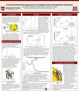
-0.05
0.05
0.15
0.25
0.35
0.45
300 400 500 600 700 800 900 1000 1100
Absorbance(AU)
Wavelength (nm)
UV-Visible Absorbance Spectrum of
AuNPs
K10A
K13A
K50A
K19A
The GB3 protein has 7 lysine residues. These
residues were changed to alanine residues
one at a time through site-directed
mutagenesis to create 7 different variants of
GB3.
K19A
Lysine side chains carry a positively charged
amine group while alanine side chains only
have a neutral methyl group. It has been
hypothesized that the electrostatic charges
between positively charged amino acid
residues and negatively charged
nanoparticles play an important role in
binding capacities between the two.
By independently reducing the charges of
theses residues, the effects of the absence of
each lysine can be analyzed, and the binding
orientation of the protein on AuNPs can be
better understood.
The protein samples’ lysine
residues were methylated
(shown in Figure 4 in blue)
in order to mark these
residues in the protein. Mass
spectrometry was used in
conjunction with HMQC
spectra (as seen in Figure 5
below) to ensure that the
desired methylations (and
therefore mutations) were
achieved. Expected mass of
the methylation of WT GB3
The “x” on the spectrum marks a significant difference in the WT
and variant data. The lack of a strong red peak seen here indicates
that a variant was created and identifies the peak to the
corresponding residue for future reference. The lack of an amine
group at the K19 location prevented methylation at this location
resulting in the absence of a strong peak. The correct mutation
was achieved.
Both the relative size and concentration of the nanoparticles in a
sample can be obtained from absorbance graphs. The max
absorbance is highlighted in red above. It has been found in
previous research that nanoparticles with diameters around 15nm
have a peak absorbance around 520nm.
(521, 0.490933418)
Known concentrations of nanoparticles were combined with
known concentrations of protein. NMR spectra of the
samples were taken after incubation in order to determine
the concentration of protein on the nanoparticles. This same
process was repeated with all 7 variants. Figures 7 and 8
show the difference in results seen. 15nm AuNPs have been
found to have a saturated binding capacity of 195 WT GB3
proteins per NP. Reductions in bound GB3 concentrations
from the WT GB3 values graphed above correlate to a
significant decrease in the average number of proteins
bound per AuNP. The K10A variant did not have much of
an effect on binding capacity while the K50A variant
showed a notable drop in binding capacity to 169 proteins.
Based on the results of the binding
capacity tests, it was found that
residues near to or residing in the
beta strands such as K13 and K50
showed significant reductions in
binding capacity when changed to
alanine residues. These findings
have lead to two conclusions:
• Electrostatic charges between
proteins and nanoparticles have
a significant effect on protein-
nanoparticle adsorption
• GB3 must be oriented on the
nanoparticle in such a way as to
allow binding between the
residues in the beta strands and
the nanoparticle.
Figure 9 : A general predicted
………….orientation of GB3
………….on AuNPs
Figure 6 : UV-Visible absorbance of a 15 nm sample of gold nanoparticles
Figure 5 : HMQC spectrum for the K19A (red) variant after methylation
.................overlaid on the wild type GB3 (blue) spectrum
Figures 7 (top) and 8 (bottom) : Binding capacities of K10A and K50A
…………………………………...compared to that of wild type GB3
Figure 4 : Liquid chromatography mass spectrum
………….of WT GB3 methylated lysine residues
Figure 2 : Chemical structures of lysine
...…...........(left) and alanine (right)²
Figure 1 : Steps to protein-adsorption¹
Figure 3 : A ribbon diagram of GB3 with
………….the different lysine residues
………….highlighted in yellow.
Further studies are needed to fully understand protein-
nanoparticle interactions. Knowledge of these interactions
will pave the way to unlocking the full potential of
nanoparticle technology making them a viable tool in modern
day biomedicine.
K10A
K50A
Intensity
+7
+6 +5
919.76
1072.89 1287.26
m/z
was 6431.8. The mass observed ((m/z)*z) was 6431.3 meaning
that the methylation process was a success.