In-vitro Interaction of αB-Crystallin on Serum Amyloid A and Serum Amyloid A ...theijes
The interactions of SAA and SAA protofibrils with protecting role of alphaB-Crystallin with hepta 1-6 cells of the mouse are dealt with in detail to study the binding of SAA protofibrils in various conditions. Specifically, interaction of serum amyloid A fibrils with a cell surface binding site/receptor might alter the local environment to cause cellular dysfunction and to be more favorable for amyloid formation and prevention with alphaB-Crystallin. This is important in relation to the activity of membrane proteins, because losing the activity of such systems will ultimately lead to malfunction or death of the cell. The interactions of Serum Amyloid A (SAA) and Serum Amyloid A protofibrils with hepta 1-6 cells of the mouse are dealt with in detail to study the binding of SAA protofibrils in various onditions. The induced fluorescence, induced circular dichroism, FACScan and MTT assay results have shown the SAA and SAA prototfibrils binding and cell toxicity with the hepta 1-6 cells with different concentrations of alphaB-Crystallin 0.15-15 nM. Specifically, cells were incubated with 1.25-6.25 M SAA-FITC and SAA protofibrils-FITC assayed. The 50% viable hepta 1-6 cells at 4–6 M with an LD50 of 3.5 M. The interaction of serum amyloid A fibrils with a cell surface binding site/receptor might alter the local environment to cause cellular dysfunction and to be more favorable for amyloid formation. In the present study, concluding that the SAA fibrils and SAA protein binding and cell cytotoxicity was reduced in the presence of alphaB-Crystallin.
A physical process by which a polypeptide chain (sequence of amino acids) folds into its characteristic & functional native structure from a random coil or a linear sequence.
Protein aggregation is the most discussed topic as it is being linked to many neurodegenerative diseases. Here, in these slides I have tried to explain about protein aggregation and its mechanism.
Histone demethylase and it srole in cell biology reviewChristopher Wynder
This document provides a scientific review of the histone demethylase enzymes; particularly the H3K4 demethlases (KDM5 family) focusing on their role in cell biology. This review was written in 2014
Collagen is made up of the repeating pattern Glycine-X-Y, where X and Y are commonly L-proline (Pro) and 4(R)-hydroxy-l-proline (Hyp), respectively. By substituting X and Y with a fluorine probe, stereoelectronic effects can be observed and compared to the effects of hydrogen bonding which has been predicted to be the main contributor to the collagen triple helix strength.
Contributed by: Alexandra Zudova, Samuel Broadbent (Undergraduates), University of Utah, 2013
Entosis is an interesting cell mechanism in which actually one cell can eat other cell and this can be helpful to combat the cancer. Future scopes are wide and lot more can be revealed in this.
“Ensemble contre le gaspillage alimentaire!” (together against food waste) on October 27 will welcome global experts to talk about the impacts of food waste on the environment and share best practices to avoid it.
Luxembourg hopes to bring an end to wasteful household practices which mean that one third of food produced ends up in bins.
The Ministry of Agriculture, Viticulture and Consumer Protection is launching a campaign to raise awareness about waste, publishing a pamphlet and organising a conference.
The International Journal of Engineering and Science (IJES)theijes
The International Journal of Engineering & Science is aimed at providing a platform for researchers, engineers, scientists, or educators to publish their original research results, to exchange new ideas, to disseminate information in innovative designs, engineering experiences and technological skills. It is also the Journal's objective to promote engineering and technology education. All papers submitted to the Journal will be blind peer-reviewed. Only original articles will be published.
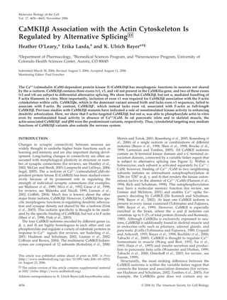
2. quences homologous to the CaMKII variable exons v1,
v3, and v4 (Figure 1), whereas the CaMKII gene does not
contain a homologue to an insert that mediates nuclear
targeting of the CaMKII␣ splice variant ␣B (Srinivasan et
al., 1994; Heist et al., 1998). Thus, we hypothesized that
sequences encoded by the exons v1, v3, and/or v4 are
involved in CaMKII-specific targeting to the F-actin cy-
toskeleton. At least CaMKII exons v1 and v4 are subject
to alternative splicing in brain. Although expression of
CaMKII dominates in mature brain, expression of CaMKIIe,
which lacks exon v1, dominates in brain around and
before birth (Brocke et al., 1995). Interestingly, alternative
splicing of exons v1 (lacking in e and eЈ) and v4 (lack-
ing in Ј and eЈ) is differentially regulated even among
individual mature hippocampal CA1 pyramidal neurons,
with most neurons expressing exclusively one splice vari-
ant (Brocke et al., 1999). Thus, tightly regulated alternative
splicing may control expression of differently actin-asso-
ciated CaMKII variants, thereby possibly affecting the
morphogenic functions of CaMKII in dendritic arboriza-
tion and/or synapse density. Results of this study sum-
marized in Figure 1 show that CaMKII and Ј, but not
e, associated with the F-actin cytoskeleton, demonstrat-
ing a developmental switch between differently targeted
splice variant.
MATERIALS AND METHODS
F-Actin Bundling Assays by Centrifugation and Electron
Microscopy
Purified actin from chicken muscle was a kind gift by Dr. R. Rock (Depart-
ment of Biochemistry, Spudich Lab, University of California, Stanford, CA).
CaMKII␣ or  was purified from a baculovirus/Sf9 expression system on a
phosphocellulose column followed by gel filtration chromatography, as de-
scribed previously (Singla et al., 2001; Bradshaw et al., 2002; Fink et al., 2003).
Actin (4 M) was polymerized on ice in F-buffer (25 mM HEPES, pH 7.4, 50
mM KCl, 2 mM MgCl2, 1 mM EGTA, 0.5 mM dithiothreitol [DTT], and 0.2
mM ATP). CaMKII (170 nM subunits), 3 M CaM, and/or 2 mM CaCl2 were
added as indicated and binding was assessed as described previously (Fink et
al., 2003). However, sedimentation was carried out at lower centrifugation
speed (10,000 ϫ g) for 20 min. Both F-actin and CaMKII were spun under the
same condition before the binding assay. Supernatants and pellets were
analyzed for actin and CaMKII␣ or  by Western blot analysis with the
specific antibodies anti-actin 20–33 (rabbit polyclonal; 1:500 in 2% bovine serum
albumin [BSA]; Sigma-Aldrich, St. Louis, MO), CB␣2 or CB1 (mouse mono-
clonals; 1:2000 and 1:1000 in 2.5% milk), respectively (Bayer et al., 1998; Fink
et al., 2003).
For electron microscopy, actin was polymerized, and kinase was added as
described above. Copper grids were coated with Formvar and carbon and
then glow discharged. Approximately 10 l of the actin/kinase mix was
applied to the copper grids for 2 min and excess liquid was wicked away with
blotting paper. Grids were stained with 1–2% uranyl acetate for 2 min and
then briefly rinsed in distilled water. Excess liquid was removed, and grids
were allowed to dry. Electron microscopy was performed on a Tecnai G2
BioTwin (at 80 kV and 49,000 magnification) in the electron microscopy core
facility of University of Colorado Health Sciences Center (Aurora, CO).
Reverse Transcription-Polymerase Chain Reaction
(RT-PCR) Expression Analysis
Pancreatic islets from Sprague Dawley rats (Harlan, Indianapolis, IN) were
isolated at the islet core facility of the Diabetes and Endocrine Research Center
at University of Colorado Health Sciences Center. Total cellular RNAs were
prepared using RNAqueous-4PCR (Ambion, Austin, TX). SuperScriptIII re-
verse transcriptase (Invitrogen, Carlsbad, CA) was used for cDNA synthesis,
primed with both random decamers and 12–18mer oligo(dT). PCR amplifi-
cation for expression analysis was done with Platinum-Taq (Invitrogen) and
CaMKII-specific primers flanking the variable region (bvf, GACAG-
GAGACTGTGGAATGTC and bvr, TCAAAGTCGCCGTTGTTGAC) for 40
cycles with 15-s denaturation at 94°C for 20 s, annealing at 55°C, and 90-s
elongation at 72°C (shorter elongation times favored shorter PCR products
from skeletal muscle cDNA, corresponding in length to  instead of M; our
unpublished data). RT-PCR products were separated on 2% agarose gels in
TAE-buffer, and stained with ethidium bromide (0.1 g/ml gel). For direct
sequencing, RT-PCR products were purified using QIAquick (QIAGEN, Va-
lencia, CA).
Cell Culture, Transfection, and Constructs
Cos-7 cells were cultured on glass-bottomed dishes (30 mm with 12-mm glass
bottom; MatTek, Ashland, MA) and transfected by the calcium phosphate
method as described previously (Bayer et al., 1998, 2001, 2006). An enhanced
green fluorescent protein [EGFP] A207K mutant with reduced dimerization
was used to create membrane-associated form of green fluorescent protein
(mGFP)-CaMKII constructs (Zacharias et al., 2002; Bayer et al., 2006). Vectors
for expression of unlabeled or hemagglutinin (HA)-tagged CaMKII splice
variants were described previously (Brocke et al., 1995; Bayer et al., 2002);
SacI/PmlI fragments were exchanged to create the mGFP fusion protein (the
SacI site in the multiple cloning site of the original mGFP-CaMKII was
deleted by religation after XhoI/EcoRI cut). mGFP-CaMKII K43R, A303R,
and T287D were created by exchanging cyan fluorescent protein in previously
described constructs (Bayer et al., 2002) with mGFP. mGFP-CaMKII⌬3 was
created by PCR mutagenesis by using mGFP-CaMKIIe as a template and
primer combination that flanked the exon v3 and v4 sequences. After se-
quencing of a positive clone, a SacI/PmlI fragment was exchanged with the
original vector as described above to avoid possible PCR-generated mutations
in the vector sequence.
F-Actin Staining and Microscopy
mGFP-CaMKII localization in Cos-7 cells was analyzed 2 d after transfection.
Scoring of actin cytoskeletal localization in live cells was done on a Nikon
TE-300 inverted microscope with a 63ϫ objective, blind of the mGFP-CaMKII
variant used. Fifty or all transfected cells per dish were scored; eight dishes
were analyzed for each construct. Alternatively, cells were fixed in 4% para-
formaldehyde in 100 mM phosphate buffer, pH 7.2, for 10 min at room
temperature, permeabilized for 20–30 min in 0.1% Triton X-100 in phosphate-
buffered saline (PBS), and F-actin was stained by 165 nM Texas Red-phalloi-
din (Invitrogen) in PBS for 30 min. Images were taken on a Zeiss Axiovert
200M microscope (Carl Zeiss, Thornwood, NY) equipped with a 63ϫ plan-
apo/1.2 numerical aperture objective, 175-W xenon light source, indepen-
dently controlled excitation and emission filter wheels, and a CoolSnapHQ
camera (Roper Scientific, Trenton, NJ). Fluorescence images were acquired
blind of the mGFP construct and analyzed for GFP/Texas Red colocalization
correlation index by using SlideBook software (Intelligent Imaging Innova-
Figure 1. CaMKII variants and their F-actin localization based on
results of this study. Schematic representation of individual iso-
forms (left) and holoenzyme structure (right). CaMKII␣ and  differ
most in the variable region; however, they are products of different
genes and thus also have minor differences in the kinase and asso-
ciation domain. Bottom, exon/intron structure of the CaMKII␣ and
 gene loci coding for the variable regions, with alternative splicing
indicated. ⌬3 is a deletion mutant, not a splice variant found in
brain (see Supplemental Figure 2). Subcellular localization of the
kinase variants (see Figure 3) implicated necessity of exon v1
(boxed) for association with the F-actin cytoskeleton.
F-Actin Localization of CaMKII Variants
Vol. 17, November 2006 4657
3. tions, Denver, CO). Six images 0.5 m apart in the z-plane were taken per cell,
deconvoluted, and maximum projected.
Fluorescence Recovery after Photobleaching (FRAP)
Analysis
Motility of mGFP-CaMKII variants in transfected Cos-7 cells was analyzed by
FRAP on the Zeiss imaging setup described above, at 30°C in 50 mM HEPES-
buffered, pH 7.4, Hank’s balanced saline. Bleaching was done using a micro-
point FRAP system (Photonic Instruments, St. Charles, IL), and images were
acquired every 5 s; the first image acquisition possible on the setup was ϳ2 s
after bleach (set to t ϭ 0 s). Background fluorescence, measured outside cells,
was subtracted. The average fluorescence intensity during 60 s before bleach
was rescaled to 1, and the intensity on the first image after bleach was rescaled
to 0. Photobleach caused by image acquisition was minimal and was not
corrected for in the data presentation. The slope (k) and maximum (ymax) of
the recovery curves was determined by curve fit to the single exponential
association function y ϭ ymax * (1 Ϫ eϪkx
) in GraphPad Prism software
(GraphPad Software, San Diego, CA). For calculating bleach efficiency, inten-
sity before bleach was rescaled to 0%, and background intensity was rescaled
to Ϫ100%. To correct the estimate of the recoverable pool for recovery
occurring even before the first test image, intensity of the first live image after
bleach was rescaled to apparent bleach efficiency in this image divided by the
real bleach efficiency as measured in fixed cells (instead of rescaling it to 0).
The estimate of the actual recoverable pool was not corrected for reduction in
the total fluorescence within cells caused by the bleach.
F-Actin Phosphorylation Assay
Purified CaMKII (100 nM subunits) was added to 4 M actin polymerized in
F-buffer as described above. The kinase/actin mix was then diluted 1:5 into
phosphorylation buffer (25 mM HEPES, pH 7.4, 50 mM KCl, 20 mM MgCl2,
0.2 mM [␥-32
P]ATP at 0.5 Ci/mmol, and 1.2 mM EGTA) and incubated for 15
min at 30°C to measure phosphorylation by basal activity. Stimulated phos-
phorylation was induced for 3 min by a mix that contained 2 mM CaCl2 and
1 M CaM instead of EGTA. CaMKII inhibitors were added as indicated. To
assess phosphorylation of kinase and actin, the reaction mixes were separated
on SDS-gels, blotted onto nitrocellulose membrane, and subjected to autora-
diography.
In a similar assay, mGFP-CaMKII wild type and K43R in extracts from
transfected Cos-7 cells were used, with extracts of mock-transfected cells as
control. Cos-7 cells were extracted in HB (50 mM PIPES, pH 7.2, 10% glycerol,
1 mM EGTA, 1 mM DTT, and 1X Boehringer protease inhibitor cocktail), and
kinase was solubilized with 150 mM sodium perchlorate before a 20-min
16,000 ϫ g centrifugation at 4°C. The extract supernatants were adjusted for
amount of kinase (as determined by GFP fluorescence) and total Cos protein
(using extracts from mock-transfected cells; determined by Bradford assay
with BSA standard). Adjusted extracts were centrifuged for 60 min at
100,000 ϫ g. The sodium perchlorate was removed from the supernatants
using Microcon YM-100 spin columns (Millipore, Billerica, MA), yielding the
final extracts used in the phosphorylation reactions.
RESULTS
F-Actin Bundling by CaMKII, but Not ␣
Based on the multimeric holoenzyme structure (Figure 1),
we hypothesized that CaMKII may be able to interact with
several F-actin filaments simultaneously. Formation of such
large F-actin complexes can be assessed in vitro by low-
speed sedimentation assays. Although individual filaments
are sedimented at 100,000 ϫ g, presence of a cross-linking
protein is required for F-actin sedimentation at 10,000 ϫ g.
Indeed, F-actin was sedimented at low speed only in pres-
ence of CaMKII (Figure 2A). As previously described for
binding, formation of large CaMKII/F-actin complexes
was disrupted by Ca2ϩ
/CaM; either Ca2ϩ
or CaM alone was
not sufficient to disrupt these complexes (Figure 2A). By
Figure 2. CaMKII, but not ␣, bundles F-actin. (A) Actin was polymerized before addition of CaMKII or ␣, as indicated. CaMKII and actin
content of supernatants (S) and pellets (P) after low-speed centrifugation (10,000 ϫ g; 20 min) were analyzed by the Western blots shown.
Left, CaMKII and actin were found in the pellet only when mixed; CaMKII␣ and actin did not cosediment. Right, Addition of Ca2ϩ
/CaM
(2 mM/3 M), but not Ca2ϩ
or CaM alone, prevented cosedimentation of CaMKII and actin. (B) F-actin filaments are visualized by electron
microscopy and do not form bundles in absence of kinase (left) or in presence of CaMKII␣ (right). (C) Addition of CaMKII to polymerized
actin resulted in bundling of the F-actin filaments, as assessed by electron microscopy. In some areas, F-actin cross-linking without bundling
was apparent (right). Bars, 50 nm.
H. O’Leary et al.
Molecular Biology of the Cell4658
4. contrast, CaMKII␣ did not result in F-actin sedimentation
under any conditions.
To examine the nature of the larger F-actin/CaMKII
complexes, electron microscopy was performed after nega-
tive staining with uranyl acetate. Without kinase or in pres-
ence of CaMKII␣, no formation of extensive actin filament
bundles was observed (Figure 1B and Supplemental Figure
1), as expected. By contrast, addition of CaMKII resulted in
formation of F-actin bundles with multiple parallel filaments
(Figure 2C). Additionally, in some areas CaMKII seemed to
cross-link filaments without bundle formation (Figure 2C,
right). In these areas, particles of CaMKII holoenzyme size
(ϳ25 nm in diameter) were observed associated with the
actin filaments. Similar particles were also seen associated
with the F-actin bundles. Although such particles also may
be present within the tightly packed bundles, they could not
be clearly distinguished in such location (Figure 2C).
CaMKII and , but Not ⌬3 or e, Associated with the
Actin Cytoskeleton
We hypothesized that the  variable exons v1, v3, and/or
v4, which are lacking in the ␣ isoform, are necessary for
F-actin association (Figure 1). CaMKIIe and Ј lack exons
v1 and v4, respectively. By contrast, no splice variant lacking
exon v3 was detected in rat brain (Supplemental Figure 2).
Thus, we generated a CaMKII mutant (⌬3) that lacks
exons v1, v3, and v4. mGFP fusion proteins of the CaMKII␣
and  isoforms, the ⌬3 mutant, and the splice variants Ј,
e, and M (Figure 1) were expressed in Cos-7 cells, and
their subcellular localization was analyzed (Figure 3). First,
apparent F-actin localization (stress fibers, cortical actin, and
membrane ruffles) of the GFP fluorescence in live cells was
scored into three categories (strong, mild, and no apparent
F-actin localization) (Figure 3A). Then, cells were fixed and
F-actin was stained with phalloidin-Texas Red, and the in-
Figure 3. Alternative splicing modulates F-actin association of CaMKII. (A) Scores of apparent F-actin localization for GFP-CaMKII
variants in live Cos-7 cells. Scoring was done blind of the variant. n ϭ 8 coverslips (50 or all transfected cells scored). There are no differences
within the groups labeled * or ** (p Ͼ 0.3; analysis of variance [ANOVA]), but each * is different from each ** (p Ͻ 0.00005; two-tailed t tests)
(tested were “strong” and “none” as independent values). (B) F-actin/GFP-CaMKII correlation of localization values determined using
SlideBook software, after staining of F-actin with phalloidin-Texas Red as shown in C. n ϭ 9 images. **, not different from each other (p Ͼ
0.2; ANOVA), but different from  and Ј (p Ͻ 0.00001) and M (p Ͻ 0.05). Error bars show SEM. (C) Examples of GFP-CaMKII variant
localization in Cos-7 cells. F-actin was stained by phalloidin-Texas Red after fixation.
F-Actin Localization of CaMKII Variants
Vol. 17, November 2006 4659
5. dex of GFP/Texas Red colocalization was determined on
deconvoluted images (Figure 3B). Examples of phalloidin-
stained transfected Cos-7 cells are shown in Figure 3C. As
hypothesized, CaMKII⌬3 did not associate with F-actin
structures. Even lack of exon v1 alone, in the splice variant
e, was sufficient to disrupt F-actin association. Localization
of both ⌬3 and of e was indistinguishable from CaMKII␣
localization in both analysis methods. By contrast, lack of
exon v4 in Ј did not reduce F-actin association seen for the
full-length CaMKII. CaMKIIM contains an additional in-
sert C-terminal of exon v5 (Figure 2) that generates putative
binding sites for Src homology 3 (SH3) domains (Bayer et al.,
1998). Localization of M, , and Ј were indistinguishable
in the scoring analysis in live cells (Figure 3A). After phal-
loidin stain, the F-actin colocalization coefficient for M was
significantly higher than for ␣, ⌬3, and e; however, it was
lower compared with  and Ј (Figure 3B). This may reflect
association of the M variant with additional subcellular
targets, as has been speculated previously based on putative
SH3 binding (Urquidi and Ashcroft, 1995; Bayer et al., 1998).
Together, the results clearly indicate that the variable region
exon v1, which is lacking in CaMKIIe, is necessary for
localization to the actin cytoskeleton within cells (for over-
view, see Figure 1).
CaMKII␣ and e Were More Mobile within Cells than 
and 
To further probe into differential subcellular anchoring of
the CaMKII isoforms and splice variants, we used FRAP of
the mGFP fusion proteins expressed in Cos-7 cells (Figure 4).
FRAP of the nonactin binding CaMKII␣ and e was indis-
tinguishable from each other and relatively fast (Figure 4A)
(k ϭ 5.79 Ϯ 0.26 and 5.12 Ϯ 0.29 minϪ1
, respectively, after
curve fit to single exponential association). Fluorescence
recovered almost completely (ϳ90%) within 2 min, indicat-
ing that ␣ and e are mobile and not persistently targeted to
any subcellular structures within Cos-7 cells. FRAP of
CaMKII and Ј was also similar to each other, but signifi-
cantly different from ␣ and e FRAP (Figure 4A). FRAP was
slower (k ϭ 1.55 Ϯ 0.08 and 2.15 Ϯ 0.09 minϪ1
, respectively)
and fluorescence recovered only partially (ϳ50%). This in-
dicates that a significant pool of  and Ј remained persis-
tently bound to F-actin and did not exchange in the unstimu-
lated Cos-7 cells. Examples of e and Ј FRAP images are
shown in Figure 4A. In addition to bleaching e in its typical
evenly distributed localization (Figure 4A, first row), we also
bleached e enriched in actin-like structures (Figure 5A,
second row), which is more typical for  or Ј localization
Figure 4. CaMKII variants differ in their mo-
tility within cells. FRAP of GFP-CaMKII was
assessed in Cos-7 cells at 30°C. (A) Top, ex-
amples of GFP-CaMKII bleach and recovery
in Cos-7 cells. Two examples are shown for
e: one bleach of its typical dispersed local-
ization and one bleach is of an F-actin–like
structure. Bottom, quantitative assessment of
bleach recovery over time, with fluorescence
before and immediately after bleach rescaled
to 1 and 0, respectively. FRAP differed be-
tween cytosolic (␣ and e) and actin-binding
( and Ј) variants (p Ͻ 0.005 in two tailed t
tests; n ϭ 10 bleaches in 5 cells for each vari-
ant). (B) Apparent bleach efficiencies differed
between cytosolic and actin-binding variants
in live but not in fixed cells. *, not different
from each other (p Ͼ 0.4), but different from
all other measurements (p Ͻ 0.005); other data
points do not differ from each other (p Ͼ 0.4;
ANOVA) (n ϭ 10). This suggests partial re-
covery even before the first picture could be
taken (ϳ2 s after bleach). This indicates that
the recoverable pool for the cytosolic variants
is even greater than indicated by the rescaled
measurements in A. Error bars show SEM.
H. O’Leary et al.
Molecular Biology of the Cell4660
6. (Figure 4A, third row). FRAP of e was equally fast and
complete in both localizations, indicating that any apparent
actin-like localization of e is different in nature from the
more extensive actin association of  or Ј.
Apparent bleach efficiencies for mGFP-CaMKII␣ and e in
live cells were only about one-half of that seen for  and Ј
(Figure 4B). This is probably because of significant diffusion
of ␣ and e even during the time between the bleach and
acquisition of the first image (ϳ2 s). Indeed, mGFP-CaMKII␣
and  showed the same bleach efficiency in fixed cells, which
was also undistinguishable from the apparent bleach effi-
ciencies for  and Ј in live cells (Figure 4B). Thus, the
recoverable pool of ␣ and e during FRAP is even higher
than initially estimated from the rescaled fluorescence re-
covery shown in Figure 4A (ϳ95% instead of ϳ90%). Nota-
bly, monomeric mGFP alone typically showed only minimal
apparent bleach efficiency but in a much larger apparent
bleach area (our unpublished data). Thus, FRAP of the sol-
uble ␣ and e is probably resolvable in our experimental
setup only because of relatively slow diffusion of the large
mGFP-CaMKII holoenzymes (Ͼ900 kDa).
Localization of CaMKII Mutants
CaMKII and several of its mutants were previously tested
for their effect on dendritic arborization of hippocampal
neurons in dissociated culture (Fink et al., 2003). We have
now quantified the actin cytoskeletal localization of these
CaMKII mutants in Cos-7 cells (Figure 5). A303R (impaired
for CaM binding) and K43R (impaired for ATP binding)
localized to F-actin similarly as wild type. However, in the
live cell scoring, A303R seemed to localize even stronger
than wild type (p Ͻ 0.05 in two-tailed t test) (Figure 5).
A303R does not disperse from F-actin upon Ca2ϩ
signals
(Shen and Meyer, 1999), likely because of impaired CaM
binding; this seems to mildly enhance localization even in
unstimulated cells. By contrast, the CaMKII mutant T287D
(constitutively active) did not show F-actin colocalization
(Figure 5). T287D mimics autophosphorylation at T287,
indicating that T287 autophosphorylation is involved in
regulation of actin binding, either directly or by increas-
ing affinity for Ca2ϩ
/CaM (Meyer et al., 1992). The T287D
mutant had significantly lower effect on dendritic ar-
borization than wild type (Fink et al., 2003), further sup-
porting functional importance of cytoskeletal localization
of CaMKII.
F-Actin Phosphorylation by Basal Activity of Bound
CaMKII
The CaM-binding impaired CaMKII mutant A303R en-
hanced dendritic arborization in hippocampal neurons,
whereas the ATP-binding impaired K43R mutant did not
(Fink et al., 2003), indicating a possible role for CaM-inde-
pendent kinase activity. Could F-actin binding directly en-
hance CaM-independent CaMKII activity? CaMKII bind-
ing to F-actin did not increase phosphorylation of a substrate
peptide, indicating that the activation state of the kinase was
not changed (Fink et al., 2003). However, CaMKII can auto-
phosphorylate at residues other than T286/T287, even by its
basal activity (Colbran, 1993), that is, by activity without
Ca2ϩ
/CaM stimulation or phosphorylation at T286/287
(Figure 6) (T286/T287 autophosphorylation requires bind-
ing of Ca2ϩ
/CaM to make T286/T287 accessible as a sub-
strate, in addition to kinase activation; Hanson et al., 1994;
Rich and Schulman, 1998). Thus, we hypothesized that basal
activity of targeted CaMKII could similarly suffice to phos-
phorylate other substrate proteins that are anchored in the
right position. Indeed, purified CaMKII phosphorylated
F-actin in vitro even by nonstimulated basal activity,
whereas the nonactin-binding CaMKII␣ isoform did not
(Figure 6). This is not because of differential substrate pref-
erence of the isoforms, because CaMKII␣ did phosphorylate
actin at least as well as CaMKII when stimulated by Ca2ϩ
/
CaM (Figure 6). The phosphorylated actin-sized band was
only observed in presence of actin, but not when actin was
omitted or substituted for BSA, or when CaMKII was omit-
Figure 6. Purified CaMKII, but not ␣, can phosphorylate actin in
vitro even by nonstimulated basal activity. Both isoforms phosphor-
ylate actin when stimulated by Ca2ϩ
/CaM. The CaM-competitive
CaMKII inhibitor KN93, but not the inactive control compound
KN92, inhibited stimulated kinase activity. Basal activity was not
affected, as expected. KN concentration was 10 M unless noted
otherwise. Phosphorylation of actin and CaMKII autophosphoryla-
tion were detected by 32
P incorporation and autoradiography.
Figure 5. F-actin localization of CaMKII mutants. GFP-CaMKII
mutants A303R (CaM binding impaired), K43R (ATP binding im-
paired), and T287D (constitutively active) were tested for F-actin
localization in Cos-7 cells by live scoring (n ϭ 8) or correlation with
phalloidin stain (n ϭ 9) as described in Figure 3. CaMKII T287D
and CaMKII␣ (**) showed significantly less F-actin localization than
any other variant (p Ͻ 0.0005 in two-tailed t test), and they did not
differ from each other.  A303R (*) showed slightly increased actin
localization in the live scoring (p Ͻ 0.05 for the strong localization
category).
F-Actin Localization of CaMKII Variants
Vol. 17, November 2006 4661
7. ted (Supplemental Figure 3). The CaMKII inhibitor KN93,
but not the inactive analogue KN92, inhibited Ca2ϩ
/CaM-
stimulated actin phosphorylation by both CaMKII␣ and .
Actin phosphorylation by basal activity of CaMKII was not
inhibited (Figure 6A), as expected, because KN93 is compet-
itive with Ca2ϩ
/CaM (Sumi et al., 1991) and thus should not
inhibit basal activity. Other inhibitors such as AIP or CaM-
KIINtide are not competitive with Ca2ϩ
/CaM, but bind to
CaMKII only in the activated and not in the basal state
(Ishida et al., 1995; Chang et al., 2001). To further test actin
phosphorylation by basal CaMKII activity, mGFP-
CaMKII wild type and the ATP-binding impaired mutant
K43R from extracts of transfected Cos-7 cells were used
(Supplemental Figure 3). The K43R extract caused phos-
phorylation above background, indicating some residual
kinase activity of this mutant; the alternative that actin bun-
dling by CaMKII activated another kinase seems less likely.
Also, wild-type kinase extracts caused a higher degree of
basal actin phosphorylation than both K43R extracts and
extracts from mock-transfected cells (Supplemental Figure
3). These results strongly support phosphorylation by basal
activity of targeted CaMKII and give a possible explana-
tion why the two activation-deficient CaMKII mutants
A303R (CaM-binding impaired) and K43R (ATP-binding im-
paired) have opposite effects on dendritic arborization (Fink
et al., 2003).
Expression of CaMKII Variants during Development and
in Different Tissues
RT-PCR analysis showed similar expression of the splice
variants CaMKII, Ј, e, and eЈ in frontal cortex of rat
brain on day 2 after birth, whereas  dominated by day 14
and was even more prominent in adult brain (Figure 7A).
This developmental switch is consistent with a previous
report (Brocke et al., 1995). However, CaMKIIe expression
is not eliminated during development (Brocke et al., 1999;
Supplemental Figure 2). Expression of CaMKII variants
was also detected in pancreatic islets and in skeletal muscle,
but not in heart (Figure 7B). The major splice variant in
skeletal muscle correspondend in length to CaMKIIM
(Bayer et al., 1998), whereas the major variant in islets cor-
responded to CaMKIIЈ. Identity of CaMKIIЈ was con-
firmed by restriction digest (Figure 7C) and by direct se-
quencing of the RT-PCR product. Thus, skeletal muscle and
islets expressed two different but F-actin–associated
CaMKII variants (cf. Figure 3). Additionally, small
amounts of  and eЈ transcripts were identified in islets,
based on the length of two minor RT-PCR products (our
unpublished data).
DISCUSSION
The isoform specificity of CaMKII function in enhancing
dendritic arborization and synapse density is thought to be
mediated by CaMKII binding to F-actin, a property not
shared by the ␣ isoform (Fink et al., 2003). Consistent with
this isoform-specific binding, the CaMKII, but not the ␣
isoform, assembled with F-actin into large complexes, in-
cluding bundles of parallel actin filaments. Most impor-
tantly, alternative splicing of CaMKII regulated its local-
ization to F-actin within cells. CaMKII and Ј, but not e,
associated with the actin cytoskeleton, indicating that the
sequence encoded by variable exon v1 is necessary for the
interaction (Figure 2). Alternative splicing of exon v1 de-
pends both on cell type and developmental stage.
CaMKIIe is the dominant splice variant before birth;
CaMKII becomes dominant between day 4 and 13 of post-
natal rat brain development, both in cortex and hippocam-
pus (Brocke et al., 1995). Thus, alternative splicing of CaMKII
provides a developmental switch from a cytoplasmic to an
actin cytoskeleton-associated isoform. The F-actin–binding
CaMKII enhances dendritic arborization and synapse for-
mation (Fink et al., 2003), and the timing of the developmen-
tal switch to this splice variant correlates with extensive
synapse formation. Notably, the e variant dominates at a
time when the other major brain isoform, CaMKII␣, is not
yet significantly expressed (Burgin et al., 1990; Bayer et al.,
1999). CaMKIIe variant transcripts also were found also in
mature rat brain. Thus, although there is a developmental
switch in  splicing, expression of e is not eliminated
during development. Indeed, e transcripts have been de-
tected previously in mature hippocampal CA1 pyramidal
neurons by single cell RT-PCR (Brocke et al., 1999). Four of
14 CaMKII-positive cells expressed e transcripts, indicat-
ing differentially regulated alternative splicing even among
mature neurons of the same cell type (Brocke et al., 1999).
Notably, splice variant expression seemed segregated, with
only one cell expressing more than one  splice variant
(Brocke et al., 1999). This segregation of expression may be
functionally important, because CaMKII coassembles also
with nonactin binding isoforms and targets such hetero-
meric holoenzymes to the actin cytoskeleton (Shen et al.,
1998). CaMKII␣ can partially escape such coassembly and
cotargeting, because its mRNA is targeted to dendrites
(Burgin et al., 1990; Mayford et al., 1996); protein synthesis in
a separate compartment then allows formation of homo-
meric holoenzymes. By contrast, coexpression of CaMKII
and e would result in actin-targeted heteromers, which can
be prevented by exclusive expression of only one variant
within individual neurons. The mechanism for differential
regulation of CaMKII splicing even among individual neu-
rons of the same cell type is presently unclear. However,
regulation by synaptic stimulation is an intriguing possibil-
ity that may provide a novel form of long-lasting neuronal
Figure 7. The actin-binding CaMKIIЈ is the dominant splice vari-
ant in rat pancreatic islets. Products from RT-PCR with CaMKII-
specific primers flanking the variable region were separated by
agarose gel electrophoresis. Grayscale-inverted images of ethidium
bromide stains are shown. (A) Cortex and whole brain was ana-
lyzed at different days after birth (pn), as indicated. Assignment of
CaMKII splice variants to the bands was based on length and
Brocke et al. (1995). (B) CaMKII splice variants were detected in
brain, skeletal muscle, and pancreatic islets, but not in heart of adult
rats. The variant detected in skeletal muscle corresponded in length
to CaMKIIM (and contained exon v1 sequences based on SacII
digest; our unpublished data); the major variant detected in islets
corresponded to CaMKIIЈ. (C) The RT-PCR product from rat pan-
creatic islets was digested with enzymes that cut in different vari-
able exons (Ϫ, no enzyme; v1, SacII; v3, BamHI; and v4, MseI).
Resistance only to MseI digest indicates lack of exon v4, correspond-
ing to CaMKIIЈ. SacII and BamHI digests yielded band of similar
length, as expected for Ј PCR amplificates (6 base pairs difference);
the same digests of  amplificates would differ by 49 base pairs.
Identity of the RT-PCR product as Ј was also confirmed by direct
sequencing (our unpublished data).
H. O’Leary et al.
Molecular Biology of the Cell4662
8. plasticity. Synaptic plasticity indeed involves F-actin dy-
namics (Kim and Lisman, 1999; Fischer et al., 2000; Krucker
et al., 2000; Star et al., 2002; Penzes et al., 2003; Okamoto et al.,
2004; for review, see Lamprecht and LeDoux, 2004), which
may be differentially modulated by CaMKII isoforms with
different actin association (Fink et al., 2003).
In addition to neurons, CaMKII gene expression was
found in pancreatic islets and in skeletal muscle, but not in
heart. The dominant  transcript in skeletal muscle,
CaMKIIM, contains exon v1 (Bayer et al., 1998) and local-
ized to the actin cytoskeleton in Cos-7 cells. However, the
F-actin colocalization index for M was lower than for  and
Ј. This may reflect additional targeting of M to other
subcellular locations, possibly mediated by the putative
binding sites for SH3 domains present in the 12-kDa M-
specific insert (Urquidi and Ashcroft, 1995; Bayer et al.,
1998).
The major CaMKII transcript found in rat pancreatic
islets was Ј, which lacks exon v4 and was found to associate
with the actin cytoskeleton. By contrast, previous studies in
rat and human found predominant expression of CaMKIIeЈ
(Rochlitz et al., 2000; Tabuchi et al., 2000), which additionally
lacks exon v1 and did not associate with F-actin. This dif-
ference may be due to use of different rat strains (Sprague-
Dawley versus Wistar), raising the possibility of variation
also among individual humans. Notably, the two rat strains
show some difference in their insulin secretion and response
(Gaudreault et al., 2001; de Groot et al., 2004). CaMKII
is thought to regulate insulin secretion and production
(Wasmeier and Hutton, 1999; Bhatt et al., 2000; Tabuchi et al.,
2000; Osterhoff et al., 2003), and it will be interesting to see
whether and how these functions depend on actin associa-
tion. CaMKII␦ may be the second major isoform in islets
(Rochlitz et al., 2000; Osterhoff et al., 2003); however, it was
not detected in all studies (Breen and Ashcroft, 1997;
Tabuchi et al., 2000). CaMKII␦ variants do not contain se-
quences homologous to exon v1, but the CaMKII␦ associa-
tion domain has been reported to mediate localization of a
GFP-fusion protein to the actin cytoskeleton (Caran et al.,
2001). However, it should be noted that actin localization of
CaMKII␦ was rather mild, and, in contrast to the CaMKII
localization observed here, became apparent only after fix-
ation of the cells (Caran et al., 2001).
CaMKII bundled and cross-linked F-actin filaments. This
suggests that CaMKII holoenzymes can bind multiple F-
actin filaments simultaneously. Inactive holoenzymes form
disk-like structures composed of two stacked hexameric
rings (Kolodziej et al., 2000; Hoelz et al., 2003; Rosenberg et
al., 2005). Thus, at least two actin filaments should be able to
bind to a holoenzyme without steric hindrance, one to each
ring and at several possible angles to each other. A previous
electron microscopic study detected binding of CaMKII to
F-actin (Ohta et al., 1986), but it did not describe cross-
linking of filaments. However, the CaMKII preparation used
in the study likely contained mostly the ␣ isoform (from a
soluble, noncytoskeletal fraction of forebrain); small amounts
of  isoform present may have been sufficient to mediate
some binding, but not bundling or cross-linking.
CaMKII dissociates from F-actin upon Ca2ϩ
/CaM stim-
ulation (Fink et al., 2003; Figure 2). Thus, cross-linking of
actin filaments by CaMKII may play a role in Ca2ϩ
-regu-
lated actin dynamics. However, such a possible direct struc-
tural function is presently unclear. Moreover, kinase activity
is required for the morphogenic function of CaMKII in
enhancing dendritic arborization, because a kinase dead
mutant (K43R) even had a dominant-negative effect (Fink et
al., 2003). By contrast, a CaM binding-deficient mutant
(A303R) still enhanced arborization (Fink et al., 2003), indi-
cating a function of CaM-independent activity. Indeed,
whereas both CaMKII and ␣ phosphorylated actin upon
Ca2ϩ
/CaM stimulation in vitro, only the F-actin–binding
CaMKII phosphorylated actin even by its nonstimulated
basal activity. Binding to F-actin does not increase CaMKII
activity toward a soluble peptide substrate (Fink et al., 2003),
suggesting that targeting generated a high local substrate
concentration that is sufficient to result in significant phos-
phorylation even by basal activity. Consistent with such
interpretation, basal CaMKII activity is sufficient for auto-
phosphorylation (at residues other than T286 in ␣ or T287 in
) (Colbran, 1993). A previous study indicated that CaMKII
can phosphorylate SynGAP also by basal activity when both
proteins are bound to the adaptor protein MUPP1 (Krapiv-
insky et al., 2004). Thus, a function of basal kinase activity
after targeting may underlie the different effects of the two
activity- or activation-impaired mutants (Fink et al., 2003).
Although the morphogenic effect of CaMKII could involve
direct phosphorylation of actin, it seems more likely that
phosphorylation of other actin-bound regulatory proteins is
required.
Previous studies showed that alternative splicing of CaMKII
modulates its Ca2ϩ
-spike frequency response (Bayer et al.,
2002). The results presented here show that alternative splicing
also regulated the association of CaMKII with the actin cy-
toskeleton, which is thought to underlie the specific morpho-
genic role of CaMKII in neurons (Fink et al., 2003). It will be
interesting to elucidate how these two biochemical properties
contribute to differential function of the CaMKII splice vari-
ants, and how the alternative splice events are regulated.
ACKNOWLEDGMENTS
We are grateful to Dot Dill and Francisco Ramirez (University of Colorado
Health Sciences Center) for excellent technical assistance with the electron
microscopy and islet preparations, respectively. We thank Dr. Mark
Dell’Acqua (University of Colorado Health Sciences Center) and laboratory
members, especially Drs. Jessica Gorski, Karen Smith, Eric Horne, and Mat-
thew Pink for critical reading of the manuscript. We thank Drs. Tobias Meyer
(University of California, Stanford) and Charles Fink (University of Wiscon-
sin, Madison, WI) for sharing constructs. Purified actin was a kind gift from
Dr. R. Rock. We thank Dr. Mike Bradshaw (Roche, Palo Alto, CA) for discus-
sions and help with CaMKII purification. The research was supported by a
Pilot and Feasibility Award P30 DK57516 from the National Institutes of
Health Diabetes and Endocrine Research Center at University of Colorado
Health Sciences Center, by National Institutes of Health Grant DK-070735,
and by American Heart Association Grant SDG 0430196N.
REFERENCES
Bayer, K. U., De Koninck, P., Leonard, A. S., Hell, J. W., and Schulman, H.
(2001). Interaction with the NMDA receptor locks CaMKII in an active con-
formation. Nature 411, 801–805.
Bayer, K. U., De Koninck, P., and Schulman, H. (2002). Alternative splicing
modulates the frequency-dependent response of CaMKII to Ca(2ϩ) oscilla-
tions. EMBO J. 21, 3590–3597.
Bayer, K. U., Harbers, K., and Schulman, H. (1998). alphaKAP is an anchoring
protein for a novel CaM kinase II isoform in skeletal muscle. EMBO J. 17,
5598–5605.
Bayer, K. U., LeBel, E., McDonald, G., O’Leary, H., Schulman, H., and De
Koninck, P. (2006). Transition from reversible to persistent binding of CaMKII
to postsynaptic sites and NR2B. J. Neurosci. 26, 1164–1174.
Bayer, K. U., Lohler, J., Schulman, H., and Harbers, K. (1999). Developmental
expression of the CaM kinase II isoforms: ubiquitous gamma- and delta-CaM
kinase II are the early isoforms and most abundant in the developing nervous
system. Brain Res. Mol. Brain Res. 70, 147–154.
Bhatt, H. S., Conner, B. P., Prasanna, G., Yorio, T., and Easom, R. A. (2000).
Dependence of insulin secretion from permeabilized pancreatic beta-cells on
the activation of Ca(2ϩ)/calmodulin-dependent protein kinase II. A re-eval-
uation of inhibitor studies. Biochem. Pharmacol. 60, 1655–1663.
F-Actin Localization of CaMKII Variants
Vol. 17, November 2006 4663
9. Bradshaw, J. M., Hudmon, A., and Schulman, H. (2002). Chemical quenched
flow kinetic studies indicate an intraholoenzyme autophosphorylation mech-
anism for Ca2ϩ/calmodulin-dependent protein kinase II. J. Biol. Chem. 277,
20991–20998.
Breen, M. A., and Ashcroft, S. J. (1997). Human islets of Langerhans express
multiple isoforms of calcium/calmodulin-dependent protein kinase II. Bio-
chem. Biophys. Res. Commun. 236, 473–478.
Brocke, L., Chiang, L. W., Wagner, P. D., and Schulman, H. (1999). Functional
implications of the subunit composition of neuronal CaM kinase II. J. Biol.
Chem. 274, 22713–22722.
Brocke, L., Srinivasan, M., and Schulman, H. (1995). Developmental and
regional expression of multifunctional Ca2ϩ/calmodulin-dependent protein
kinase isoforms in rat brain. J. Neurosci. 15, 6797–6808.
Burgin, K. E., Waxham, M. N., Rickling, S., Westgate, S. A., Mobley, W. C.,
and Kelly, P. T. (1990). In situ hybridization histochemistry of Ca2ϩ/calmod-
ulin-dependent protein kinase in developing rat brain. J. Neurosci. 10, 1788–
1798.
Caran, N., Johnson, L. D., Jenkins, K. J., and Tombes, R. M. (2001). Cytosolic
targeting domains of gamma and delta calmodulin-dependent protein kinase
II. J. Biol. Chem. 276, 42514–42519.
Chang, B. H., Mukherji, S., and Soderling, T. R. (2001). Calcium/calmodulin-
dependent protein kinase II inhibitor protein: localization of isoforms in rat
brain. Neuroscience 102, 767–777.
Colbran, R. J. (1993). Inactivation of Ca2ϩ/calmodulin-dependent protein
kinase II by basal autophosphorylation. J. Biol. Chem. 268, 7163–7170.
Colbran, R. J., and Brown, A. M. (2004). Calcium/calmodulin-dependent
protein kinase II and synaptic plasticity. Curr. Opin. Neurobiol. 14, 318–327.
de Groot, M., de Haan, B. J., Keizer, P. P., Schuurs, T. A., van Schilfgaarde, R.,
and Leuvenink, H. G. (2004). Rat islet isolation yield and function are donor
strain dependent. Lab. Anim. 38, 200–206.
De Koninck, P., and Schulman, H. (1998). Sensitivity of CaM kinase II to the
frequency of Ca2ϩ oscillations. Science 279, 227–230.
Easom, R. A. (1999). CaM kinase II: a protein kinase with extraordinary talents
germane to insulin exocytosis. Diabetes 48, 675–684.
Erondu, N. E., and Kennedy, M. B. (1985). Regional distribution of type II
Ca2ϩ/calmodulin-dependent protein kinase in rat brain. J. Neurosci. 5, 3270–
3277.
Fink, C. C., Bayer, K. U., Myers, J. W., Ferrell, J. E., Jr., Schulman, H., and
Meyer, T. (2003). Selective regulation of neurite extension and synapse for-
mation by the beta but not the alpha isoform of CaMKII. Neuron 39, 283–297.
Fischer, M., Kaech, S., Wagner, U., Brinkhaus, H., and Matus, A. (2000).
Glutamate receptors regulate actin-based plasticity in dendritic spines. Nat.
Neurosci. 3, 887–894.
Gaudreault, N., Santure, M., Pitre, M., Nadeau, A., Marette, A., and Bachelard, H.
(2001). Effects of insulin on regional blood flow and glucose uptake in Wistar
and Sprague-Dawley rats. Metabolism 50, 65–73.
Giese, K. P., Fedorov, N. B., Filipkowski, R. K., and Silva, A. J. (1998).
Autophosphorylation at Thr286 of the alpha calcium-calmodulin kinase II in
LTP and learning. Science 279, 870–873.
Griffith, L. C. (2004). Calcium/calmodulin-dependent protein kinase II: an
unforgettable kinase. J. Neurosci. 24, 8391–8393.
Hain, J., Onoue, H., Mayrleitner, M., Fleischer, S., and Schindler, H. (1995).
Phosphorylation modulates the function of the calcium release channel of
sarcoplasmic reticulum from cardiac muscle. J. Biol. Chem. 270, 2074–2081.
Hanson, P. I., Meyer, T., Stryer, L., and Schulman, H. (1994). Dual role of
calmodulin in autophosphorylation of multifunctional CaM kinase may un-
derlie decoding of calcium signals. Neuron 12, 943–956.
Heist, E. K., Srinivasan, M., and Schulman, H. (1998). Phosphorylation at the
nuclear localization signal of Ca2ϩ/calmodulin-dependent protein kinase II
blocks its nuclear targeting. J. Biol. Chem. 273, 19763–19771.
Hoelz, A., Nairn, A. C., and Kuriyan, J. (2003). Crystal structure of a tetra-
decameric assembly of the association domain of Ca2ϩ/calmodulin-depen-
dent kinase II. Mol. Cell 11, 1241–1251.
Hudmon, A., and Schulman, H. (2002). Neuronal CA2ϩ/calmodulin-depen-
dent protein kinase II: the role of structure and autoregulation in cellular
function. Annu. Rev. Biochem. 71, 473–510.
Huntley, G. W., Benson, D. L., and Colman, D. R. (2002). Structural remod-
eling of the synapse in response to physiological activity. Cell 108, 1–4.
Ishida, A., Kameshita, I., Okuno, S., Kitani, T., and Fujisawa, H. (1995). A
novel highly specific and potent inhibitor of calmodulin-dependent protein
kinase II. Biochem. Biophys. Res. Commun. 212, 806–812.
Kim, C. H., and Lisman, J. E. (1999). A role of actin filament in synaptic
transmission and long-term potentiation. J. Neurosci. 19, 4314–4324.
Kolodziej, S. J., Hudmon, A., Waxham, M. N., and Stoops, J. K. (2000).
Three-dimensional reconstructions of calcium/calmodulin-dependent (CaM)
kinase II␣ and truncated CaM kinase II␣ reveal a unique organization for its
structural core and functional domains. J. Biol. Chem. 275, 14354–14359.
Krapivinsky, G., Medina, I., Krapivinsky, L., Gapon, S., and Clapham, D. E.
(2004). SynGAP-MUPP1-CaMKII synaptic complexes regulate p38 MAP ki-
nase activity and NMDA receptor-dependent synaptic AMPA receptor po-
tentiation. Neuron 43, 563–574.
Krucker, T., Siggins, G. R., and Halpain, S. (2000). Dynamic actin filaments are
required for stable long-term potentiation (LTP) in area CA1 of the hippocam-
pus. Proc. Natl. Acad. Sci. USA 97, 6856–6861.
Lamprecht, R., and LeDoux, J. (2004). Structural plasticity and memory. Nat.
Rev. Neurosci. 5, 45–54.
Lantsman, K., and Tombes, R. M. (2005). CaMK-II oligomerization potential
determined using CFP/YFP FRET. Biochim. Biophys. Acta 1746, 45–54.
Lisman, J., Schulman, H., and Cline, H. (2002). The molecular basis of CaMKII
function in synaptic and behavioural memory. Nat. Rev. Neurosci. 3, 175–190.
Lisman, J. E., and McIntyre, C. C. (2001). Synaptic plasticity: a molecular
memory switch. Curr. Biol. 11, R788–R791.
Malenka, R. C., and Nicoll, R. A. (1999). Long-term potentiation–a decade of
progress? Science 285, 1870–1874.
Malinow, R., Schulman, H., and Tsien, R. W. (1989). Inhibition of postsynaptic
PKC or CaMKII blocks induction but not expression of LTP. Science 245,
862–866.
Mayford, M., Baranes, D., Podsypanina, K., and Kandel, E. R. (1996). The
3Ј-untranslated region of CaMKII alpha is a cis-acting signal for the localiza-
tion and translation of mRNA in dendrites. Proc. Natl. Acad. Sci. USA 93,
13250–13255.
McGee, A. W., and Bredt, D. S. (2003). Assembly and plasticity of the gluta-
matergic postsynaptic specialization. Curr. Opin. Neurobiol. 13, 111–118.
Meyer, T., Hanson, P. I., Stryer, L., and Schulman, H. (1992). Calmodulin
trapping by calcium-calmodulin-dependent protein kinase. Science 256,
1199–1202.
Morris, E. P., and Torok, K. (2001). Oligomeric structure of alpha-calmodulin-
dependent protein kinase II. J. Mol. Biol. 308, 1–8.
Ohta, Y., Nishida, E., and Sakai, H. (1986). Type II Ca2ϩ/calmodulin-depen-
dent protein kinase binds to actin filaments in a calmodulin-sensitive manner.
FEBS Lett. 208, 423–426.
Okamoto, K., Nagai, T., Miyawaki, A., and Hayashi, Y. (2004). Rapid and
persistent modulation of actin dynamics regulates postsynaptic reorganiza-
tion underlying bidirectional plasticity. Nat. Neurosci. 7, 1104–1112.
Osterhoff, M., Mohlig, M., Schwanstecher, M., Seufert, J., Ortmann, J., Schatz,
H., and Pfeiffer, A. F. (2003). Ca2ϩ/calmodulin-dependent protein kinase II
delta2 regulates gene expression of insulin in INS-1 rat insulinoma cells. Cell
Calcium 33, 175–184.
Penzes, P., Beeser, A., Chernoff, J., Schiller, M. R., Eipper, B. A., Mains, R. E.,
and Huganir, R. L. (2003). Rapid induction of dendritic spine morphogenesis
by trans-synaptic ephrinB-EphB receptor activation of the Rho-GEF kalirin.
Neuron 37, 263–274.
Rich, R. C., and Schulman, H. (1998). Substrate-directed function of calmod-
ulin in autophosphorylation of Ca2ϩ/calmodulin-dependent protein kinase
II. J. Biol. Chem. 273, 28424–28429.
Rochlitz, H., Voigt, A., Lankat-Buttgereit, B., Goke, B., Heimberg, H., Nauck,
M. A., Schiemann, U., Schatz, H., and Pfeiffer, A. F. (2000). Cloning and
quantitative determination of the human Ca2ϩ/calmodulin-dependent pro-
tein kinase II (CaMK II) isoforms in human beta cells. Diabetologia 43,
465–473.
Rosenberg, O. S., Deindl, S., Sung, R. J., Nairn, A. C., and Kuriyan, J. (2005).
Structure of the autoinhibited kinase domain of CaMKII and SAXS analysis of
the holoenzyme. Cell 123, 849–860.
Rosenberg, O. S., Deindl, S., Comolli, L. R., Hoelz, A., Downing, K. H., Nairn,
A. C., and Kuriyan, J. (2006). Oligomerization states of the association domain
and the holoenyzme of Ca/CaM kinase II. FEBS J. 273, 682–694.
Segal, M. (2005). Dendritic spines and long-term plasticity. Nat. Rev. Neuro-
sci. 6, 277–284.
Shen, K., and Meyer, T. (1999). Dynamic control of CaMKII translocation and
localization in hippocampal neurons by NMDA receptor stimulation. Science
284, 162–166.
H. O’Leary et al.
Molecular Biology of the Cell4664
10. Shen, K., Teruel, M. N., Subramanian, K., and Meyer, T. (1998). CaMKIIbeta
functions as an F-actin targeting module that localizes CaMKIIalpha/beta
heterooligomers to dendritic spines. Neuron 21, 593–606.
Silva, A. J., Stevens, C. F., Tonegawa, S., and Wang, Y. (1992). Deficient
hippocampal long-term potentiation in alpha-calcium-calmodulin kinase II
mutant mice. Science 257, 201–206.
Singla, S. I., Hudmon, A., Goldberg, J. M., Smith, J. L., and Schulman, H.
(2001). Molecular characterization of calmodulin trapping by calcium/cal-
modulin-dependent protein kinase II. J. Biol. Chem. 276, 29353–29360.
Soderling, T. R., Chang, B., and Brickey, D. (2001). Cellular signaling through
multifunctional Ca2ϩ/calmodulin-dependent protein kinase II. J. Biol. Chem.
276, 3719–3722.
Srinivasan, M., Edman, C. F., and Schulman, H. (1994). Alternative splicing
introduces a nuclear localization signal that targets multifunctional CaM
kinase to the nucleus. J. Cell Biol. 126, 839–852.
Star, E. N., Kwiatkowski, D. J., and Murthy, V. N. (2002). Rapid turnover of
actin in dendritic spines and its regulation by activity. Nat. Neurosci. 5,
239–246.
Sumi, M., Kiuchi, K., Ishikawa, T., Ishii, A., Hagiwara, M., Nagatsu, T., and
Hidaka, H. (1991). The newly synthesized selective Ca2ϩ/calmodulin depen-
dent protein kinase II inhibitor KN-93 reduces dopamine contents in PC12h
cells. Biochem. Biophys. Res. Commun. 181, 968–975.
Tabuchi, H., Yamamoto, H., Matsumoto, K., Ebihara, K., Takeuchi, Y., Fukunaga,
K., Hiraoka, H., Sasaki, Y., Shichiri, M., and Miyamoto, E. (2000). Regulation
of insulin secretion by overexpression of Ca2ϩ/calmodulin-dependent pro-
tein kinase II in insulinoma MIN6 cells. Endocrinology 141, 2350–2360.
Tobimatsu, T., and Fujisawa, H. (1989). Tissue-specific expression of four
types of rat calmodulin-dependent protein kinase II mRNAs. J. Biol. Chem.
264, 17907–17912.
Tombes, R. M., Faison, M. O., and Turbeville, J. M. (2003). Organization and
evolution of multifunctional Ca(2ϩ)/CaM-dependent protein kinase genes.
Gene 322, 17–31.
Urquidi, V., and Ashcroft, S. J. (1995). A novel pancreatic beta-cell isoform of
calcium/calmodulin-dependent protein kinase II (beta 3 isoform) contains a
proline-rich tandem repeat in the association domain. FEBS Lett. 358, 23–26.
Wang, J., and Best, P. M. (1992). Inactivation of the sarcoplasmic reticulum
calcium channel by protein kinase. Nature 359, 739–741.
Wasmeier, C. and Hutton, J. C. (1999) Secretagogue-dependent phosphoryla-
tion of phogrin, an insulin granule membrane protein tyrosine phosphatase
homologue. Biochem. J. 341, 563–569.
Xu, A., Hawkins, C., and Narayanan, N. (1993). Phosphorylation and activa-
tion of the Ca(2ϩ)-pumping ATPase of cardiac sarcoplasmic reticulum by
Ca2ϩ/calmodulin-dependent protein kinase. J. Biol. Chem. 268, 8394–8397.
Zacharias, D. A., Violin, J. D., Newton, A. C., and Tsien, R. Y. (2002). Parti-
tioning of lipid-modified monomeric GFPs into membrane microdomains of
live cells. Science 296, 913–916.
F-Actin Localization of CaMKII Variants
Vol. 17, November 2006 4665
![quences homologous to the CaMKII variable exons v1,
v3, and v4 (Figure 1), whereas the CaMKII gene does not
contain a homologue to an insert that mediates nuclear
targeting of the CaMKII␣ splice variant ␣B (Srinivasan et
al., 1994; Heist et al., 1998). Thus, we hypothesized that
sequences encoded by the exons v1, v3, and/or v4 are
involved in CaMKII-specific targeting to the F-actin cy-
toskeleton. At least CaMKII exons v1 and v4 are subject
to alternative splicing in brain. Although expression of
CaMKII dominates in mature brain, expression of CaMKIIe,
which lacks exon v1, dominates in brain around and
before birth (Brocke et al., 1995). Interestingly, alternative
splicing of exons v1 (lacking in e and eЈ) and v4 (lack-
ing in Ј and eЈ) is differentially regulated even among
individual mature hippocampal CA1 pyramidal neurons,
with most neurons expressing exclusively one splice vari-
ant (Brocke et al., 1999). Thus, tightly regulated alternative
splicing may control expression of differently actin-asso-
ciated CaMKII variants, thereby possibly affecting the
morphogenic functions of CaMKII in dendritic arboriza-
tion and/or synapse density. Results of this study sum-
marized in Figure 1 show that CaMKII and Ј, but not
e, associated with the F-actin cytoskeleton, demonstrat-
ing a developmental switch between differently targeted
splice variant.
MATERIALS AND METHODS
F-Actin Bundling Assays by Centrifugation and Electron
Microscopy
Purified actin from chicken muscle was a kind gift by Dr. R. Rock (Depart-
ment of Biochemistry, Spudich Lab, University of California, Stanford, CA).
CaMKII␣ or  was purified from a baculovirus/Sf9 expression system on a
phosphocellulose column followed by gel filtration chromatography, as de-
scribed previously (Singla et al., 2001; Bradshaw et al., 2002; Fink et al., 2003).
Actin (4 M) was polymerized on ice in F-buffer (25 mM HEPES, pH 7.4, 50
mM KCl, 2 mM MgCl2, 1 mM EGTA, 0.5 mM dithiothreitol [DTT], and 0.2
mM ATP). CaMKII (170 nM subunits), 3 M CaM, and/or 2 mM CaCl2 were
added as indicated and binding was assessed as described previously (Fink et
al., 2003). However, sedimentation was carried out at lower centrifugation
speed (10,000 ϫ g) for 20 min. Both F-actin and CaMKII were spun under the
same condition before the binding assay. Supernatants and pellets were
analyzed for actin and CaMKII␣ or  by Western blot analysis with the
specific antibodies anti-actin 20–33 (rabbit polyclonal; 1:500 in 2% bovine serum
albumin [BSA]; Sigma-Aldrich, St. Louis, MO), CB␣2 or CB1 (mouse mono-
clonals; 1:2000 and 1:1000 in 2.5% milk), respectively (Bayer et al., 1998; Fink
et al., 2003).
For electron microscopy, actin was polymerized, and kinase was added as
described above. Copper grids were coated with Formvar and carbon and
then glow discharged. Approximately 10 l of the actin/kinase mix was
applied to the copper grids for 2 min and excess liquid was wicked away with
blotting paper. Grids were stained with 1–2% uranyl acetate for 2 min and
then briefly rinsed in distilled water. Excess liquid was removed, and grids
were allowed to dry. Electron microscopy was performed on a Tecnai G2
BioTwin (at 80 kV and 49,000 magnification) in the electron microscopy core
facility of University of Colorado Health Sciences Center (Aurora, CO).
Reverse Transcription-Polymerase Chain Reaction
(RT-PCR) Expression Analysis
Pancreatic islets from Sprague Dawley rats (Harlan, Indianapolis, IN) were
isolated at the islet core facility of the Diabetes and Endocrine Research Center
at University of Colorado Health Sciences Center. Total cellular RNAs were
prepared using RNAqueous-4PCR (Ambion, Austin, TX). SuperScriptIII re-
verse transcriptase (Invitrogen, Carlsbad, CA) was used for cDNA synthesis,
primed with both random decamers and 12–18mer oligo(dT). PCR amplifi-
cation for expression analysis was done with Platinum-Taq (Invitrogen) and
CaMKII-specific primers flanking the variable region (bvf, GACAG-
GAGACTGTGGAATGTC and bvr, TCAAAGTCGCCGTTGTTGAC) for 40
cycles with 15-s denaturation at 94°C for 20 s, annealing at 55°C, and 90-s
elongation at 72°C (shorter elongation times favored shorter PCR products
from skeletal muscle cDNA, corresponding in length to  instead of M; our
unpublished data). RT-PCR products were separated on 2% agarose gels in
TAE-buffer, and stained with ethidium bromide (0.1 g/ml gel). For direct
sequencing, RT-PCR products were purified using QIAquick (QIAGEN, Va-
lencia, CA).
Cell Culture, Transfection, and Constructs
Cos-7 cells were cultured on glass-bottomed dishes (30 mm with 12-mm glass
bottom; MatTek, Ashland, MA) and transfected by the calcium phosphate
method as described previously (Bayer et al., 1998, 2001, 2006). An enhanced
green fluorescent protein [EGFP] A207K mutant with reduced dimerization
was used to create membrane-associated form of green fluorescent protein
(mGFP)-CaMKII constructs (Zacharias et al., 2002; Bayer et al., 2006). Vectors
for expression of unlabeled or hemagglutinin (HA)-tagged CaMKII splice
variants were described previously (Brocke et al., 1995; Bayer et al., 2002);
SacI/PmlI fragments were exchanged to create the mGFP fusion protein (the
SacI site in the multiple cloning site of the original mGFP-CaMKII was
deleted by religation after XhoI/EcoRI cut). mGFP-CaMKII K43R, A303R,
and T287D were created by exchanging cyan fluorescent protein in previously
described constructs (Bayer et al., 2002) with mGFP. mGFP-CaMKII⌬3 was
created by PCR mutagenesis by using mGFP-CaMKIIe as a template and
primer combination that flanked the exon v3 and v4 sequences. After se-
quencing of a positive clone, a SacI/PmlI fragment was exchanged with the
original vector as described above to avoid possible PCR-generated mutations
in the vector sequence.
F-Actin Staining and Microscopy
mGFP-CaMKII localization in Cos-7 cells was analyzed 2 d after transfection.
Scoring of actin cytoskeletal localization in live cells was done on a Nikon
TE-300 inverted microscope with a 63ϫ objective, blind of the mGFP-CaMKII
variant used. Fifty or all transfected cells per dish were scored; eight dishes
were analyzed for each construct. Alternatively, cells were fixed in 4% para-
formaldehyde in 100 mM phosphate buffer, pH 7.2, for 10 min at room
temperature, permeabilized for 20–30 min in 0.1% Triton X-100 in phosphate-
buffered saline (PBS), and F-actin was stained by 165 nM Texas Red-phalloi-
din (Invitrogen) in PBS for 30 min. Images were taken on a Zeiss Axiovert
200M microscope (Carl Zeiss, Thornwood, NY) equipped with a 63ϫ plan-
apo/1.2 numerical aperture objective, 175-W xenon light source, indepen-
dently controlled excitation and emission filter wheels, and a CoolSnapHQ
camera (Roper Scientific, Trenton, NJ). Fluorescence images were acquired
blind of the mGFP construct and analyzed for GFP/Texas Red colocalization
correlation index by using SlideBook software (Intelligent Imaging Innova-
Figure 1. CaMKII variants and their F-actin localization based on
results of this study. Schematic representation of individual iso-
forms (left) and holoenzyme structure (right). CaMKII␣ and  differ
most in the variable region; however, they are products of different
genes and thus also have minor differences in the kinase and asso-
ciation domain. Bottom, exon/intron structure of the CaMKII␣ and
 gene loci coding for the variable regions, with alternative splicing
indicated. ⌬3 is a deletion mutant, not a splice variant found in
brain (see Supplemental Figure 2). Subcellular localization of the
kinase variants (see Figure 3) implicated necessity of exon v1
(boxed) for association with the F-actin cytoskeleton.
F-Actin Localization of CaMKII Variants
Vol. 17, November 2006 4657](data:image/gif;base64,R0lGODlhAQABAIAAAAAAAP///yH5BAEAAAAALAAAAAABAAEAAAIBRAA7)