
Recommended
More Related Content
What's hot
What's hot (20)
Similar to Retinal complications of LASIK- DR AJAY DUDANI
Similar to Retinal complications of LASIK- DR AJAY DUDANI (20)
More from AjayDudani1
More from AjayDudani1 (20)
Recently uploaded
Recently uploaded (20)
Retinal complications of LASIK- DR AJAY DUDANI
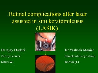
- 1. Retinal complications after laser assisted in situ keratomileusis (LASIK). Dr Ajay Dudani Zen eye center Khar (W) Dr Yashesh Maniar Shreekrishna eye clinic Borivli (E)
- 2. Refractive surgery Has been accepted for correcting ametropias. Lasik has become one of the most popular options for the correction of low to moderate myopia world wide.
- 3. Complications of lasik •Undercorrections •Overcorrections •Flap displacement •Epithelial ingrowth •Flap melting •Retinal hemorrhages •Macular holes •Choroidal neovascular membrane •Keratitis •Retinal tears •Retinal detachments •Corneosceral perforation •Macular holes •Choroidal neovascular membrane •Irregular astigmatism
- 4. Retinal detachments and breaks •Very little has been reported in the literature regarding retinal detachments after LASIK. •Ozdamar et al. have reported a case of bilateral retinal detachment associated wi8th giant retinal tear after LASIK. • Ruiz-Moreno et al. reported four retinal detachments(an incidence of 0.25%) in myopic eyes after LASIK. •Arevalo et al. reported 0.08% developed RRD after LASIK.
- 5. Retinal detachment characteristics and retinal breaks distribution Arevalo et al evaluated the fundus drawings of 33 eyes: Inferotemporal 14% Inferonasal 9% Superotemporal 8% Superonasal 7% 8 total 25 subtotal
- 6. Macular hemorrhage Very few reports have been published regarding macular hemorrhage after LASIK. Kim and Jung reported one eye lost more than 2 lines of preoperative BCVA because of macular hemmorrhage.
- 7. Lacquer cracks Lacquer cracks in Pathological myopia CNV and macular atrophy Poor visual outcome Lacquer cracks have been found to be associated to CNV in up to 82% of cases with myopia.
- 8. Choroidal neovacular membranes The incidence of of CNV after LASIK seems to be very low. Very few cases have been studied. Choroidal neovascularization is related to myopia itself and its incidence varies from 4% to 11% in patient with high myopia.
- 9. Choroidal neovacular membranes Break in bruch’s membrane
- 10. Choroidal neovacular membranes Break in bruch’s membrane Neovascular complexes
- 11. The increase in IOP to levels more than 60 mmHg during suction with the microkeratome suction ring Excimer laser is responsible for a shock wave transmitted to eye
- 12. PDT with verteporfin for subfoveal CNVM after LASIK Arevalo et al. has reported success in stabilizing or improving vision in patients with subfoveal CNV from pathologic myopia after LASIK with PDT.
- 13. Macular hole Vitreo retinal interface changes Macular hole may develop in myopic eyes after LASIK Ruiz-Moreno reported PVD was not present before and was documented after LASIK on 80% of eyes.
- 14. Corneoscleral perforation This complication may occur with microkeratome when a corneal flap is performed. Severe cases may be associated with posterior segment damage.
- 15. Corneoscleral perforation Some cases may be treated by Therapeutic soft contact lens •Topical antibiotics •Oral CAIs •Eye patching •Be meticulous in properly assembling the microkeratome to creat a corneal flap during lasik. •The use of currently availabledisposable microkeratomes may help to avoid this complication in future.
- 16. Displacement of corneal flap during vitrectomy Dislocated corneal flap may occur from corneal epithelial debridement during vitrectomy after lasik. Displacement of a corneal flap after lasik is a serious complication due to •Losing the flap •Epithelial ingrowth •Interface particles •Striae in the flap
- 17. Management of displaced corneal flap during vitrectomy Avoid debridement of corneal epithelium If necessary, Start nasally and advance temporally.(most cases – nasal hinge) Displaced corneal flap Reposition of flap, patching and topical steroids, BCL Refracory cases Suture fixation
- 18. Anatomy of vitreous base • 3-4 mm wide zone straddling ora serrata • Strong adhesion of cortical vitreous • Anterior limit of posterior vitreous detachment Vitreous base Pars Plicata Pars Plana Mechanism of acute vitreoretinal traction at the vitrous base
- 19. Indirect ophthalmoscopy • Keep lens parallel to patient’s iris plane • Avoid tendency to move towards patient • Ask the patient to move eyes and head into optimal positions for examination
- 20. Morphology of tears a. Complete U-tear b. Linear c. Incomplete L-shaped d. Operculated e. Dialysis
- 21. Scleral indentation Retinal breaks in detached retina without indentation Enhanced visualization of breaks with indentation
- 22. PROPHYLAXIS OF RHEGMATOGENOUS RETINAL DETACHMENT • Lattice • Snailtrack • White-without-pressure 1. Retinal breaks • Laser photocoagulation • Cryotherapy 3. Treatment modalities 2. Predisposing degenerations 4. Benign peripheral degenerations
- 23. Retinal breaks a - Large U-tear with ‘ subclinical RD ’ - treat b - Large symptomatic U-tear - treat c - Operculated tear bridged by blood vessel - treat d - Asymptomatic operculated tear - do not treat
- 24. Retinal breaks not requiring treatment e - Asymptomatic dialysis surrounded by pigment f - Breaks in both layers of retinoschisis g - Small asymptomatic holes near ora serrata h - Small inner layer holes in retinoschisis
- 25. Typical lattice degeneration • Present in about 8% of general population • Present in about 40% of eyes with RD • Spindle-shaped islands of retinal thinning • Network of white lines within islands • Variable associated RPE changes • Small round holes within lesions are common • Overlying vitreous liquefaction • Exaggerated attachments around margin of lesion Retina Vitreous
- 26. Complications of lattice degeneration Indications for prophylaxis • No complications - in most cases • RD associated with atropic holes, particularly in young myopes • RD associated with tractional tears in eyes with acute PVD • RD in fellow eye • Extensive lattice in high myopia
- 27. Snailtrack degeneration Indications for prophylaxis - presence of holes Sharply demarcated, frost-like bands which are longer than lattice Large round holes which carry high risk of RD
- 28. White-without-pressure Indications for prophylaxis - giant tear in other eye Translucent grey appearance of retina Occasional giant tear formation along posterior margin of lesion
- 29. Benign peripheral retinal degenerations
- 30. Rhegmatogenous - caused by a retinal break
- 31. PRINCIPLES OF RETINAL DETACHMENT SURGERY 1. Scleral buckling 2. Pneumatic retinopexy • Configuration of buckles • Preliminary steps • Localization of breaks • Cryotherapy • Insertion of local explant • Encircling procedure • Drainage of subretinal fluid • Causes of early failure 3. Vitrectomy • Giant tears • Proliferative vitreoretinopathy (PVR) • Diabetic tractional RD
- 32. Configuration of scleral buckles Radial Segmental circumferential Encircling augmented by radial sponge Encircling augmented by solid silicone tyre
- 33. Preliminary steps Peritomy Insertion of squint hook under rectus muscle Insertion of bridle suture Inspection of sclera for thinning or anomalous vortex veins
- 34. Localization of breaks • Insert 5/0 Dacron scleral suture at site of apex of break • Grasp cut suture with curved mosquito forceps close to knot • While viewing with indirect ophthalmoscope check position of indentation in relation to break
- 35. While viewing with indirect ophthalmoscope indent sclera gently with tip of cryoprobe Freeze break until sensory retina just turns white Cryotherapy
- 36. Technique of cryotherapy • Surround lesion with single row of cryo-applications • Preferred for treatment of large areas
- 37. Insertion of local explant Distance separating sutures measured and marked Ends trimmed Sutures tightened over explant Insertion of mattress-type suture
- 38. Encircling procedure Strap fed under four recti Ends secured with Watzke sleeve Strap slid posteriorly and secured in each quadrant Strap tightened to produce required amount of internal indentation
- 39. Drainage of subretinal fluid Indications Haemorrhage • Difficulty in localizing break • Immobile retina • Longstanding RD • Inferior RD Retinal incarceration Complications Technique
- 40. Causes of early failure May be associated with communicating radial retinal fold Insert additional radia buckle Buckle failure ‘ Fishmouthing ’ of retinal tear Buckle inadequate size or height Buckle incorrectly positioned
- 41. Technique (a) Cryotherapy Pneumatic retinopexy Indications RD with superior breaks (b) Gas injection (c) Postoperative positioning (d) Flat retina
- 42. Vitrectomy for giant tears Unrolling of flap with light pipe and probe Completion of unrolling Injection of silicone oil or heavy liquid
- 43. Mechanism of acute vitreoretinal traction at the vitrous base and post. Pole (A) Suction ring- deforms AP axis Increased AP diameter Closed system- contract along horizontal axis Decreased equatorial diameter
- 44. Mechanism of acute vitreoretinal traction at the vitrous base and post. pole (B) Suction stops and ring released- decompression dynamic overshoot Equatorial elongation and AP contaction
- 45. Mechanism of acute vitreoretinal traction at the vitrous base and post. Pole (C) Excimer laser Shock waves Pulsed energy
- 46. Prophylaxis of retinal detachment before LASIK Based on published data indication of prophylactic treatment can not be determined. It is not possible to determine scientifically whether peripheral retinal lesions should be treated in a way different from standard practice just because a patient is to undergo LASIK.
- 47. Prophylaxis of retinal detachment before LASIK Careful examination with IO and scleral depression Normal Go for lasik No obvious lesion and very high myope Explain to patient Go for lasik Obvious lesion Treat aggressively Go for lasik
- 48. Lasik in scleral buckled cases •Change of ap and axial length. •How to decide parameters. •Pre op precautions. •Post of care.
- 49. Relative contraindications •Macular disease •High myopia and lacquer cracks Macular hemmorhage and CNV •Eyes at risk of needing vitreoretinal surgery •Angioid streaks •Tramatic choroidal ruptures
- 50. Conclusion Serious complications after lasik are infrequent. Inform patient not only for lasik but also the vitreoretinal abnormalities and complications. Dilated fundus examination with IO and scleral depression.