1. Anatomy Primer Section
Foot drop
roots (L4 and L5), plexus, sciatic nerve, peroneal nerve
T extbooks of neurology are just like cookery or garden-
ing books. It is all so easy on paper. With a good work-
ing knowledge of neurological anatomy, it seems, almost
and severe generalised neuropathies can all present in this
way (Table 2). The first step in clinical evaluation is to
any problem can be precisely localised. A couple of of exclude cord or other CNS pathology and examine for
hours yomping through a busy out patients leads to a other peripheral nervous system involvement. In sciatic
rapid re-evaluation of that view. Foot drop, however, is a neuropathy there may also be weakness in a tibial nerve
common neurological problem that is particularly distribution also, however it is possible to have selective
amenable to an anatomical approach, so this month I will involvement of the common peroneal fasicles only.
briefly outline the anatomy of peroneal nerve and discuss In L5 radiculopathy both ankle dosrsiflexion and inver- Dr Brian McNamara is
Consultant
the clinical and neurophysiological approach to foot drop. sion/eversion will be affected while in a pure common Neurophyisologist at
The common peroneal nerve is a branch of the sciatic peroneal neuropathy inversion will be spared. There is a Cork University Hospital.
nerve. The sciatic nerve is formed in the pelvis by fibres slight caveat however. If the foot is tested in the dropped He was SHO and
Registrar at Cork
from the lumbosacral trunk (L4,5) and by fibres from position inversion may appear to be weak so inversion University Hospital, and
S1,2,3. The nerve immediately leaves the pelvis through should be tested in a passively dorsiflexed position. In an SpR at Addenbrooke's
the greater sciatic notch, below the piriformis muscle. The isolated superficial peroneal neuropathy eversion will be Hospital in Cambridge.
nerve may divide immediately, or may pass either above weak and dorsiflexion spared while in an isolated deep His interests include
magnetic stimulation,
or through the piriformis. In the gluteal region the nerve peroneal neuropathy there will be weakness of dorsiflex- cellular electrophysiology,
lies deep to gluteus maximus, between the greater ion with sparing of eversion. In a common peroneal neu- and all aspects of clinical
trochanter and the ischial tuberosity. The nerve then pass- ropathy sensation over the lateral foot (sural territory), neurophysiology.
es down the back of the thigh to the apex of the popliteal sole of foot (plantar nerves) and medial calf and foot will
fossa. In the thigh the nerve divides into lateral common be spared. Finally ankle jerks will be spared in a pure com-
peroneal and medial tibial divisions. The common per- mon peroneal neuropathy.
oneal division supplies fibres to the short head of biceps
femoris. Neurophysiological Evaluation
The common peroneal nerve leaves the popliteal fossa The neurophysiological evaluation of foot drop nicely
between the tendon of biceps femoris and the lateral head illustrates the old maxim that electrophsyiology is an
of gastrocnemius. It crosses behind the head of the fibula extension of clinical assessment. The first step is to deter-
and passes laterally around the neck of the fibula, where it mine if the pathology is restricted to the common per-
is particularly vulnerable to compression or blunt trauma. oneal nerve only, so where possible it is worth studying-
The nerve gives off the sural communicating branch to doing the works in both lower limbs (Bilateral peroneal
the sural nerve, and the lateral cutaneous nerve of the calf. and tibial motor studies, bilateral superficial peroneal and
The nerve pierces the peroneus longus muscle to divide sural sensory studies). If a common peroneal mono-neu-
into deep and superficial branches. The deep peroneal ropathy is confirmed, the next objective is to determine
nerve supplies the muscles of the anterior compartment - any site of injury or compression and give an estimate of
(table 1). The superficial peroneal nerve supplies the mus- severity. This can be achieved with a combination of nerve
cles in the lateral compartment (table 1) and the skin over conduction studies and EMG. Segmental conduction
the anterior lower leg and dorsum of the foot. studies around the fibular head should be performed-
focal slowing or conduction block is a sign of compres-
Clinical Evaluation of Foot Drop sion or neuropraxia and also in an isolated deep peroneal
The manner in which the common peroneal nerve snakes neuropathy superficical peroneal sensory studies will be
around the fibular head exposes it to injury and external normal. EMG should be performed in one L4/L5 muscle
compression and this can sometimes occur in bizarre and not innervated by the common peroneal nerve (tibialis
quite unexpected ways (table 2). Common peroneal neu- posterior is often used), one deep peroneal muscle
ropathy presents with foot drop; foot drop is due to weak- (Tibialsis anterior) and one superficial (peroneus longus).
ness of the muscles in the anterior and lateral compart- The degree of dennervation and the presence or absence
ments of the leg. Since it is these compartments that work of voluntary activity will give pointers as to severity of the
against gravity, pathology in the spinal chord, lumbar neuropathy.
Figure 1 Table 1: Muscles supplied by the two divisions of the
common peroneal nerve.
Deep peroneal Superficial Peroneal
Tibialis Anterior Peroneus Longus
Extensor Digitorum Longus Peroneus Brevis
Extensor Digitorum Brevis
Peroneus Tertius
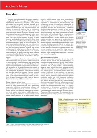
Figure 1: Course of the common, deep (blue) and superficial peroneal nerve, sites of
stimulation for motor nerve conduction studies are shown by green discs.
24 ACNR • VOLUME 3 NUMBER 1 MARCH/APRIL 2003
2. Anatomy Primer
Table 2: Some peripheral causes of foot drop
Generalised Neuropathy Motor Neuronopathy
Motor Neuropathy
Motor and Sensory Polyneuropathy
HMSN
Mononeuritis Multiplex
Localised Neuropathy L4/L5 Radiculopathy
Lumbosacral Plexopathy
Sciatic Neuropathy eg. Buttock Figure 2: Normal peroneal motor study, note there is no slowing of conduction
across the fibular head.
injection
Common Peroneal
Neuropathy Trauma at fibular head
Forcible stretch
External Compression eg.
Casts stockings etc.
Prolonged immobility eg. During
anaesthesia
Occupational eg. gardening
Figure 3: The same normal study superimposed on abnormal study (opposite
Habitual Leg crossing leg). This study was taken from a patient who developed foot drop after having
Weight loss his leg in a plaster of Paris cast for 9 weeks, note the reduced amplitude and
slowing across the fibular head.
P o w e r To R e n e w A L i f e
Clinically proven therapy for
long-term seizure control and quality-of-life benefits
Daily Seizures Seizure-Free Months
Four Medications One Medication EUROPEAN INDICATION FOR USE:
Lethargic Energetic The VNS Therapy System is indicated for use as an adjunctive therapy in
reducing the frequency of seizures in patients whose epileptic disorder is
Isolated Fun With Friends dominated by partial seizures (with or without secondary generalisation)
or generalised seizures, which are refractory to antiepileptic medications.
Powerless In Control Cyberonics Europe S.A./N.V.
ADBF0802-11-1000-EC
Belgicastraat 9, 1930 Zaventem
Disoriented Alert Belgium
Tel: +32 2 720 95 93
Inattentive Achieving At School Fax: +32 2 720 60 53
w w w . v n s t h e r a p y. c o m / i n t e r n a t i o n a l
ACNR • VOLUME 3 NUMBER 1 MARCH/APRIL 2003 25