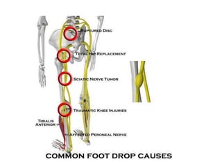
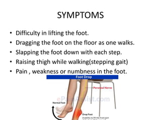
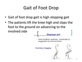
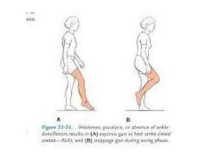
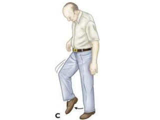
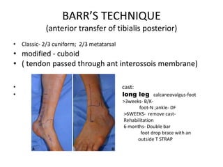
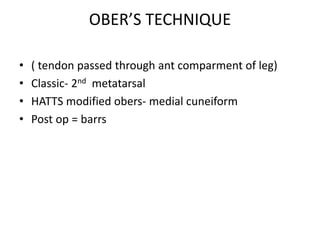
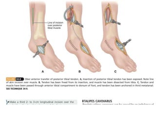
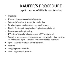
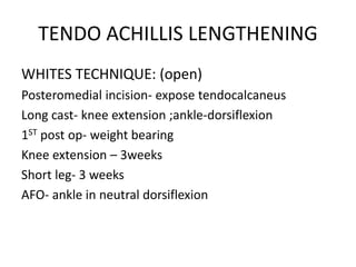
FOOT DROP
Dr.A.Supraja
PG II YEAR
Gandhi Medical College
Inability to actively dorsiflex and
evert the foot
Definition
I. Traumatic:
• Tendon injuries to dorsiflexors of foot
• Neurogenic
A)At or below the level of common peroneal nerve
Direct injuries: incised and penetrating injuries
Fracture and dislocations:
Fracture of lateral condyle of tibia
Fracture/ dislocation of head/neck of fibula
Dislocation of knee
compound fracture of upper 1/3rd of tibia
4
Causes of Foot Drop
 Iatrogenic :
High tibial skeletal traction
Tight plaster around knee
High tibial osteotomy
Total knee replacement
B) Above the level of common peroneal nerve
Fracture of shaft of femur
Posterior dislocation of hip
Deep intra muscular injection
PIVD
Spina bifida
If any cerebral tumors and space occupying lesions of
CNS
5
II. infective
• leprosy
• Poliomyelitis
Guillain-Barré Syndrome
• Syphilis
III. Metabolic:
• Diabetes mellitus
• Beri beri
• Alcoholic neuritis
IV. Exogenous toxin:
• Lead
• Arsenic
• Mercury
6

COURSE OF THE COMMON PERONEAL
NERVE
COURSE OF THE COMMON PERONEAL
NERVE
• Fasiculi of the peroneal nerve - larger and have
less connective tissue
• Fewer autonomic fibers, so in any injury, motor
and sensory fibers bear the brunt of the trauma.
• More superficial course, especially at the fibular
neck
• Adheres closely to the periosteum of the
proximal fibula
Vulnerability of Peroneal Nerve
• Cutaneous sensation is impaired over the lateral aspect
of the lower leg and ankle and dorsum of the foot.
• Reduced dorsiflexion and eversion of the foot and of
toe extension
– The patients will compensate by having a steppage gait.
• N.B Inversion and plantar flexion are normal.
Signs
• Progressive weakness of the peronei and
tibialis anterior muscles which result in foot
drop.
peroneus longus , tibialis anterior and the
extensor digitorum wasting
• N.B The paresis results in ankle weakness and
predispose to ankle sprains
• Difficulty in lifting the foot.
• Dragging the foot on the floor as one walks.
• Slapping the foot down with each step.
• Raising thigh while walking(stepping gait)
• Pain , weakness or numbness in the foot.
SYMPTOMS
Types of foot drop
• Type I – High
above the level of fibular head
deep peroneal nerve
• Type II- Low
below the level of fibular head
superficial peroneal nerve
• High lesion : total foot drop
• Unable to dorsiflex and invert foot
• Able to do eversion
• Wasting of ant group of muscles
• Loss of sensation over the 1st web space
16
Clinical features of Type 1 foot drop
• Low lesion : incomplete foot drop
• Unable to do eversion
• Able to do dorsiflexion and inversion of the
foot
• Wasting of outer half of leg
• Sensation lost over outer leg and foot
17
Clinical features of type 2 foot drop
• Gait of foot drop gait is high stepping gait
• The patients lift the knee high and slaps the
foot to the ground on advancing to the
involved side
18
Gait of Foot Drop
X-Ray
Post-Traumatic - tibia/injuryfibula and ankle
- any bony.
Anatomic dysfunction (eg. Charcot joint)
• Ultrasonography
If bleeding is suspected in a patient with a
hip or knee prosthesis
• Magnetic Resonance Neurography
Tumor or a compressive mass lesion to
the peroneal nerve
DIAGNOSIS
– This study can confirm the type of neuropathy, establish
the site of the lesion, estimate extent of injury, and
provide a prognosis.
– Sequential studies are useful to monitor recovery of acute
lesions.
Electromyelogram
• Depends on the underlying cause.
• If cause is successfully treated foot drop
may improve or even disappear.
• Medical treatment - painful paresthesia
amitriptyline
nortriptyline
pregabalin
TREATMENT
• Assistive and adaptive devices and equipment.
– Canes, crutches, or walkers may be used to help
prevent falling, normalize gait patterns, or unload
a painful weight-bearing limb.
• Electrical Stimulation.
– Transcutaneous electrical nerve stimulation
(TENS) for the reduction or obliteration of pain.
• Positioning.
– Correct positioning of limb
• Protective Devices and Equipment eg splints,
orthoses
• Cryotherapy, massage
• Conservative treatment : shows high incidence
of recovery
• Splintage – splint knee in 20° of flexion and
ankle in 90° for night time
• In day time, walking is allowed by using ‘foot-
drop appliance’
• Varieties of foot drop appliances:
i) dynamic-spring shoe
ii) static- back stop shoe
26
Treatment
• Ankle foot orthotics (AFO)
-support the foot with light-weight leg braces
and shoe inserts
• Exercises
-strengthen the muscle, help to maintain
range of motion (ROM) and improve gait
• Electrical Functional Stimulations
-electrically stimulate the peroneal nerve
27
Stimulating the nerve (peroneal nerve)
improves foot drop especially if it caused by a
stroke.
Nerve Stimulation
30
• Surgery – done if conservative management
fails
• Repairs or decompression of a damaged
nerve, fusion of the foot and ankle joint or
transfers tendons from stronger leg muscles
31
SURGICAL MANAGEMENT
Points to be considered
age
i. Mobility of joints
ii. Availability of muscles and tendons for transfer
A. Soft tissue and muscle contractures
B. Bony changes
TENDON TRANSFER SURGERIES
• Objectives:
• Provide active motor power – paralysed
muscle
• Eliminate deforming effect of muscle-
antagonist paralysed
• Improve stability- muscle balance
PRINCIPLES
• Muscle
• transferred-strong—good/better
• Nerve and blood supply-not impaired
• Free end of tendon-
• close to insertion of paralysed muscle
• Retained in its own sheath/ another tendon
• Routed direct in line bet muscle origin and new insertion
• Contractures near the joint on which muscle acts- released
• Agonists> antagonists
• Tendon =range of excursion=one reinforcing /replacing
BARR’S TECHNIQUE
(anterior transfer of tibialis posterior)
• Classic- 2/3 cuniform; 2/3 metatarsal
• modified - cuboid
• ( tendon passed through ant interossois membrane)
• cast:
• long leg calcaneovalgus-foot
• >3weeks- B/K-
• foot-N ;ankle- DF
• >6WEEKS- remove cast-
• Rehabilitation
• 6 months- Double bar
• foot drop brace with an
• outside T STRAP
OBER’S TECHNIQUE
• ( tendon passed through ant comparment of leg)
• Classic- 2nd metatarsal
• HATTS modified obers- medial cuneiform
• Post op = barrs
KAUFER’S PROCEDURE
( split transfer of tibialis post tendon)
• Incision-
• 1st- curvilinear- navicular tuberosity
• Extend-inf and post to medial malleolus
• Proximal- post midline over tendocalcaneus
• Tibialis Post –split longitudinally-plantar and dorsal
• Tendocalneus lengthening
• 2nd- tip of lateral malleolus-base of 5th metatarsal
• Peronius brevis- pass tendon carrier –proximally –just post to
lat malleolus- t.post tendon- foot in corrected position-
• t.post sutured to brevis under tension.
• Post op:
• long leg cast- 2months
• short leg cast – 2 months
SRINIVASAN TECHNIQUE
( two tailed transfer of tibialis post)
• Position- supine –
• passive dorsiflexion of ankle- knee extension
• 1st-Short transverse incision- navicular-tibialis post endon
exposed
• Flexar retinaculum split
• 2nd-Medial aspect of L/3rd of leg-incision-FDL retracted-
tibialis post hooked up-split
• 3rd& 4th -2 curvilinear – dorsum of foot-
• medial- tendon of EHL-LOWER SLIP(anderson tunneler)
• Lateral- tendon of EDL- UPPER SLIP- laced up
• Post op:
• B/K- foot in 70 deg dorsiflexion
• >3weeks- non-weight bearing reeducative exercises
• Bivalve POP cast- crutches
• 7th post op week- weight bearing
TENDO ACHILLIS LENGTHENING
WHITES TECHNIQUE: (open)
Posteromedial incision- expose tendocalcaneus
Long cast- knee extension ;ankle-dorsiflexion
1ST post op- weight bearing
Knee extension – 3weeks
Short leg- 3 weeks
AFO- ankle in neutral dorsiflexion
• HAUSER TECHNIQUE:
• Posteromedial incision- expose tendocalcaneus
• Plantaris tendon- incised-beneficial
• Cast- mid thigh to toe
knee-full flexion; ankle-neutral dorsiflexion
skin blanching- little equinus (1st cast change-N)
>6WEEKS- AFO
PERCUTANEOUS LENGTHENING OF
TENDOCALCANEUS
• Position: prone
knee-E; ankle- DF
3 partial tenotomies
Heel-varus-2 incisions medially
valgus- 2 incisions laterally
After suregery Mx = White technique
Semiopen sliding tenotomy of
tendocalcaneus
• Position-prone
• 2 long incision 2cm along the tendocalcaneus
• Plantaris tendon- tenotomy
• Post op-=Hauser technique
BONE SURGERIES
LAMBRINUDI ARTHRODESIS
• Indiactions:Isolated fixed equinus deformity
> 10 years
• CI: flail foot
hip& knee instability requiring brace
Lateral x-ray- ant subluxation of talus- 2 stage
plantar arthodesis
Complications-1. ankle instability
2.residual varus/valgus due to muscle imbalance
3.Pseudoarthrosis of talonavicular joint
• S/R – 10-14 days
• Short leg cast- x –ray satisfactory
• Weight bearing- > 6weeks
• Short leg walking cast- fusion complete(3mon)
TRIPLE ARTHODESIS
• Most effective ; age > 12 years
• Subtalar-calcaneocuboid-talonavicular joints
• Indications: weakness and deformity of subtalar
& mid tarsal joints
stable& static realignment
remove deforming forces
arrest progression of deformity
Eliminate pain
eliminate use of short leg brace/ provide sufficient correction for
fitting long leg brace
obtain near normal correction of foot
After Rx: walk with crutches / walker
CAMPBELLS post bone block
• Permits lengthening of tendocalcaneus & ankylosing both ankle and
subtalar joints
• Incision: medial and parallel to TA
• FHL retracted to capsule of ankle joint is exposed
• Post part of talus and articular surfaces of ankle and subtalar joints-
excised
• Ilium- large bony bridge across the ankle and subtalar joints
• post op:
ankle- plaster cast- A/K- foot at right angle
>4 weeks- boot cast ( snugly fitting)
full weight bearing delayed > 8-12 weeks
cast immobilisation- until fusion
walking on irregular surfaces difficult
ANKLE ARTHODESIS
• BARR & BONE
• Severe paralytic equinus deformity in adults
• Subcutaneous plantar fasciotomy- lenthening
of TA – ankle arthodesis
Foot drop

Foot drop

  • 1.
    FOOT DROP Dr.A.Supraja PG IIYEAR Gandhi Medical College
  • 2.
    Inability to activelydorsiflex and evert the foot Definition
  • 4.
    I. Traumatic: • Tendoninjuries to dorsiflexors of foot • Neurogenic A)At or below the level of common peroneal nerve Direct injuries: incised and penetrating injuries Fracture and dislocations: Fracture of lateral condyle of tibia Fracture/ dislocation of head/neck of fibula Dislocation of knee compound fracture of upper 1/3rd of tibia 4 Causes of Foot Drop
  • 5.
     Iatrogenic : Hightibial skeletal traction Tight plaster around knee High tibial osteotomy Total knee replacement B) Above the level of common peroneal nerve Fracture of shaft of femur Posterior dislocation of hip Deep intra muscular injection PIVD Spina bifida If any cerebral tumors and space occupying lesions of CNS 5
  • 6.
    II. infective • leprosy •Poliomyelitis Guillain-Barré Syndrome • Syphilis III. Metabolic: • Diabetes mellitus • Beri beri • Alcoholic neuritis IV. Exogenous toxin: • Lead • Arsenic • Mercury 6 
  • 8.
    COURSE OF THECOMMON PERONEAL NERVE
  • 9.
    COURSE OF THECOMMON PERONEAL NERVE
  • 10.
    • Fasiculi ofthe peroneal nerve - larger and have less connective tissue • Fewer autonomic fibers, so in any injury, motor and sensory fibers bear the brunt of the trauma. • More superficial course, especially at the fibular neck • Adheres closely to the periosteum of the proximal fibula Vulnerability of Peroneal Nerve
  • 11.
    • Cutaneous sensationis impaired over the lateral aspect of the lower leg and ankle and dorsum of the foot. • Reduced dorsiflexion and eversion of the foot and of toe extension – The patients will compensate by having a steppage gait. • N.B Inversion and plantar flexion are normal. Signs
  • 12.
    • Progressive weaknessof the peronei and tibialis anterior muscles which result in foot drop. peroneus longus , tibialis anterior and the extensor digitorum wasting • N.B The paresis results in ankle weakness and predispose to ankle sprains
  • 13.
    • Difficulty inlifting the foot. • Dragging the foot on the floor as one walks. • Slapping the foot down with each step. • Raising thigh while walking(stepping gait) • Pain , weakness or numbness in the foot. SYMPTOMS
  • 14.
    Types of footdrop • Type I – High above the level of fibular head deep peroneal nerve • Type II- Low below the level of fibular head superficial peroneal nerve
  • 16.
    • High lesion: total foot drop • Unable to dorsiflex and invert foot • Able to do eversion • Wasting of ant group of muscles • Loss of sensation over the 1st web space 16 Clinical features of Type 1 foot drop
  • 17.
    • Low lesion: incomplete foot drop • Unable to do eversion • Able to do dorsiflexion and inversion of the foot • Wasting of outer half of leg • Sensation lost over outer leg and foot 17 Clinical features of type 2 foot drop
  • 18.
    • Gait offoot drop gait is high stepping gait • The patients lift the knee high and slaps the foot to the ground on advancing to the involved side 18 Gait of Foot Drop
  • 21.
    X-Ray Post-Traumatic - tibia/injuryfibulaand ankle - any bony. Anatomic dysfunction (eg. Charcot joint) • Ultrasonography If bleeding is suspected in a patient with a hip or knee prosthesis • Magnetic Resonance Neurography Tumor or a compressive mass lesion to the peroneal nerve DIAGNOSIS
  • 22.
    – This studycan confirm the type of neuropathy, establish the site of the lesion, estimate extent of injury, and provide a prognosis. – Sequential studies are useful to monitor recovery of acute lesions. Electromyelogram
  • 23.
    • Depends onthe underlying cause. • If cause is successfully treated foot drop may improve or even disappear. • Medical treatment - painful paresthesia amitriptyline nortriptyline pregabalin TREATMENT
  • 24.
    • Assistive andadaptive devices and equipment. – Canes, crutches, or walkers may be used to help prevent falling, normalize gait patterns, or unload a painful weight-bearing limb. • Electrical Stimulation. – Transcutaneous electrical nerve stimulation (TENS) for the reduction or obliteration of pain.
  • 25.
    • Positioning. – Correctpositioning of limb • Protective Devices and Equipment eg splints, orthoses • Cryotherapy, massage
  • 26.
    • Conservative treatment: shows high incidence of recovery • Splintage – splint knee in 20° of flexion and ankle in 90° for night time • In day time, walking is allowed by using ‘foot- drop appliance’ • Varieties of foot drop appliances: i) dynamic-spring shoe ii) static- back stop shoe 26 Treatment
  • 27.
    • Ankle footorthotics (AFO) -support the foot with light-weight leg braces and shoe inserts • Exercises -strengthen the muscle, help to maintain range of motion (ROM) and improve gait • Electrical Functional Stimulations -electrically stimulate the peroneal nerve 27
  • 29.
    Stimulating the nerve(peroneal nerve) improves foot drop especially if it caused by a stroke. Nerve Stimulation
  • 30.
  • 31.
    • Surgery –done if conservative management fails • Repairs or decompression of a damaged nerve, fusion of the foot and ankle joint or transfers tendons from stronger leg muscles 31
  • 33.
    SURGICAL MANAGEMENT Points tobe considered age i. Mobility of joints ii. Availability of muscles and tendons for transfer A. Soft tissue and muscle contractures B. Bony changes
  • 34.
    TENDON TRANSFER SURGERIES •Objectives: • Provide active motor power – paralysed muscle • Eliminate deforming effect of muscle- antagonist paralysed • Improve stability- muscle balance
  • 35.
    PRINCIPLES • Muscle • transferred-strong—good/better •Nerve and blood supply-not impaired • Free end of tendon- • close to insertion of paralysed muscle • Retained in its own sheath/ another tendon • Routed direct in line bet muscle origin and new insertion • Contractures near the joint on which muscle acts- released • Agonists> antagonists • Tendon =range of excursion=one reinforcing /replacing
  • 36.
    BARR’S TECHNIQUE (anterior transferof tibialis posterior) • Classic- 2/3 cuniform; 2/3 metatarsal • modified - cuboid • ( tendon passed through ant interossois membrane) • cast: • long leg calcaneovalgus-foot • >3weeks- B/K- • foot-N ;ankle- DF • >6WEEKS- remove cast- • Rehabilitation • 6 months- Double bar • foot drop brace with an • outside T STRAP
  • 37.
    OBER’S TECHNIQUE • (tendon passed through ant comparment of leg) • Classic- 2nd metatarsal • HATTS modified obers- medial cuneiform • Post op = barrs
  • 39.
    KAUFER’S PROCEDURE ( splittransfer of tibialis post tendon) • Incision- • 1st- curvilinear- navicular tuberosity • Extend-inf and post to medial malleolus • Proximal- post midline over tendocalcaneus • Tibialis Post –split longitudinally-plantar and dorsal • Tendocalneus lengthening • 2nd- tip of lateral malleolus-base of 5th metatarsal • Peronius brevis- pass tendon carrier –proximally –just post to lat malleolus- t.post tendon- foot in corrected position- • t.post sutured to brevis under tension. • Post op: • long leg cast- 2months • short leg cast – 2 months
  • 41.
    SRINIVASAN TECHNIQUE ( twotailed transfer of tibialis post) • Position- supine – • passive dorsiflexion of ankle- knee extension • 1st-Short transverse incision- navicular-tibialis post endon exposed • Flexar retinaculum split • 2nd-Medial aspect of L/3rd of leg-incision-FDL retracted- tibialis post hooked up-split • 3rd& 4th -2 curvilinear – dorsum of foot- • medial- tendon of EHL-LOWER SLIP(anderson tunneler) • Lateral- tendon of EDL- UPPER SLIP- laced up
  • 43.
    • Post op: •B/K- foot in 70 deg dorsiflexion • >3weeks- non-weight bearing reeducative exercises • Bivalve POP cast- crutches • 7th post op week- weight bearing
  • 44.
    TENDO ACHILLIS LENGTHENING WHITESTECHNIQUE: (open) Posteromedial incision- expose tendocalcaneus Long cast- knee extension ;ankle-dorsiflexion 1ST post op- weight bearing Knee extension – 3weeks Short leg- 3 weeks AFO- ankle in neutral dorsiflexion
  • 46.
    • HAUSER TECHNIQUE: •Posteromedial incision- expose tendocalcaneus • Plantaris tendon- incised-beneficial • Cast- mid thigh to toe knee-full flexion; ankle-neutral dorsiflexion skin blanching- little equinus (1st cast change-N) >6WEEKS- AFO
  • 47.
    PERCUTANEOUS LENGTHENING OF TENDOCALCANEUS •Position: prone knee-E; ankle- DF 3 partial tenotomies Heel-varus-2 incisions medially valgus- 2 incisions laterally After suregery Mx = White technique
  • 49.
    Semiopen sliding tenotomyof tendocalcaneus • Position-prone • 2 long incision 2cm along the tendocalcaneus • Plantaris tendon- tenotomy • Post op-=Hauser technique
  • 51.
  • 52.
    LAMBRINUDI ARTHRODESIS • Indiactions:Isolatedfixed equinus deformity > 10 years • CI: flail foot hip& knee instability requiring brace Lateral x-ray- ant subluxation of talus- 2 stage plantar arthodesis Complications-1. ankle instability 2.residual varus/valgus due to muscle imbalance 3.Pseudoarthrosis of talonavicular joint
  • 54.
    • S/R –10-14 days • Short leg cast- x –ray satisfactory • Weight bearing- > 6weeks • Short leg walking cast- fusion complete(3mon)
  • 55.
    TRIPLE ARTHODESIS • Mosteffective ; age > 12 years • Subtalar-calcaneocuboid-talonavicular joints • Indications: weakness and deformity of subtalar & mid tarsal joints stable& static realignment remove deforming forces arrest progression of deformity Eliminate pain eliminate use of short leg brace/ provide sufficient correction for fitting long leg brace obtain near normal correction of foot After Rx: walk with crutches / walker
  • 59.
    CAMPBELLS post boneblock • Permits lengthening of tendocalcaneus & ankylosing both ankle and subtalar joints • Incision: medial and parallel to TA • FHL retracted to capsule of ankle joint is exposed • Post part of talus and articular surfaces of ankle and subtalar joints- excised • Ilium- large bony bridge across the ankle and subtalar joints • post op: ankle- plaster cast- A/K- foot at right angle >4 weeks- boot cast ( snugly fitting) full weight bearing delayed > 8-12 weeks cast immobilisation- until fusion walking on irregular surfaces difficult
  • 61.
    ANKLE ARTHODESIS • BARR& BONE • Severe paralytic equinus deformity in adults • Subcutaneous plantar fasciotomy- lenthening of TA – ankle arthodesis