Radius Bone Anatomy and Articular Surfaces
•Download as PPTX, PDF•
2 likes•2,556 views

anatomy of the radius bone osteology myology

Recommended
More Related Content
What's hot
What's hot (20)
Similar to Radius Bone Anatomy and Articular Surfaces
Similar to Radius Bone Anatomy and Articular Surfaces (20)
More from Dr Adnan Sami
More from Dr Adnan Sami (15)
Recently uploaded
Recently uploaded (20)
Radius Bone Anatomy and Articular Surfaces
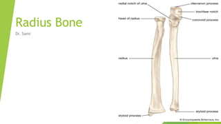
- 4. Upper Extremity of the radius (Proximal Extremity) Head, Neck Tuberosity.
- 5. Posterior view of Radial Head
- 7. Head: The radial head has a cylindrical form, and on its upper surface is a shallow cup or fovea for articulation with the capitulum (or capitellum) of the humerus. The circumference (outer Boundry) of the head is smooth; it is broad medially where it articulates with the radial notch of the ulna, narrow in the rest of its extent, which is embraced by the annular ligament. The deepest point in the fovea is not axi-symmetric with the long axis of the radius, creating a cam effect during pronation and supination.
- 9. Neck: The head is supported on a round, smooth, and constricted portion called the neck, on the back of which is a slight ridge for the insertion of part of the supinator muscle.
- 11. Tuberosity: Beneath the neck of the radius, on the medial side, is an eminence, the radial tuberosity; its surface is divided into: a posterior, rough portion, for the insertion of the tendon of the biceps brachii. an anterior, smooth portion, on which a bursa is interposed between the tendon and the bone.
- 16. Distal End of the Radius The distal end of the radius is large and of quadrilateral form. Joint surfaces It is provided with two articular surfaces – one below, for the carpus, and another at the medial side, for the ulna. The carpal articular surface is triangular, concave, smooth, and divided by a slight antero-posterior ridge into two parts. Of these, the lateral, triangular, articulates with the scaphoid bone; the medial, quadrilateral, with the lunate bone. The articular surface for the ulna is called the ulnar notch (sigmoid cavity) of the radius; it is narrow, concave, smooth, and articulates with the head of the ulna. These two articular surfaces are separated by a prominent ridge, to which the base of the triangular articular disk is attached; this disk separates the wrist- joint from the distal radioulnar articulation.
- 19. Other Surfaces: This end of the bone has three non-articular surfaces – volar, Dorsal lateral.
- 20. Volar: The volar surface, rough and irregular, affords attachment to the volar radiocarpal ligament. The palmar radiocarpal ligament (anterior ligament, volar radiocarpal ligament) is a broad membranous band, attached above to the distal end of the radius, and passing downward to the scaphoid, lunate, triquetrum and capitate of the carpal bones in the wrist.
- 23. Dorsal: The dorsal surface is convex, affords attachment to the dorsal radiocarpal ligament, and is marked by three grooves. Enumerated from the lateral side: The first groove is broad, but shallow, and subdivided into two by a slight ridge: the lateral of these two, transmits the tendon of the extensor carpi radialis longus muscle; the medial, the tendon of the extensor carpi radialis brevis muscle. The second is deep but narrow, and bounded laterally by a sharply defined ridge; it is directed obliquely from above downward and lateralward, and transmits the tendon of the extensor pollicis longus muscle. The third is broad, for the passage of the tendons of the extensor indicis proprius and extensor digitorum communis.
- 25. Lateral The lateral surface is prolonged obliquely downward into a strong, conical projection, the styloid process, which gives attachment by its base to the tendon of the brachioradialis, and by its apex to the radial collateral ligament of wrist joint. The lateral surface of this process is marked by a flat groove, for the tendons of the abductor pollicis longus muscle and extensor pollicis brevis muscle.
- 26. Shaft: The body of the radius (or shaft of radius) is prismoid in form, narrower above than below, and slightly curved, so as to be convex lateralward. It presents three borders and three surfaces.
- 27. Borders The volar border (margo volaris; anterior border; palmar;) It extends from the lower part of the tuberosity above to the anterior part of the base of the styloid process below, and separates the volar from the lateral surface. Its upper third is prominent, and from its oblique direction has received the name of the oblique line of the radius; it gives origin to the flexor digitorum superficialis muscle (also flexor digitorum sublimis) and flexor pollicis longus muscle; the surface above the line gives insertion to part of the supinator muscle. The middle third of the volar border is indistinct and rounded. The lower fourth is prominent, and gives insertion to the pronator quadratus muscle, and attachment to the dorsal carpal ligament; it ends in a small tubercle, into which the tendon of the brachioradialis muscle is inserted.
- 30. The dorsal border (margo dorsalis; posterior border) begins above at the back of the neck, and ends below at the posterior part of the base of the styloid process; it separates the posterior from the lateral surface. is indistinct above and below, but well-marked in the middle third of the bone.
- 32. The interosseous border (internal border; crista interossea; interosseous crest;) begins above, at the back part of the tuberosity, and its upper part is rounded and indistinct; it becomes sharp and prominent as it descends, and at its lower part divides into two ridges which are continued to the anterior and posterior margins of the ulnar notch. To the posterior of the two ridges the lower part of the interosseous membrane is attached, while the triangular surface between the ridges gives insertion to part of the pronator quadratus muscle. This crest separates the volar from the dorsal surface, and gives attachment to the interosseous membrane. The connection between the two bones is actually a joint referred to as a syndesmosis joint.
- 34. Surfaces: The volar surface (facies volaris; anterior surface) is concave in its upper three-fourths, and gives origin to the flexor pollicis longus muscle; it is broad and flat in its lower fourth, and affords insertion to the Pronator quadratus. A prominent ridge limits the insertion of the Pronator quadratus below, and between this and the inferior border is a triangular rough surface for the attachment of the volar radiocarpal ligament. At the junction of the upper and middle thirds of the volar surface is the nutrient foramen, which is directed obliquely upward.
- 35. The dorsal surface (facies dorsalis; posterior surface) is convex, and smooth in the upper third of its extent, and covered by the Supinator. Its middle third is broad, slightly concave, and gives origin to the Abductor pollicis longus above, and the extensor pollicis brevis muscle below. Its lower third is broad, convex, and covered by the tendons of the muscles which subsequently run in the grooves on the lower end of the bone.
- 36. The lateral surface (facies lateralis; external surface) is convex throughout its entire extent and is known as the convexity of the radius, curving outwards to be convex at the side. Its upper third gives insertion to the supinator muscle. About its center is a rough ridge, for the insertion of the pronator teres muscle. Its lower part is narrow, and covered by the tendons of the abductor pollicis longus muscle and extensor pollicis brevis muscle.