

Recommended
More Related Content
What's hot
What's hot (20)
Similar to Placental circulation
Similar to Placental circulation (20)
More from Abhilasha verma
More from Abhilasha verma (20)
Recently uploaded
Recently uploaded (20)
Placental circulation
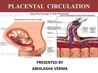
- 4. UTERO-PLACENTAL CIRCULATION ( MATERNAL CIRCULATION) It is concerned with circulation of maternal blood through intervillous spaces. Amount of blood- Mature placenta= 500 ml Occupied in villi system= 350 ml In intervillous space= 150 ml Intervillous blood flow at term= 500-600 ml/min. Blood in intervillous space completely replaced about 3-4 minutes. Pressure within interillous space is about 10-15 mmhg during uterine relaxation and 30-50 mmhg during uterine contraction. Villi provide nutrition depends on their maternal blood. Fetal capillary pressure in villi is 23-40 mmhg.
- 6. UTEROPLACENTAL CIRCULATION ARTERIAL CIRCULATION VENOUS CIRCULATION CIRCULATION IN INTER VILLOUS SPACE
- 7. ARTERIAL & VENOUS CIRCULATION • About 120-200 spiral arteries opens into intervillous space by piercing the basal plate. • Normally there is cytotrophoblastic invasion into spiral arteries up to inter decidual portion within 12 week of pregnancy. • There is secondary invasion of trophoblast between 12 weeks extending up to radial arteries within myometrium. • Thus spiral arteries are converted to large bore utereoplacental arteries. • So funneling of arteries reduce the pressure of blood before it reaches the intervillous space. Venous blood of intervillous space drains through uterine vein which pierce basal plate.
- 8. CIRCULATION IN INTERVILLOUS SPACE • Arterial blood enters the space under pressure. • Lateral dispersion occurs after reaches chorionic plate. • Villi helps in mixing and slowing of blood by villi pulsation process. • Uterine contraction helps in migration of blood towards basal plate and uterine vessels. • During uterine contraction, veins are occluded but arterial blood forced into intervillous space. • Uterine relaxation facilitates venous drainage. • Spiral arteries are perpendicular and veins are parallel to uterine wall, so during contraction larger volume of blood is available for exchange even through rate flow decreased. • Blood in intervillous space are protected from clotting by fibrinolytic enzyme activity of trophoblast.
- 9. FETO PLACENTAL CIRCULATION • The two umbilical arteries carry impure blood from fetus. • They enters the chorionic plate underneath amnion, each supplying half of placenta. • Arteries breaks up into small branches which enters stem of chorionic villi. • Each divide into primary, secondary and tertioary vessel corresponding to villi. • Blood flows into corresponding venous channel either through terminal capillary network or through shunts. • Maternal and fetal blood stream flow side by side but in opposite direction, this countercurrent flow facilitates maternal exchange between fetus and mother. • Fetal blood flow to placenta is about 400 ml/min, maily facilitated by pimping action of heart.
- 10. PLACENTAL BARRIER • Placental barriers are the partition between fetal and maternal circulation. • This barrier is not perfect barrier because fetal blood cells are found in maternal circulation and maternal blood cells are found in fetal circulation. Placenta barrier separate fetal and placental circulation. • Thickness- 0.025 mm • Placental barrier consisting of following- 1. Syntiotrophoblasts. 2. Cytotrophoblasts. 3. Basement membrane. 4. Stromal tissues. 5. Endothelium of fetal capillary.
- 11. POINTS TO BE REMEMBER • Fetal blood flows through placenta= 400 ml/min • Pressure in umbillical artery= 60 mmHg • Pressure in umbillical vein= 10 mm Hg • Fetal capillary pressure in villi= 20-40 mmHg • Placenta=1/6th of newborn weight • Volume of blood in mature placenta= 500ml • Volume of blood in Inter Villous space= 150 ml • Blood flow in Inter Villous space = 500-600 ml/min • Pressure in Inter Villous space During contraction- 30-50 mmHg During relaxation- 10-15 mmHg • Oxygen saturation Umb. Artery < Umbilical vein (50-60%) < (70-80%)
Editor's Notes
- 5