2. Only the eutherian mammals posses
the placenta. The human placenta is
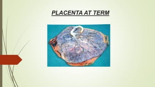
discoid, because of its shape;
hemochorial because of direct contact
of the chorion with the maternal blood
and deciduate, because some maternal
tissue is shed at parturition.
Placenta is attached to the uterine wall
and establishes connection between
the mother and fetus through the
umbilical cord.
6. DEVELOPMENT OF PLACENTA
Placenta is developed from two sources. The principle component is fetal which develops from
the chorion frondosum and the maternal component consists of decidua basalis.
When the interstitial implantation is completed on 11th day ,the blastocyst is surrounded on all
sides by lacunar spaces around cords of syncytial cells, called trabeculae.
From the trabeculae develops the stem villi on 13th day which connect the chorionic plate with
the basal plate.
Primary, secondary, and tertiary villi are successively developed from the stem villi.
Arterio capillary venous system in the mesenchymal core of each villus is completed on 21st
day.
This ultimately makes connection with the intraembryonic vascular system through the body
stalk.
8. DEVELOPMENT OF PLACENTA Cont.
These two i.e., chorion frondosum and the decidua basalis form the discrete placenta. It
begins at 6th week and is completed by 12th week.
Until the end of 16th week ,the placenta grows both in thickness and circumference due to
growth of chorionic villi with accompanying expansion of the intervillous space.
The human hemochorial placenta derived its name from hemo ( blood) that is in contact
with the syncytiotrophoblasts of chorionic tissue.
19. PLACENTAL CIRCULATION
Placental circulation consists of independent circulation of blood in
two systems :
Uteroplacental circulation & fetoplacental
circulation
UTEROPLACENTAL CIRCULATION :
A mature placenta has a volume of about 500 ml of blood ,350ml
being occupied in the villi system and 150 ml lying in the
intervillous space.
Intervillous blood flow at the term is estimated to be 500-
600ml/min ,blood in the intervillous space is completely
replaced about 3-4times /min
Intervillous space pressure is about 10-15 mm hg during uterine
relaxation and 30-50mmhg during uterine contraction.
In contrast ,the fetal capillary pressure in the villi is 20-40mmhg
ARTERIAL CIRCULATION
About 120-200 spiral arteries open into the intervillous
space.
Normally there is cytotrophoblastic invasion into the spiral
arteries initially upto the intra decidual portion within 12wks
of pregnancy.
There is a secondary invasion of trophoblast between 12-16
wks extending upto radial arteries within the myometrium
,thus spiral arteries are converted to large bore
uteroplacental arteries.
The net effect is funneling of arteries which reduce the
pressure of the blood to 70-80mmhg before it reaches the
intervillous space.
VENOUS DRAINAGE :
Through the uterine veins which pierce the basal plate
randomly like the arteries
20. CIRCULATION OF THE INTERVILLOUS SPACE :
The arterial blood enters the space
under pressure. lateral dispersion
occurs after it reaches the chorionic
plate .villi help in mixing and slowing
of blood flow .mild stirring by the villi
pulsation aided by uterine contraction
help migration of the blood towards
the basal plate and to uterine veins
27. CHANGES OCCURE AS FOLLOWING
1.Closure of the umbilical arteries-
The distal parts form the lateral umbilical ligaments & proximal parts remain open
as superior vesicle arteries.
Actual obliteration takes about 2-3months.
2.Closure of the umbilical vein-
Obliteration occurs little later than the arteries.
This forms the ligamentum teres & ductus venosus becomes ligamentum
venosum.
3.Closure of the ductus arteriosus—
Functional closure of the ducts may occure soon but anatomical obliteration takes
about 1-3 months & becomes ligamentum arteriosum.
4.Closure of the foramen ovale–
Anatomical closure takes time about 1year.
28. PLACENTAL FUNCTION
Transfer of nutrients and waste products between the mother and fetus .it attributes to the following functions—
respiratory ,excretory and nutritive.
The mechanisms involved in the transfer of substances across the placenta are :
Simple diffusion (based on concentration gradient or electrical gradient)
Facilitated diffusion (transporter mediated) using transporter proteins in syncytiotrophoblast (glucose, amino acids)
Active transfer ( against concentration gradient, energy ATPase mediated)
Endocytosis ( Invagination of the cell membrane to form an intracellular vesicle)
Exocytosis (release of molecule from within vesicle to the extracellular space)
Leakage (break in the placental membranes maternal or fetal red blood cells)
29. PLACENTAL FUNCTION
RESPIRATORY FUNCTION : Although fetal respiratory movements are observed as early as 11 wks ,there is no
gaseous exchange.
Intake of oxygen & output of carbon dioxide take place by simple diffusion.
The oxygen supply to the fetus is at the rate of 8ml/kg/min and this is achieved with cord blood flow of 165-
330ml/min
EXCRETORY FUNCTION Waste products like urea, uric acid and creatinine are excreted to maternal blood by
simple diffusion.
The main substance excreted from the fetus is co2 also bilirubin will be excreted.
Endocrine function – placenta is an endocrine gland ,produces both steroid and peptide hormones to maintain
pregnancy.
Barrier function -- placental membrane acts as a barrier for noxious substances.
Immunological function– The fetus & the placenta contains paternally determined antigens, which are foreign to
the mother. still, no evidence of graft rejection. so, placenta probably offers immunological protection against
rejection.
30. Placental function cont.
Nutritive function –
LIPID- Direct transport
Amino acids active transport (ATPase)
Water & electrolytes
Na ,k, cl -simple diffusion
Water soluble & fat soluble vitamins also transferred slowly.
31. PLACENTAL AGING
As the placenta has got a limited life
span ,it is likely to undergo degenerative
changes as a mark of senescene. the
aging process involves both the fetal and
maternal components.
33. UMBILICAL CORD
MEASUREMENT –
LENGTH- It is about 40 cm with usual variation 30-100cm
DIAMETER- Average 1.5cm with variation of 1-2.5cm.
Thickness is not uniform but presents nodes or swelling at places, these swellings (false
knots) may be due to kinking of umbilical vessels or local collections of wharton’s jelly.
True knots are rare .
36. AMNIOTIC FLUID
ORIGIN OF AMNIOTIC FLUID—
Still not well understood, probably
of mixed maternal & fetal origin.
CIRCULATION—
Amniotic fluid is completely
changed & replaced in every 3hrs as
clearance of radioactive sodium
injected directly into the amniotic
cavity.
VOLUME
At 12wks it is about 50ml
At 20 weeks -400ml
At 36-38 weeks 1lit.
Thereafter the amount diminishes,
at term it measures about 600-
800ml.
At post term, further reduction
occurs, 200ml at 43weeks
37. AMNIOTIC FLUID Cont.
PHYSICAL FEATURES—
Fluid is alkaline
pH 7.0-7.5
Specific gravity- 1.010
Highly hypotonic to maternal serum.
Osmolarity-250mOsmol/lit ; it falls with advancing gestation.
38. AMNIOTIC FLUID Cont.
COLOR-
In early pregnancy, it is
colorless,
Near term, it becomes pale,
straw colored
It may look turbid due to the
presence of vernix caseosa.
ABNORMAL COLOR—
Meconium stained ( green)
Golden color ( Rh incompatibility)
Greenish yellow (saffron) in post
maturity
Dark colored in concealed accidental
hemorrhage.
Dark brown (tobacco juice) in case
of IUD
40. FUNCTIONS OF AMNIOTIC FLUID –
During pregnancy—
It acts as a shock absorber, protecting
the fetus from possible extraneous
injury.
Maintains an even temperature.
Fluid distends the amniotic sac and
thereby allows for growth and free
movement of the fetus &
Prevents adhesion between the fetal
part & amniotic sac.
During Labor—
Helps in dilatation of cervix.
During uterine contraction, it prevents
marked interference with placental
circulation so long as the membranes
remain intact.
It guards against umbilical cord
compression.
It flushes the birth canal at the end of 1st
stage of labor .
by its aseptic & bactericidal action it
protects the fetus & prevents ascending
infection to the uterine cavity.
41.
42. AMNIOTIC FLUID INDEX
Maternal abdomen is divided into
quadrants taking the umbilicus
,symphysis pubis, and the fundus
.with ultrasound the largest
vertical pocket in each quadrant
is measured.
The sum of the four
measurements is AFI.
It is measured to diagnose the
clinical condition of poly
hydromnios or oligohydromnios
respectively.
44. PLACENTA SUCCENTURIATA
MORPHOLOGY
One or more small lobes of placenta ,size of a
cotyledon ,may be placed at varying distances
from the main placental margin .
A leash of vessels connecting the main to the
small lobe traverse through the membranes.
Incase, absense of communicating blood vessels
,called placenta spuria.
The incidence of placenta succenturiata is about
3%
45. PLACENTA SUCCENTURIATA
CLINICAL SIGNIFICANCE –If the succenturiate lobe is retained following birth
of the placenta ,it may lead to—
1. post partum hemorrhage, primary or secondary
2. Sub involution ,
3. uterine sepsis &
4. polyp formation
TREATMENT –Whenever the diagnosis of missing lobe is made ,exploration of
the uterus & removal of the lobe under general anesthesia is to be done.
46. PLACENTA EXTRACHORIALIS
CIRCUMVALLATE PLACENTA
MORPHOLOGY –
The fetal surface is divided into a central depressed
zone surrounded by a thickend white complete ring.
The ring is situated at varying distances from the
margin of placenta.
The ring is composed of a double fold of amnion &
chorion with degenerated decidua(vera) and fibrin in
between ,
1. Vessels radiate from the cord insertion as far as the
ring & then disappear from view
2. The peripheral zone outside the ring is thicker & the
edge is elevated and rounded.
47. PLACENTA EXTRACHORIALIS
PLACENTA MARGINATA
A thin fibrous ring is present at the margin of the
chorionic plate where the fetal vessels appear to
terminate.
CLINICAL SIGNIFICANCE—There is increased
chance of ,
1. Abortion
2. Hydrorrhea gravidarum (excessive watery vaginal
discharge)
3. Antepartum hemorrhage
4. Growth retardation of the baby
5. Preterm delivery
6. Retained placenta or membranes.
48. PLACENTA MEMBRANECCEA
The placenta is unduly large and thin, not only
develops from the chorion frondosum but also
from the chorion leave so that the whole
ovum is practically covered by the placenta.
CLINICAL SIGNIFICANCE –
1. Encroachment of some part over the
lower segment leads to placenta previa.
2. Imperfect separation in the 3rd stage ,leads
to post partum hemorrhage.
3. Chance of retained placenta is more and
manual removal becomes difficult.
49. PLACENTA BILOBATE
Rarely placenta may develop as separate
and nearly equally sized discs.
The umbilical cord is attached into a
connecting chorionic bridge or into the
intervening membranes in between two
placental lobes.
Multilobed placenta may develop having
three or more lobes of equal size.
CLINICAL SIGNIFICANCE—Similar to placenta
succenturiate . placenta previa & accreta are
high.
50. CORD ABNORMALITIES
BATTLEDORE PLACENTA—Cord is
attached to the margin of the placenta .
If associated with the low implantation
of the placenta ,there is a chance of
cord compression in vaginal delivery
leading to fetal anoxia or even death.
51. VELAMENTOUS PLACENTA
Cord is attached to the membranes
The branching vessels traverse between the membranes for
a varying distance before they rich & supply the placenta.
If the leash of blood vessels happens to traverse through the
membranes overlying the internal os , in front of the
presenting part, the condition is called vasa previa.
Rupture of the membranes involving the overlying vessels
leads to vaginal bleeding.
As it is entirely fetal blood ,this may result in fetal
exsanguination and even death.
MANAGEMENT
Urgent delivery is needed either vaginally or cesarean
section.
infants hemoglobin should be estimated ,if necessary, blood
transfusion be carried out.
If the baby is dead, vaginal delivery is awaited.
52. ABNORMAL LENGTH
SHORT CORD— May be less than 20cm
CLINICAL SIGNIFICANCE-It may cause,
failure to external version.
Prevent descent of the presenting part
during labour
Separation of a normally situated placenta
Malpresentation & fetal distress.
LONG CORD—It may cause,
Cord prolapse
Cord entanglement round the neck or the
body. This condition may produce
sufficient compression on the vessels so as
to produce fetal distress or rarely death.
True knot is rare. Even with true knot fetal
vessels are protected from compression by
Wharton’s jelly.
False knots are the result of wharton’ s
jelly or due to varices.
54. SINGLE UMBILICAL ARTERY
It is present in about 1-2% cases; may be due
to failure of development of one artery or due
to its atrophy in later month,
More common in twin babies & the babies
born of women with diabetes, epilepsy,
oligohydromnios, pre eclampsia and
antepartum hemorrhage.
It is frequently associated with congenital
malformation of the fetus 20-25%
Renal & genital anomalies ,trisomy 18 are
common.
Also increased chance of abortion, IUGR and
increased perinatal mortality.