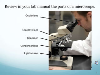
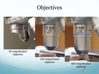
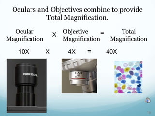
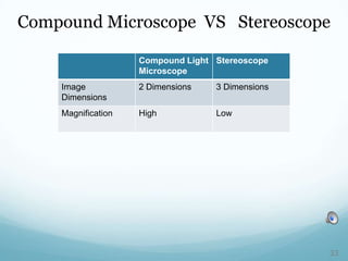
Review in your lab manual the parts of a microscope.
Ocular lens
Objective lens
Specimen
Condenser lens
Light source
1
Compound Binocular Light Microscope
Compound microscopes have more than one lens.
Ocular Lens
Objective Lens
2
Your microscope is also binocular.
Two
Oculars
3
Identical images pass to both oculars.
4
Prism
Binocular microscopes split light into two paths.
Light Path
5
Interpupilary Distance
Your microscope is binocular.
6
Your microscope is binocular.
Adjust the Interpupilary Distance
7
Adjustable Ocular
Adjustment Ring
Microscope oculars are adjustable.
8
Microscope oculars are adjustable.
Begin with a slide in place.
9
Open Eye
Closed
Eye
Focus Using
Fine
Adjustment
Knob
Focus while viewing through the non-
adjustable ocular.
10
Open Eye
Closed Eye
Focus Using
Ring of
Adjustable
Ocular
View only through the adjustable ocular.
11
Light microscopes
use visible light to
form an image.
12
Electron microscopes
use radiation of small
wavelengths to make
sharper and higher
resolution images.
Electron Microscope
13
Magnification allows us to see beyond our
limited senses.
14
1X magnification 10X magnification
Magnification
15
10X magnification
objective
4X magnification
objective
40X magnification
objective
Objectives
16
Oculars are typically 10x magnifiers.
17
10X X 4X = 40X
Ocular
Magnification
Objective
Magnification
Total
Magnification
X =
Oculars and Objectives combine to provide
Total Magnification.
18
1X Total Mag.
40X Total Mag.
4x Objective with Ocular provides
40x Total Magnification
4X Objective
19
1X Total Mag.
40X Objective
40x Objective with Ocular provides
400x Total Magnification
400X Total Mag.
20
Resolution is the ability to distinguish between two
points that are close together.
Good resolution depends on both magnification and lens
quality.
RESOLUTION
Low Magnification High Magnification
Higher Resolution of
individual legs
21
Lens Quality Improves Resolution
40X Magnification
Poor Quality Lens
40X Magnification
High Quality Lens
Better Resolution
22
Field of View (FOV)
FOV is the area of the specimen seen through the
microscope.
Field of
View
23
Field of View
Low Magnification
Large FOV
High Magnification
Small FOV
24
Field of View
Measure field of view with low magnification.
Field of View is about 2.5 mm diameter at 40X total
magnification.
25
FOV low mag. X Mag. low = FOV high mag. X Mag. high
Calculate Field of View at High Magnification
For example, if the FOV diameter is 2.5 mm at
4X then at 100X the FOV diameter is 0.1 mm.
2.5 mm X 4X = FOV high mag. X 100X
0.1 mm = FOV high mag.
26
Depth of Field (DOF)
DOF refers to the thickness of the specimen that is all
in focus.
The higher the magnification the thinner the DOF.
Chloroplast within
the DOF is in focus
Chloroplast
below the DOF is
out of focus
27
Working Distance
Working distance is the distance between the
tip of the objective and the specimen.
Working Distance []
28
Stereoscope
29
Normal vision has depth perception.
We see the world in 3 dimensions.
Height
Width
Depth
Width
Height
2 Dimensions 3 Dimensions
30
Light Path
Light Path
Stereoscope combine magnification
with 3 Dimensional images.
Two aligned light
paths
Objective
31
Dissection or surgery requires 3D vision.
32
Compound Light
Microscope
Stereoscope
Image
Dimensions
2 Dimensions 3 Dimensions
Magnification High Low
Compound Microscope VS Stereoscope
33
Working Distance
Stereoscope provide a greater
working distance.
34
Compound
Light
Microscope
Stereoscope
Working Distance Low High
Magnification High Low
Image Dimensions 2 - Dimension 3 - Dimension
Zoom Magnification No Yes
Compound Microscope vs Stereoscope
35
Ocular
Stage
Zoom Magnification
Focus Knob
Objective
Specimen
Parts of a stereoscope
36
37
Photo Credits
Slides 1-3: © Darrell Vodopich; 4 (top): © Darrell Vodopich; © The McGraw-Hill Companies, Inc./Don Rubbelke, photographer; 5-12: © Darrell
Vodopich; 13: © Centers for Disease Control/James Gathany; 14-17: © Darrell Vodopich; 18: © Steven P. Lynch RF; 18-36: © Darrell
Vodopich
Line Art Credits
Slide 2: Sylvia Mader, Biology 7e © 2001 reproduced with permission of McGraw-Hill Education

#3 Microscope (Lab Thursday 9/12/13)