
Recommended
More Related Content
What's hot
What's hot (20)
Similar to Pulmonary embolism notes 2021
Similar to Pulmonary embolism notes 2021 (20)
More from Best Doctors
More from Best Doctors (17)
Recently uploaded
Recently uploaded (20)
Pulmonary embolism notes 2021
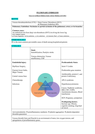
- 1. PULMONARY EMBOLISM Notes: by Col Bharat Malhotra Senior Advisor Medicine (JAN 2021) DEFINE Venous thromboembolism (VTE) = Deep Venous Thrombosis (DVT) & Pulmonary Embolism (PE) Pulmonary Embolism: Occlusion or partial occlusion of the pulmonary artery or its branches Common cause: An embolized clot from deep vein thrombosis (DVT) involving the lower leg. Less common causes: a) Tissue fragments b) Fat embolism c) Air embolism d) Amniotic fluid e) Tumor embolism EPIDEMIOLOGY PE is the most common preventable cause of death among hospitalized patients ETIOLOGY Stasis Immobilization, Paralytic stroke Venous obstruction, Venous insufficiency, CHF Endothelial injury Hip/Knee Surgery, Fracture lower limbs, Major Trauma Central venous lines Chemotherapy Infections Prothrombotic States factor V Leiden Prothrombin gene mutation Antithrombin, protein C, and protein S deficiency APLA syndrome Hyperhomocysteinemia Cancer, Nephrotic syndrome, IBD, CHF, COPD, Autoimmune diseases, Blood transfusions OCP, Pregnancy, postpartum Predisposing factors: Increase age, Obesity, cigarette smoking, long-haul air travel Activated platelets → proinflammatory mediators → platelet aggregation → platelet-dependent thrombin generation Venous thrombi form and flourish in an environment of stasis, low oxygen tension, and upregulation of proinflammatory genes.
- 2. PATHOPHYSIOLOGY Embolize Deep venous thrombi detach from their site of formation they embolize to the vena cava, right atrium, and right ventricle, and lodge in the pulmonary arterial circulation, thereby causing acute PE. Many patients with PE have no evidence of DVT because the clot has already embolized to the lungs. Pulmonary Hypertension, Right Ventricular (RV) Dysfunction, and RV Microinfarction Pulmonary artery obstruction and neurohumoral mediators cause a rise in pulmonary artery pressure and in pulmonary vascular resistance. RV wall tension rises → RV dilation and dysfunction ensue, with release of the cardiac biomarker leading to: • Compresses the right coronary artery → RV ischemia • Underfilling of the LV → fall in LV cardiac output and BP PHYSIOLOGICAL CHANGES • Arterial hypoxemia • Increased alveolar-arterial O2 tension gradient, which represents the inefficiency of O2 transfer across the lungs. o Increased pulmonary vascular resistance o Impaired gas exchange o Alveolar hyperventilation, Increased airway resistance o Decreased pulmonary compliance due to lung edema, lung hemorrhage, or loss of surfactant .
- 3. CLASSIFICATION OF PULMONARY EMBOLISM AND DEEP VENOUS THROMBOSIS Pulmonary Embolism Massive PE 5-10% Extensive thrombosis affecting at least half of the pulmonary vasculature Syncope & collapse, Severe Dyspnea, Central Chest pain, Cyanosis and hypoxemia Hypotension Cardiogenic shock Die from multisystem organ failure Sub-massive PE 20-25% Characterized by RV dysfunction despite normal systemic arterial pressure Worsening dyspnoea, Anginal chest pain, Syncope, Haemoptysis Features of RV dysfunction Low-risk PE 65-75% No RV dysfunction No hypotension Pulmonary infarction usually indicates a small PE. This condition is exquisitely painful because the thrombus lodges peripherally, near the innervation of pleural nerves. Deep Venous Thrombosis: Lower extremity DVT usually begins in the calf and propagates proximally to the popliteal vein, femoral vein, and iliac veins. DIAGNOSIS Clinical Evaluation “the Great Masquerader” Diagnosis is difficult because symptoms and signs are nonspecific In some cases, PE may be asymptomatic or discovered incidentally during diagnostic workup for another disease in predisposed individuals. The most common symptom is unexplained breathlessness. When occult PE occurs concomitantly with overt congestive heart failure or pneumonia, clinical improvement often fails to ensue despite standard medical treatment of the concomitant illness. This scenario presents a clinical clue to the possible coexistence of PE. Asymptomatic or discovered incidentally Worsening dyspnea Cough, Sputum Hemoptysis Syncope Chest pain – Anginal, Pleuritic Fever, Diaphoresis Cardiogenic shock → multiorgan dysfunction Evidence of DVT Normal Exam with sinus tachycardia Sinus Tachycardia Tachypnoea Loud S2, RV dysfunction Crackles, Pleural rub Hypotension, Cardiogenic shock in massive PE Evidence of DVT
- 4. WELLS SCORE – CLINICAL DECISION RULES DVT Interpretation of score: High probability if 3 points or more, moderate probability if 1 or 2 points, and low probability if 0 points or less PULMONARY EMBOLISM Interpretation of total score: 0-1 point: low probability; 2-6 points: moderate probability; 7 or more points: high probability DIFFERENTIAL DIAGNOSIS Not all leg pain is due to DVT, and not all dyspnea is due to PE Deep Venous Thrombosis (DVT) Ruptured Baker’s cyst, Muscle strain/injury, Cellulitis, Acute post thrombotic syndrome/venous insufficiency Pulmonary Embolism (PE) Costochondritis, Musculoskeletal discomfort, Rib fracture Pleurisy, Pneumothorax, Pneumonia, Severe asthma, chronic obstructive pulmonary disease Pericarditis, Congestive heart failure, Acute coronary syndrome, Aortic dissection Anxiety
- 5. ALGORITHM OF PE DIAGNOSIS - INTEGRATED DIAGNOSTIC APPROACH DVT PULMONARY EMBOLISM Wells score for DVT, ECG, CXR Wells score for Pulmonary Embolism, ECG, CXR The Great Masquerader – suspect and assess for PE D Dimer D Dimer Normal → No PE High → Imaging USS Doppler venous Leg → CT Angio LL ECHO Sub-massive/Massive PE (RV dysfunction) CECT Chest → CT Venous angiography for PE Lung Scan (Second line) Do USS Doppler venous Leg for DVT ProBNP: for RV dysfunction Troponin T: for RV microinfarctions EVALUATION NONIMAGING DIAGNOSTIC MODALITIES plasma D-dimer (ELISA) (> 500 ng/ml or 0.5 mcg/ml) Rises in the presence of DVT or PE because of the breakdown of fibrin by plasmin due to endogenous although often clinically ineffective thrombolysis. A normal D-dimer is a useful “rule out” test. However, the D- dimer assay is not specific. Levels increase in patients due to systemic illness. Serum troponin – due to RV microinfarction NT-pro-BNP – myocardial stretch leads to release of Pro-brain natriuretic peptide.
- 6. ELECTROCARDIOGRAM Frequent - sinus tachycardia RV strain – RBBB, T-wave inversion in leads V1 to V4 S1Q3T3 sign: S wave in lead I, Q wave in lead III, and an inverted T wave in lead III CHEST ROENTGENOGRAPHY A normal or nearly normal chest x-ray often occurs in PE. Pulmonary opacities Peripheral wedged-shaped density usually located at the pleural base (Hampton’s hump) Horizontal linear opacities Pleural effusion Focal oligemia (Westermark’s sign) Enlarged right descending pulmonary artery (Palla’s sign). Elevated hemidiapharm USS LOWER LIMB VENOUS DOPPLER Relies on loss of vein compressibility Loss of normal respiratory variation thrombus is directly visualized
- 7. ECHOCARDIOGRAPHY Echocardiography is not a reliable diagnostic imaging tool for acute PE because most patients with PE have normal echocardiograms. However, echocardiography is a very useful diagnostic tool for detecting conditions that may mimic PE Indirect sign of PE: McConnell’s sign: hypokinesis of the RV free wall with normal or hyperkinetic motion of the RV apex CHEST CT WITH VENOUS ANGIOGRAPHY In patients without PE, the lung parenchymal images may establish alternative diagnoses not apparent on chest x-ray that explain the presenting symptoms and signs CT of the chest with intravenous contrast is the principal imaging test for the diagnosis of PE. Sixth-order branches can be visualized. When imaging is extended distally below the chest to the knee, pelvic and proximal leg DVT also can be diagnosed by CT scanning. Criteria Arterial occlusion with failure to enhance the entire lumen due to a large filling defect; the artery may be enlarged compared with adjacent patent vessels A partial filling defect surrounded by contrast material, producing the “polo mint” sign on images acquired perpendicular to the long axis of a vessel and the “railway track” sign on longitudinal images of the vessel The diagnosis of PE is very unlikely in patients with normal and nearly normal scans and, in contrast, is about 90% certain in patients with high-probability scans
- 8. LUNG SCANNING (second line) Lung scanning has become a second-line diagnostic test for PE, used mostly for patients who cannot tolerate intravenous contrast. INVASIVE PULMONARY ANGIOGRAPHY (not done) Chest CT with contrast has virtually replaced invasive pulmonary angiography TREATMENT Deep Venous Thrombosis PRIMARY THERAPY Primary therapy Clot dissolution- catheter-directed thrombolysis Reserved for patients with extensive femoral, iliofemoral, or upper extremity DVT leading to less long-term damage to venous valves, with consequent lower rates of post thrombotic syndrome
- 9. SECONDARY PREVENTION Anticoagulation Effective anticoagulation is the foundation for successful treatment of DVT and PE Inferior vena caval (IVC) filter FDA approved a new retrievable IVC filter that is inserted at the bedside with ultrasound visualization of the femoral or internal jugular vein (Angel® Filter) but without the need for any fluoroscopic or other radiological imaging. Indications: Contraindication to anticoagulation, Complication of anticoagulation necessitating cessation, Propagation/progression of DVT during therapeutic anticoagulation, Iliocaval or large free-floating proximal DVT, limited cardiopulmonary reserve, Recurrent PE Below-knee graduated compression stockings Swelling of the legs when acute DVT is diagnosed, may be prescribed, usually with pressure of 30–40 mmHg, to lessen patient discomfort. They should be replaced every 3 months because they lose their elasticity. Pulmonary Embolism RISK STRATIFICATION High risk of an adverse clinical outcome Hemodynamic instability, RV dysfunction on echocardiography, RV enlargement on chest CT, or elevation of the troponin level due to RV microinfarction Good clinical outcome RV function remains normal ANTICOAGULATION Effective anticoagulation is the foundation for successful treatment of DVT and PE. There are three major strategies: 1 S/C anticoagulation with (UFH), or (LMWH), or fondaparinux “bridged” 5d → to warfarin 2 S/C anticoagulation with (UFH), or (LMWH), or fondaparinux “bridged” 5d → novel oral anticoagulant such as dabigatran (a direct thrombin inhibitor) or apixaban (an anti-Xa agent) 3 Oral anticoagulation monotherapy with rivaroxaban (3week) or apixaban (1 week) (both are anti-Xa agents) loading dose, followed by a maintenance dose without parenteral anticoagulation. For patients with VTE in the setting of suspected or proven heparin-induced thrombocytopenia, one can choose between two parenteral direct thrombin inhibitors: argatroban and bivalirudin
- 10. Mechanism of Action PARENTRAL ANTICOAGULANTS Unfractionated Heparin Action UFH anticoagulates by binding to and accelerating the activity of antithrombin, thus preventing additional thrombus formation Aim UFH is dosed to achieve a target activated partial thromboplastin time (aPTT) of 60–80 s. Dose an initial bolus of 80 U/kg, followed by an initial infusion rate of 18 U/kg per h in patients with normal liver function Benefit short half-life, which is especially useful in patients in whom hour-to- hour control of the intensity of anticoagulation Heparin also has pleiotropic effects that may decrease systemic and local inflammation. Low-Molecular-Weight Heparins These fragments of UFH exhibit less binding to plasma proteins and endothelial cells and consequently have greater bioavailability, a more predictable dose response, and a longer half-life than does UFH. No monitoring or dose adjustment is needed unless the patient is markedly obese or has chronic kidney disease
- 11. Fondaparinux Fondaparinux, an anti-Xa Penta saccharide, is administered as a weight-based once-daily subcutaneous injection in a prefilled syringe. No laboratory monitoring is required. Fondaparinux is synthesized in a laboratory and, unlike LMWH or UFH, is not derived from animal products. It does not cause heparin-induced thrombocytopenia. The dose must be adjusted downward for patients with renal dysfunction. ORAL ANTICOAGULANTS Warfarin Anticoagulation (Unfractionated heparin, LMWH, and fondaparinux are the usual immediately effective “bridging agents” used when initiating warfarin) Requires 5–10 days of administration to achieve effectiveness Warfarin Action This vitamin K antagonist prevents carboxylation activation of coagulation factors II, VII, IX, and X Bridge therapy The full effect of warfarin requires at least 5 days, even if the prothrombin time, used for monitoring, becomes elevated more rapidly If warfarin is initiated as monotherapy during an acute thrombotic illness, a paradoxical exacerbation of hypercoagulability increases the likelihood of thrombosis. Overlapping UFH, LMWH, fondaparinux, or parenteral direct thrombin inhibitors with warfarin for at least 5 days will nullify the early procoagulant effect of warfarin Dose Monitor Usual start dose is 5 mg Titrate to international normalized ratio (INR), target 2.0–3.0 Continue parenteral anticoagulation for a minimum of 5 days and until two sequential INR values, at least 1 day apart, achieve the target INR range. Problems The warfarin dose is usually titrated empirically to achieve the target INR. Proper dosing is difficult because hundreds of drug-drug and drug-food interactions affect warfarin metabolism. Warfarin can cause major hemorrhage, including intracranial hemorrhage, even when the INR remains within the desired therapeutic range. Warfarin can cause “off target” side effects such as alopecia or arterial vascular calcification. Some patients complain that warfarin makes them feel cold or fatigued CYP2C9 variant alleles impair the hydroxylation of S-warfarin, thereby lowering the dose requirement ...
- 12. Novel Oral Anticoagulants Novel oral anticoagulants (NOACs) are administered in a fixed dose, establish effective anticoagulation within hours of ingestion, require no laboratory coagulation monitoring, and have few of the drug-drug or drug-food interactions Rivaroxaban and apixaban, direct factor Xa inhibitors, are approved as monotherapy for acute and extended treatment of DVT and PE, without a parenteral “bridging” anticoagulant. Dabigatran, a direct thrombin inhibitor, and edoxaban, a factor Xa inhibitor, are approved for treatment of VTE after an initial 5-day course of parenteral anticoagulation. Betrixaban, a direct factor Xa inhibitor, was approved by the FDA in 2017 for VTE prophylaxis in acutely ill medical patients during hospitalization and continuing for a total duration of 5 to 6 weeks. Non-Warfarin Anticoagulation Unfractionated heparin, bolus and continuous infusion, to achieve activated partial thromboplastin time (aPTT) 2–3 times the upper limit of the laboratory normal, or Enoxaparin 1 mg/kg twice daily with normal renal function, or Dalteparin 200 U/kg once daily or 100 U/kg twice daily, with normal renal function, or Tinzaparin 175 U/kg once daily with normal renal function, or Fondaparinux weight-based once daily; adjust for impaired renal function Direct thrombin inhibitors: argatroban or bivalirudin (with suspected or proven heparin-induced thrombocytopenia) Rivaroxaban 15 mg twice daily for 3 weeks, followed by 20 mg once daily with the dinner meal thereafter Apixaban 10 mg twice daily for 1 week, followed by 5 mg twice daily thereafter Dabigatran 5 days of unfractionated heparin, low-molecular-weight heparin (LMWH), or fondaparinux followed by dabigatran 150 mg twice daily Edoxaban 5 days of unfractionated heparin, LMWH, or fondaparinux followed by edoxaban 60 mg once daily with normal renal function, weight >60 kg, in the absence of potent P-glycoprotein inhibitors Complications of Anticoagulants The most serious adverse effect of anticoagulation is hemorrhage. For life-threatening or intracranial hemorrhage following can be administered Heparin or LMWH → protamine sulphate Dabigatran → Idarucizumab (NA) Fondaparinux or factor Xa inhibitors → No reversal Universal anti-Xa antidote → Andexanet (NA)
- 13. Major bleeding from warfarin is best managed with prothrombin complex concentrate. With less serious bleeding, fresh-frozen plasma or intravenous vitamin K can be used. Duration of Anticoagulation INTERMEDIATE RISK (3-8%) HIGH RISK FACTOR (> 8 %) Major surgery Lower limb plaster cast > 3 days Short-term immobilization for >3 days Hormonal contraception, pregnancy Acute infectious disease leading to confinement > 3 days Inflammatory bowel disease Active autoimmune disease Active Cancer Antiphospholipid syndrome Previous episodes of VTE Myeloproliferative disorders Thrombophilia For DVT isolated to an upper extremity or calf that has been provoked by surgery, trauma, estrogen, or an indwelling central venous catheter or pacemaker, 3 months of anticoagulation usually suffice For an initial episode of provoked proximal leg DVT or PE, 3–6 months of anticoagulation used to be the classic teaching For patients with cancer and VTE, prescribe LMWH as monotherapy without warfarin and continue anticoagulation indefinitely unless the patient is rendered cancer-free. Patients with antiphospholipid antibody syndrome may warrant indefinite-duration anticoagulation, even if the initial VTE was provoked by trauma or surgery. Among patients with idiopathic, unprovoked VTE, the recurrence rate is high after cessation of anticoagulation. VTE that occurs during long- haul air travel is considered unprovoked. Unprovoked VTE may be caused by an exacerbation of an underlying inflammatory state and can be conceptualized as a chronic illness, with latent periods between flares of recurrent episodes. American College of Chest Physicians (ACCP) guidelines recommend considering anticoagulation for an indefinite duration with a target INR between 2 and 3 for patients with idiopathic VTE and a low bleeding risk. An alternative approach after the first 6 months of anticoagulation is to reduce the intensity of anticoagulation and to lower the target INR range to between 1.5 and 2. Another approach for patients at lower risk of recurrence, especially if there is an important reason to avoid long-term anticoagulation, is to consider low-dose aspirin after completing the initial period of standard anticoagulation.
- 14. INFERIOR VENA CAVA FILTERS (Reserve therapy) The two principal indications for insertion of an IVC filter are (1) active bleeding that precludes anticoagulation and (2) recurrent venous thrombosis despite intensive anticoagulation. Prevention of recurrent PE in patients with right heart failure who are not candidates for fibrinolysis and prophylaxis of extremely high-risk patients are “softer” indications for filter placement. Retrievable filters can now be placed for patients with an anticipated temporary bleeding disorder or for patients at temporary high risk of PE, such as individuals undergoing bariatric surgery who have a prior history of perioperative PE. The filters can be retrieved for months after insertion, unless thrombus forms and is trapped within the filter. The retrievable filter becomes permanent if it remains in place or if, for technical reasons such as rapid endothelialization, it cannot be removed. The filter itself may fail by permitting the passage of small- to medium-size clots. Large thrombi may embolize to the pulmonary arteries via collateral veins that develop. Paradoxically, by providing a nidus for clot formation, filters increase the DVT rate, even though they usually prevent PE. Therefore, a common complication is recurrent DVT or caval thrombosis with marked leg swelling.
- 15. MANAGEMENT OF MASSIVE PE For patients with massive PE and hypotension, replete volume with 500 mL of normal saline. Additional fluid should be infused with extreme caution because excessive fluid administration exacerbates RV wall stress, causes more profound RV ischemia, and worsens LV compliance and filling by causing further interventricular septal shift toward the LV. Dopamine and dobutamine are first-line inotropic agents for treatment of PE-related shock. Maintain a low threshold for initiating these pressors. Often, a “trial-and-error” approach works best; other agents that may be effective include norepinephrine, vasopressin. FIBRINOLYSIS The only Food and Drug Administration–approved indication for PE fibrinolysis is massive PE. For patients with submassive PE, who have preserved systolic blood pressure but moderate or severe RV dysfunction, use of fibrinolysis remains controversial Successful fibrinolytic therapy and may result in a lower rate of death and recurrent PE by . Rapidly reverses right heart failure and lower rate of death (1) dissolving much of the anatomically obstructing pulmonary arterial thrombus, (2) preventing the continued release of serotonin and other neurohumoral factors that exacerbate pulmonary hypertension, and (3) lysing much of the source of the thrombus in the pelvic or deep leg veins, thereby decreasing the likelihood of recurrent PE Dose 100 mg of recombinant tissue plasminogen activator (tPA) prescribed as a continuous peripheral intravenous infusion over 2 h A popular off-label dosing regimen is 50 mg of TPA administered over 2 h. This lower dose is widely perceived to be associated with fewer bleeding complications Contraindication Intracranial disease, recent surgery, and trauma. The overall major bleeding rate is about 10%, including a 2–3% risk of intracranial hemorrhage PHARMACOMECHANICAL CATHETER-DIRECTED THERAPY Many patients have relative contraindications to full-dose thrombolysis. Pharmaco-mechanical catheter-directed therapy usually combines physical fragmentation or pulverization of thrombus with catheter-directed low-dose thrombolysis.
- 16. EMOTIONAL SUPPORT PREVENTION OF VTE Prevention of DVT and PE is of paramount importance because VTE is difficult to detect and poses a profound medical and economic burden. • High Risk orthopedic surgery • Major Orthopedic Surgery • Cancer Surgery • Medically ill patients during hospitalization Low-dose UFH or LMWH is the most common form of in-hospital prophylaxis. S/C ENOXAPARIN 40 MG OD Extended-duration prophylaxis with the novel anti-Xa agent, BETRIXABAN, appears to be both effective and safe in medically ill patients during hospitalization, after hospital discharge, and is undergoing FDA review. NOTES: BY COL BHARAT MALHOTRA SENIOR ADVISOR MEDICINE (JAN 2021) REFERENCES: DAVIDSON’S PRINCIPLES AND PRACTICE OF MEDICINE 23RD (2018) CHAPTER 17 HARRISON’S PRINCIPLES OF INTERNAL MEDICINE 20TH (2018) CHAPTER 273 2019 ESC GUIDELINES FOR THE DIAGNOSIS AND MANAGEMENT OF ACUTE PULMONARY EMBOLISM.