SlideShare a Scribd company logo
1 of 14
ANKYLOSING SPONDYLITIS
Notes by Col Bharat Malhotra Senior Advisor Medicine
DEFINE
AS is an inflammatory disorder of unknown cause that
primarily affects
• the axial skeleton;
• peripheral joints and extraarticular structures are
also frequently involved
It has striking correlation to HLA-B27
Non-HLA susceptibility alleles are also identified
The disease usually begins in the second or third decade.
WOMEN: Has Peripheral arthritis >> Axial involvement
Prevalence USA 0.9 – 1.4% INDIA 0.7- 0.9%
Spectrum
CRITERIA
The term axial spondyloarthritis (ax-SpA) is now in common use, supported by ASAS Criteria for
Classification of AS formulated in 2009. This classification includes definite AS, early stages that will
progress to meet classical criteria for AS, and one or more non progressing phenotypes. Patients with
ax-SpA that do not have radiologic criteria for AS are said to have non-radiographic axial SpA
(nr-ax-SpA). The prevalence of HLA-B27 in these patients is similar to that in AS (Female > Male).
BACK PAIN > 3 MONTHS
AGE OF ONSET < 45 YEARS
SACROILIITIS ON IMAGING
PLUS > 1 SpA Feature OR
HLA-B27
PLUS >2 SpA Features
Sacroiliitis on imaging
Active (acute) inflammation on MRI highly
suggestive of SpA associated sacroiliitis
(Bone marrow edema + osteitis on STIR or
gadolinium enhanced T1 image)
And/or
Definite radiographic sacroiliitis according to
modified New York Criteria
(Bilateral grade >2 or Unilateral grade 3 or 4)
SpA Features
Inflammatory back pain (>4 diagnostic)
(1) age of onset <40 years;
(2) insidious onset;
(3) improvement with exercise;
(4) no improvement with rest; and
(5) pain at night with improvement upon getting up;
(6) morning stiffness >30 min;
(7) awakening from back pain during only the second half of the night; and
(8) alternating buttock pain.
Arthritis -Past or present diagnosed by physician
Enthesitis (heel) – Past or present pain or tenderness on
exam at calcaneus insertion of Achilles tendon or plantar fascia
Dactylitis- Past or present diagnosed by physician
Anterior Uveitis - Past or present confirmed by eye spl
Psoriasis- Past or present diagnosed by physician
Chron’s disease or Ulcerative colitis - Past or
present diagnosed by physician
Family history of SpA- First or second-degree relatives
HLAB27
Elevated CRP- exclude other causes
Good response to NSAID- relief in 24-48h
Enthesitis -Inflammation of attachment of
tendon & ligament to bone.
Dactylitis- Severe Inflammation of Finger and
toes giving appearance like sausages.
PATHOGENESIS – IT IS IMMUNE MEDIATED (Auto Inflammatory pathogenesis)
HLA B27 plays direct role, Association of AS with ERAP1 which influences MHC class 1 peptide
This causes B27 heavy chain to unusual tendency to misfold → process that can be proinflammatory
Unlike RA synovium, citrullinated proteins and cartilage gp39 peptide-MHC are ABSENT
BRIEF OVERVIEW
IL-23 / IL-17
PATHWAY
New bone formation – WNT signaling
pathways
SYNDESMOPHYTES
PATHOLOGY
Sacroiliitis
Macrophages, T cells, plasma
cells, and osteoclasts are
prevalent.
• Sacroiliitis is often an early manifestation of AS and nr-ax-SpA.
• Synovitis and myxoid marrow represent the earliest changes
• Followed by pannus and subchondral granulation tissue
• Marrow edema, enthesitis, and chondroid differentiation found.
• If the process continues to progress, eventually the eroded joint
margins are gradually replaced by fibrocartilage regeneration and
then by ossification.
Spine • Inflammatory granulation tissue in the paravertebral connective
tissue at the junction of annulus fibrosus and vertebral bone, and in
some cases along the entire outer annulus.
• The outer annular fibers are eroded and eventually replaced by bone,
forming the beginning of a syndesmophyte.
• It then grows by continued endochondral ossification, ultimately
bridging the adjacent vertebral bodies. Ascending progression of
this process can lead to the “bamboo spine.”
• Other lesions in the spine
o Diffuse osteoporosis (loss of trabecular bone despite
accretion of periosteal bone),
o Erosion of vertebral bodies at the disk margin,
o Inflammation and destruction of the disk-bone border.
Inflammatory
arthritis
• Inflammatory arthritis of the apophyseal (facet) joints is common,
• With synovitis, inflammation at the bony attachment of the joint
capsule, and subchondral bone marrow granulation tissue.
• Erosion of joint cartilage by pannus is often followed by bony
ankylosis.
• Formation of syndesmophytes bridging the adjacent disks.
• Bone mineral density is diminished in the spine and proximal femur
early in the disease course.
The characteristics of peripheral arthritis in AS and other forms of
SpA are similar, and distinct from those of RA.
Fibrocartilaginous
enthesis
Inflammation in the fibrocartilaginous enthesis, the region where a
tendon, ligament, or joint capsule attach to bone, is a characteristic
lesion in AS and other SpAs, both at axial and peripheral sites.
Enthesitis is associated with prominent edema of the adjacent bone
marrow and is often characterized by erosive lesions that eventually
undergo ossification.
Subclinical intestinal inflammation has been found in the colon or
distal ileum in a majority of patients with SpA
CLINICAL MANIFESTATIONS
Age Late adolescence or early adulthood
Usually < 45 years (in 5% symptoms begin after 40 years)
Initial
symptoms
LBA
• Usually dull pain, Insidious in onset
• Felt deep in the lower lumbar or gluteal region,
• Accompanied by low-back morning stiffness of up to a few hours’
duration that improves with activity and returns following inactivity
• Within a few months, the pain usually become persistent and bilateral.
• Nocturnal exacerbation of pain often forces the patient to rise and move
around.
Enthesitis or
Osteitis
Dactylitis
In some patients, bony tenderness (presumably reflecting enthesitis or
osteitis) may accompany back pain or stiffness.
Common sites are Costo-sternal junctions, spinous processes, iliac crests,
greater trochanters, ischial tuberosities, tibial tubercles and heels
Dactylitis may be seen due to severe inflammation of fingers and toes.
Asymmetric
Oligoarthrits
Arthritis of peripheral joints other than the hips and shoulders, usually
asymmetric, may occur at any point in the disease course
Hip &
Shoulder joint
Hip and shoulder (“root” joint) arthritis is considered part of axial
disease. Hip arthritis occurs in 25–35% of patients.
Shoulder involvement may be at least as common, but is usually less
symptomatic
Late –
Cervical Spine
Neck pain and stiffness from involvement of the cervical spine are usually
relatively late manifestations, but are occasionally dominant symptoms.
Juvenile onset In juvenile onset spondylarthritis, peripheral arthritis and enthesitis
predominate, with axial symptoms supervening in late adolescence.
It should be emphasized that in early mild, or atypical cases, the symptoms and/or physical
findings may be subtle and/or nonspecific.
Axial physical findings mirror the inflammatory process.
Limitation of motion >> ankylosing spondylosis (Due to pain & spasm)
Loss of spinal
mobility
imitation of anterior
and lateral flexion
limited extension of
the lumbar spine
Limited
chest expansion
Pain in the sacroiliac
joints
tenderness on
posterior spinous
processes
symptomatic bony
tenderness
Enthesitis
Modified
Schober test
The patient stands erect, with heels together, and marks are made
on the spine at the lumbosacral junction (identified by a
horizontal line between the posterosuperior iliac spines) and
10 cm above. The patient then bends forward maximally with
knees fully extended, and the distance between the two marks is
measured.
This distance increases by ≥5 cm in the case of normal mobility
and by <4 cm in the case of decreased mobility.
Wall Occiput Distance Perpendicular distance between
• bony prominence of the occiput
• and the wall in a standing position
Lateral bending Lateral bending measures the distance the patient’s middle finger
travels down the leg with maximal lateral bending.
Normal is >10 cm.
Chest expansion Chest expansion is measured as the difference between maximal
inspiration and maximal forced expiration in the fourth intercostal
space in males or just below the breasts in females, with the
patient’s hands resting on or just behind the head.
Normal chest expansion is ≥5 cm
Hips or Shoulders Limitation or pain with motion of the hips or shoulders is usually
present if these joints are involved.
OUTCOME MEASURES EVALUATION SCALE
Schober Test
Oswestry Disability Index (ODI)
Neck Disability Index (NDI)
Visual Analogue Scale
Patient Specific Functional Scale
Bath Ankylosing Spondylitis Metrology Index (BASMI)
Revised Leeds Disability Questionnaire (RLDQ)
European Quality of Life (EuroQoL)
Bath AS Disease Activity Index (BASDAI; 0-100)
AMOR criteria for Spondylarthritis
BASFI index (Bath Ankylosing Spondylitis Functional Index)
BAS-G index (Bath Ankylosing Spondylitis Global Index)
The course of ax-SpA is extremely variable
Mild stiffness and normal radiographs → totally fused spine and severe bilateral hip arthritis,
accompanied by severe peripheral arthritis and extraarticular manifestations.
Pain tends to be persistent early in the disease and intermittent later, with alternating
exacerbations and quiescent periods.
Patient’s posture undergoes characteristic changes, with obliterated
lumbar lordosis, buttock atrophy, and accentuated thoracic kyphosis.
There may be a forward stoop of the neck or flexion contractures at
the hips, compensated by flexion at the knees.
Disease progression can be estimated clinically from loss of height,
limitation of chest expansion and spinal flexion, and occiput-to-wall
distance.
EXTRAARTICULAR MANIFESTATION
Acute Anterior Uveitis 40% of patients
Can antedate the spondylitis
Attacks are typically unilateral, causing pain, photophobia,
and increased lacrimation.
These tend to recur, often in the opposite eye.
Cataracts and secondary glaucoma may ensue.
Inflammation in the Colon or
Ileum
60% of patients
usually asymptomatic, but frank IBD occurs in 5–10%
Psoriasis 10% of patients
Acne fulminans or hidradenitis suppurativa can occur
There is an apparently increased risk of ischemic heart disease
Aortic insufficiency occurs in a small percentage of patients.
Third-degree heart block may occur alone or together with aortic insufficiency, and
association with lesser degrees of heart block has been described.
Cauda equina syndrome and upper pulmonary lobe fibrosis are rare late complications.
Prostatitis has been reported
Amyloidosis is rare
COMPLICATION
The most serious complication of the spinal disease is spinal fracture, which can occur with
even minor trauma to the rigid, osteoporotic spine
lower cervical spine Its most commonly involved. These fractures are often
displaced, causing spinal cord injury.
thoracolumbar spine Occasionally, fracture through a disk vertebral junction and
adjacent neural arch, termed pseudoarthrosis, most common
in the thoracolumbar spine, can be an unrecognized source of
persistent localized pain and/or neurologic dysfunction.
thoracic vertebrae Wedging of thoracic vertebrae is common and correlates with
accentuated kyphosis.
Mortality attributable to AS is largely the result of spinal trauma, aortic insufficiency,
respiratory failure, amyloid nephropathy, or complications of therapy such as upper
gastrointestinal hemorrhage. The impact of anti-TNF therapy on outcome and mortality is not
yet known, except for significantly improved work productivity.
LABORATORY FINDINGS
No laboratory test is diagnostic of AS.
Patients are rheumatoid factor (RF) negative, hence seronegative AS.
HLA-B27 is present in 75–90% of patients
In most ethnic groups, HLA-B27 is present in 75–90% of AS patients.
HLA B27 is present in 5% healthy individuals
10–15% of patients with AS may be HLA B27 negative
HLA-B27 positivity, in general, confers a greater risk of disease severity and also a concentration of
disease in the spine and the eye. HLA-B27-negative patients are more likely to develop peripheral
arthritis, skin, and nail disease, or IBD.
In the absence of any preventative treatment, asymptomatic children of AS patients should not be
tested. Testing is recommended only in symptomatic children.
Erythrocyte sedimentation rate (ESR) and C-reactive protein (CRP) are often, but not
always, elevated.
Mild anemia may be present.
Patients with severe disease may show an elevated alkaline phosphatase level.
Elevated serum IgA levels are common. Rheumatoid factor, anti-cyclic citrullinated peptide
(CCP), and antinuclear antibodies (ANAs) are largely absent unless caused by a coexistent
disease, although ANAs may appear with anti-TNF therapy.
Synovial fluid from peripheral joints in AS is nonspecifically inflammatory.
Restricted chest wall motion causes decreased vital capacity, but ventilatory function is
usually well maintained.
HLA-B27
HLA-B27 is a MHC class I molecule consisting of an alpha chain encoded in the MHC region on chromosome
6 and a non-MHC encoded beta chain, β2 microglobulin. The development of ankylosing spondylitis may be
promoted by increased expression of HLA-B27 on the surface of peripheral blood mononuclear cell.
The arthritogenic peptide hypothesis suggests that HLA-B27 has a unique ability to bind antigens from a
microorganism that trigger a CD8 T-cell response that then cross-reacts with a HLA-B27/self-peptide pair.
Furthermore, it has been shown that HLA-B27 can bind peptides at the cell surface.
RADIOGRAPHIC FINDINGS
The diagnosis of AS is clinico-radiologic. Sacroiliitis is considered the radiographic hallmark of AS.
Xray S I Joint
Radiographically demonstrable
sacroiliitis, usually symmetric.
• Blurring of the cortical margins of the
subchondral bone
• Followed by erosions and sclerosis.
• Progression of the erosions leads to
“pseudo-widening” of the joint space
• As fibrous and then bony ankylosis
supervene, the joints may become
obliterated
Sacroiliitis grading – New York criteria
Grade 0: normal
Grade I: suspicious changes (some blurring of the joint
margins)
Grade II: minimum abnormality (small localized areas
with erosion or sclerosis, with no alteration in the joint
width)
Grade III: unequivocal abnormality (moderate or
advanced sacroiliitis with erosions, evidence of
sclerosis, widening, narrowing, or partial ankylosis)
Grade IV: severe abnormality (complete ankylosis)
MRI SI Joint – with contrast or STIR image
MRI is thus much more useful for the timely diagnosis of ax-SpA.
Active sacroiliitis is best visualized by dynamic MRI
• Either T2-weighed (STIR) image: bone marrow edema
• Or T1-weighted images with contrast enhancement- synovitis & capsulitis.
These techniques identify early intraarticular inflammation, cartilage changes, and
underlying bone marrow edema in sacroiliitis.
Active inflammation of the sacroiliac joints as determined by dynamic MRI is considered
equivalent to definite radiographic sacroiliitis.
X RAY Lumbosacral Spine
In the lumbar spine, progression of the disease can lead
to
• Loss of lordosis,
• Osteitis of the anterior corners of the vertebral
bodies
with subsequent erosion: Romanus lesions
• Leading to “squaring” or even “barreling” of one or
more vertebral bodies.
• Linear ossification along central spine: Dagger spine
• Progressive ossification leads to eventual formation
of marginal syndesmophytes: Bamboo spine
Hatchet Sign (Humeral Head)
refers to the limited erosion of
the lateral aspect of the humeral
head that produces a hatchet-
shaped deformity. This finding is
typically associated with
ankylosing spondylitis.
Shiny Corner Sign (Spine)
is a spinal finding in ankylosing
spondylitis, representing reactive
sclerosis secondary to inflammatory
erosions at the superior and inferior
endplates (corners on lateral radiograph)
of the vertebral bodies which are known
as Romanus lesions. Eventually, the
vertebral bodies become Squared.
Dagger Sign (Spine)
is a radiographic feature seen in
ankylosing spondylitis as a single
central radio dense line on frontal
radiographs related to ossification
of the supraspinous and
interspinous ligaments secondary
to enthesitis.
Bamboo spine
Diffuse
syndesmophytic
ankylosis.
DEXA SCAN
Reduced bone mineral density can be detected by dual-energy x-ray absorptiometry of
the femoral neck and the lumbar spine. Use of a lateral projection of the L3 vertebral body
can prevent falsely elevated readings related to spinal ossification.
DIAGNOSIS
It is important to recognize ax-SpA before the development of irreversible deformity.
This goal is challenging for several reasons:
(1) only a minority of back pain patients have ax-SpA;
(2) an early diagnosis often relies on clinical grounds and/or an appropriate MRI protocol
requiring considerable expertise;
(3) young individuals with symptoms of ax-SpA often do not seek medical care;
(4) reliance on definite radiographic sacroiliitis causes early or mild cases to be missed.
Low back ache > 3 months
Age of onset < 45 years
Defined
radiological
sacroiliitis
Presence of spondyloarthritis features – Inflammatory back pain,
asymmetric arthritis, enthesitis, dactylitis, anterior uveitis, IBD, psoriasis,
family history of SpA, Elevated ESR/CRP, good response to NSAID.
Confirm as AS
> 4 features 2-3 Features 0-1 Features
Compelling
clinical picture
suggest AS
HLA-B27
If positive → AS
If negative do
MRI SI Joint
HLA-B27
If positive → AS
If negative do
MRI SI Joint
HLA-B27
If positive also do
MRI SI Joint
DIFFERENTIAL DIAGNOSIS
• Less-common metabolic, infectious, and malignant causes of back pain must also be
differentiated from AS, including infectious spondylitis, spondylodiskitis, and sacroiliitis,
and primary or metastatic tumor.
• Ochronosis can produce a phenotype similar to AS.
• Calcification and ossification of paraspinous ligaments occur in diffuse idiopathic skeletal
hyperostosis (DISH), which occurs in the middle-aged and elderly and is usually not
symptomatic. Ligamentous calcification gives the appearance of “flowing wax” on the
anterior bodies of the vertebrae. Intervertebral disk spaces are preserved, and sacroiliac and
apophyseal joints appear normal, helping to differentiate DISH from spondylosis and from
AS, respectively.
• Both primary and secondary hyperparathyroidism can cause subchondral bone resorption
around the SI joints, with bilateral widened and ill-defined joints on radiographs, but
without joint space narrowing.
MANAGEMENT
TREATMENT PLAN
EXERSIZE
Exercise program to maintain posture and range of motion.
NSAID
Nonsteroidal anti- inflammatory drugs (NSAIDs) are the first line of pharmacologic therapy.
They reduce pain and tenderness and increase mobility in many patients. Continuous high-dose
NSAID therapy may slow radiographic progression, particularly in patients who are at higher
risk for progression.
ANTI-TNF-Α THERAPY
However, many patients have continued symptoms despite NSAID therapy and are likely to
benefit from anti-TNF-α therapy. Patients with AS treated with infliximab (chimeric
human/mouse anti-TNF-α monoclonal antibody), etanercept (soluble p75 TNF-α receptor–IgG
fusion protein), adalimumab, or golimumab (human anti-TNF-α monoclonal antibodies, or
certolizumab pegol [humanized mouse anti-TNF-α monoclonal antibody]) have shown rapid,
profound, and sustained reductions in all clinical and laboratory measures of disease activity.
Indications of anti-TNF-α therapy
Because of the expense, potentially serious side effects, and unknown long-term effects of these
agents, their use should be restricted to patients with a definite diagnosis and active disease
(BASDAI ≥4 out of 10 and expert opinion) that is inadequately responsive to therapy with at
least two different NSAIDs. Before initiation of anti-TNF therapy, all patients should be tested
for tuberculin (TB) reactivity, and reactors (≥5 mm on PPD testing or a positive quantiferon
test) should be treated with anti-TB agents.
Biologics are indicated for a disease that is active and refractory. Refractory axial disease is
defined as one that has not responded to at least 2 NSAIDs tried in full doses over 4 weeks.
Refractory peripheral disease is one that is unresponsive to intra-articular steroids (if indicated)
and sulfasalazine (2–3 Gm/day) given for 3 months after reaching the target dose. Active AS
is defined by BASDAI score >4/10 or ASDAS >2.1
Dose of anti-TNF-α therapy
Infliximab IV 5mg/kg Stat, 2 weeks, 6 weeks
Then 6-8 weeks interval
Etanercept S/C 50mg Once weekly
Adalimumab S/C 40mg Biweekly
Golimumab S/C 50-100mg Every 4 weeks
Certolizumab
pegol
S/C 400mg Every 4 weeks
Side effects of anti-TNF-α therapy
These potent immunosuppressive agents are relatively safe, but patients are at increased risk
for serious infections, including disseminated tuberculosis. Hypersensitivity infusion or
injection site reactions are not uncommon. Cases of anti-TNF-induced psoriasis have been
increasingly recognized. Rare cases of systemic lupus erythematosus (SLE)–related disease
have been reported, as have hematologic disorders such as pancytopenia, demyelinating
disorders, exacerbation of congestive heart failure, and severe liver disease. The overall
incidence of malignancy does not appear to be increased in AS patients treated with anti-TNF
therapy, but isolated cases of hematologic malignancy have occurred shortly after the start of
treatment.
Contraindications
Contraindications include active infection or high risk of infection; malignancy or
premalignancy; and history of SLE, multiple sclerosis, or related autoimmunity. Pregnancy and
breast-feeding are no longer considered contraindications if appropriate precautions are taken.
Infants exposed to anti-TNF in utero should not be given live vaccines before age 6 months.
Response
Continuation beyond 12 weeks of therapy requires either a 50% reduction in BASDAI or
absolute reduction of ≥2 out of 10, and favorable expert opinion. Switching to a second anti-
TNF agent may be effective, especially if there was a response to the first that was lost rather
than primary failure.
ASDAS CUT OFF CRITERIA ASDAS IMPROVEMENT CRITERIA
In a good response, there is significant improvement in both objective and subjective
indicators of disease activity and function, including morning stiffness, pain, spinal mobility,
peripheral joint swelling, CRP, ESR, and bone mineral density. MRI studies indicate
substantial resolution of bone marrow edema, enthesitis, and joint effusions in the sacroiliac
joints, spine, and peripheral joints. About one-half of the patients achieve a ≥50% reduction in
the BASDAI. The response tends to persist over time, and partial or full remissions are
common.
Predictors of the best responses include younger age, shorter disease duration, higher
baseline inflammatory markers, and lower baseline functional disability. Nonetheless, some
patients with long-standing disease and even spinal ankylosis can obtain significant benefit.
Syndesmophyte formation may continue despite the therapy, but this may apply mainly during
the early years of therapy. Although less well studied, the response of patients with nr-ax-SpA
to anti-TNF therapy is generally similar to that of patients with AS.
HUMAN MONOCLONAL ANTIBODY TO IL-17A
Secukinumab, a human monoclonal antibody to IL-17A, shows dramatic efficacy in AS,
similar to that seen with TNF inhibitors, and is effective in some patients who have failed or
not tolerated anti-TNF therapy.
Most recommendations place the use of TNFi ahead of IL-17 inhibitors. This is primarily
because of the earlier introduction and greater familiarity with these agents.
In primary nonresponders, to TNFi, secukinumab or ixekizumab are preferred over treatment
with a different TNFi. In secondary nonresponders, a different TNFi can also be tried.
Secukinumab S/C 150mg Weekly x 4 weeks
Then 4-week interval
Ixekizumab
Precautions regarding infection are similar to those for anti-TNF agents. An additional concern
is potential exacerbation of underlying IBD, whether previously recognized or not, and careful
monitoring is advised.
ORAL JAK INHIBITOR, TOFACITINIB
The oral Jak inhibitor, tofacitinib, showed efficacy in AS, with reduction of inflammation
evident on MRI, in a 16-week phase 2 study.
DMARD- SULFASALAZINE
Sulfasalazine, in doses of 2–3 g/d, has modest benefit, primarily for peripheral arthritis.
Methotrexate, although widely used, has not been shown to be of benefit in AS, nor has any
therapeutic role for gold or oral glucocorticoids been documented.
LOCAL STEROIDS
Unlike RA, the response to systemic corticosteroids is not as good in AS. Prolonged oral
corticosteroid therapy does more harm than good and should be avoided. Intra-articular
corticosteroids for the persistently active joint are helpful, provided septic arthritis has been
ruled out. Painful enthesopathy or refractory plantar fasciitis may also benefit from local
corticosteroid injection. Direct injection into tendons is best avoided because of risk of tendon
rupture.
SURGERY
The most common indication for surgery in patients with AS is severe hip joint arthritis, the
pain and stiffness of which are usually dramatically relieved by total hip arthroplasty. Rare
patients may benefit from surgical correction of extreme flexion deformities of the spine or of
atlantoaxial subluxation.
EXTRAARTICULAR MANIFESTATIONS TREATMENT
Attacks of uveitis are usually managed effectively with local glucocorticoids and mydriatic
agents, although systemic glucocorticoids, immunosuppressive drugs, or anti-TNF therapy
may be required. TNF inhibitors reduce the frequency of attacks of uveitis in patients with ax-
SpA, and adalimumab has recently been approved by the FDA for treating uveitis. Cases of
new or recurrent uveitis after use of a TNF inhibitor have been observed, especially with
etanercept. Anti-IL-17 does not appear as effective for uveitis as anti-TNF therapy.
Management of axial osteoporosis is at present similar to that used for primary osteoporosis,
since data specific for AS are not available.
Notes from: Harrison’s Principle of Internal medicine 20th
Ed (2018) chapter 255
Few points from
• Clinical Rheumatology (2021) by Rohini Handa
• Firestein Kelley’s Textbook of Rheumatology, 11th
Ed (2020)
• Targeting inflammatory pathways in axial spondyloarthritis. Arthritis Research & Therapy volume 21,
Article number: 135 (2019) open access
NOTES PREPARED BY COL BHARAT MALHOTRA SR ADV MEDICINE (Jun 2021)
PATHOGENESIS
CLINICAL FINDINGS
TREATMENT

More Related Content

What's hot

Dr tarek spondyloarthropathy
Dr tarek spondyloarthropathyDr tarek spondyloarthropathy
Dr tarek spondyloarthropathyal azhar universty
 
Ankylosing spondylitis pathogenesis
Ankylosing spondylitis pathogenesisAnkylosing spondylitis pathogenesis
Ankylosing spondylitis pathogenesisSitanshu Barik
 
Axial sp a and its management
Axial sp a and its managementAxial sp a and its management
Axial sp a and its managementDR RML DELHI
 
Ankylosing spondylitis,Causes,symptoms,diagnosis,management
Ankylosing spondylitis,Causes,symptoms,diagnosis,managementAnkylosing spondylitis,Causes,symptoms,diagnosis,management
Ankylosing spondylitis,Causes,symptoms,diagnosis,managementDr.Md.Monsur Rahman
 
Psoriatic arthritis
Psoriatic arthritisPsoriatic arthritis
Psoriatic arthritishamidreza227
 
Ankylosing Spondylitis
Ankylosing SpondylitisAnkylosing Spondylitis
Ankylosing SpondylitisEneutron
 
Spondyloarthropathy:An update
Spondyloarthropathy:An updateSpondyloarthropathy:An update
Spondyloarthropathy:An updateRafiqul Islam
 
Cervical radiculopathy
Cervical radiculopathyCervical radiculopathy
Cervical radiculopathymrinal joshi
 
Ankylosing spondylitis
Ankylosing spondylitisAnkylosing spondylitis
Ankylosing spondylitisorthoprince
 
Ankylosing spondylitis
Ankylosing spondylitisAnkylosing spondylitis
Ankylosing spondylitisRatan Khuman
 
Student's Elbow (Olecranon Bursitis) - Dr Rohit Bhaskar
Student's Elbow (Olecranon Bursitis) - Dr Rohit BhaskarStudent's Elbow (Olecranon Bursitis) - Dr Rohit Bhaskar
Student's Elbow (Olecranon Bursitis) - Dr Rohit BhaskarDr Rohit Bhaskar, Physio
 
Osteoarthritis knee
Osteoarthritis  kneeOsteoarthritis  knee
Osteoarthritis kneeNarula Gandu
 

What's hot (20)

Dr tarek spondyloarthropathy
Dr tarek spondyloarthropathyDr tarek spondyloarthropathy
Dr tarek spondyloarthropathy
 
Ankylosing spondylitis pathogenesis
Ankylosing spondylitis pathogenesisAnkylosing spondylitis pathogenesis
Ankylosing spondylitis pathogenesis
 
Axial sp a and its management
Axial sp a and its managementAxial sp a and its management
Axial sp a and its management
 
Ankylosing spondylitis,Causes,symptoms,diagnosis,management
Ankylosing spondylitis,Causes,symptoms,diagnosis,managementAnkylosing spondylitis,Causes,symptoms,diagnosis,management
Ankylosing spondylitis,Causes,symptoms,diagnosis,management
 
Psoriatic arthritis
Psoriatic arthritisPsoriatic arthritis
Psoriatic arthritis
 
Ankylosing Spondylitis
Ankylosing SpondylitisAnkylosing Spondylitis
Ankylosing Spondylitis
 
Low Back Pain
Low Back Pain Low Back Pain
Low Back Pain
 
Spondyloarthropathy:An update
Spondyloarthropathy:An updateSpondyloarthropathy:An update
Spondyloarthropathy:An update
 
Psoriatic arthropathy
Psoriatic arthropathyPsoriatic arthropathy
Psoriatic arthropathy
 
SERO-NEGATIVE ARTHRITIS
SERO-NEGATIVE ARTHRITISSERO-NEGATIVE ARTHRITIS
SERO-NEGATIVE ARTHRITIS
 
Ankylosing spondylitis. (ben)
Ankylosing spondylitis. (ben)Ankylosing spondylitis. (ben)
Ankylosing spondylitis. (ben)
 
Polyarteritis nodosa
Polyarteritis nodosaPolyarteritis nodosa
Polyarteritis nodosa
 
Cervical radiculopathy
Cervical radiculopathyCervical radiculopathy
Cervical radiculopathy
 
Ankylosing spondylitis
Ankylosing spondylitisAnkylosing spondylitis
Ankylosing spondylitis
 
psoriatic arthritis
 psoriatic  arthritis psoriatic  arthritis
psoriatic arthritis
 
Ankylosing spondylitis
Ankylosing spondylitisAnkylosing spondylitis
Ankylosing spondylitis
 
Student's Elbow (Olecranon Bursitis) - Dr Rohit Bhaskar
Student's Elbow (Olecranon Bursitis) - Dr Rohit BhaskarStudent's Elbow (Olecranon Bursitis) - Dr Rohit Bhaskar
Student's Elbow (Olecranon Bursitis) - Dr Rohit Bhaskar
 
Osteoarthritis knee
Osteoarthritis  kneeOsteoarthritis  knee
Osteoarthritis knee
 
Pseudogout
PseudogoutPseudogout
Pseudogout
 
Osteoarthritis
OsteoarthritisOsteoarthritis
Osteoarthritis
 

Similar to Ankylosing Spondylitis - Notes 2021

ANKYLOSING SPONDYLITIS.pptx
ANKYLOSING SPONDYLITIS.pptxANKYLOSING SPONDYLITIS.pptx
ANKYLOSING SPONDYLITIS.pptxPraneethaNouduri1
 
Ankylos ing spondylitis
Ankylos ing spondylitisAnkylos ing spondylitis
Ankylos ing spondylitisRavi Kumar Shah
 
Seronegative spondyloarthropathy
Seronegative spondyloarthropathySeronegative spondyloarthropathy
Seronegative spondyloarthropathyAmol Gaikwad
 
Artritis-sle tifa.pptx
Artritis-sle tifa.pptxArtritis-sle tifa.pptx
Artritis-sle tifa.pptxameliavirshany
 
Seronegative arthropathies
Seronegative arthropathiesSeronegative arthropathies
Seronegative arthropathiesMuhammad Eimaduddin
 
Ankylosing Spondylitis .pptx
Ankylosing Spondylitis .pptxAnkylosing Spondylitis .pptx
Ankylosing Spondylitis .pptxSudaisKhattak3
 
Seronegative arthritis 111226134709-phpapp02
Seronegative arthritis 111226134709-phpapp02Seronegative arthritis 111226134709-phpapp02
Seronegative arthritis 111226134709-phpapp02Mandar Borde
 
Paediatric msk problems
Paediatric msk problemsPaediatric msk problems
Paediatric msk problemsmedicostest
 
Arthritis and arthroplasty- dr. Mahmoud Abdel Kareem
Arthritis and arthroplasty- dr. Mahmoud Abdel KareemArthritis and arthroplasty- dr. Mahmoud Abdel Kareem
Arthritis and arthroplasty- dr. Mahmoud Abdel KareemAhmed-shedeed
 
Ankylosing spondylitis clinical feature and diagnosis
Ankylosing spondylitis clinical feature and diagnosisAnkylosing spondylitis clinical feature and diagnosis
Ankylosing spondylitis clinical feature and diagnosisdattasrisaila
 
Ankylosing Spondylitis PPT pharmacotherapeutics II
Ankylosing Spondylitis PPT pharmacotherapeutics IIAnkylosing Spondylitis PPT pharmacotherapeutics II
Ankylosing Spondylitis PPT pharmacotherapeutics IIjesmitha2740
 
Differential diagnosis of hip
Differential diagnosis of hipDifferential diagnosis of hip
Differential diagnosis of hipRutuja Patharkar
 
Approach towards a case of musculoskeletal disorder.#
Approach towards a case of musculoskeletal disorder.#Approach towards a case of musculoskeletal disorder.#
Approach towards a case of musculoskeletal disorder.#sirmohit
 
SERONEGATIVE SPONDYLOARTHROPATHY.pptx
SERONEGATIVE SPONDYLOARTHROPATHY.pptxSERONEGATIVE SPONDYLOARTHROPATHY.pptx
SERONEGATIVE SPONDYLOARTHROPATHY.pptxNihal Ahmed
 
seronegative arthropathies.pptx
seronegative arthropathies.pptxseronegative arthropathies.pptx
seronegative arthropathies.pptxZOHAIB57
 
Rheumatoid arthritis
Rheumatoid arthritisRheumatoid arthritis
Rheumatoid arthritisJulie Jane
 
Ankylosing spondylitis
Ankylosing spondylitisAnkylosing spondylitis
Ankylosing spondylitisPonnilavan Ponz
 

Similar to Ankylosing Spondylitis - Notes 2021 (20)

ANKYLOSING SPONDYLITIS.pptx
ANKYLOSING SPONDYLITIS.pptxANKYLOSING SPONDYLITIS.pptx
ANKYLOSING SPONDYLITIS.pptx
 
Spondyloarthropathy
SpondyloarthropathySpondyloarthropathy
Spondyloarthropathy
 
Ankylos ing spondylitis
Ankylos ing spondylitisAnkylos ing spondylitis
Ankylos ing spondylitis
 
Seronegative spondyloarthropathy
Seronegative spondyloarthropathySeronegative spondyloarthropathy
Seronegative spondyloarthropathy
 
Artritis-sle tifa.pptx
Artritis-sle tifa.pptxArtritis-sle tifa.pptx
Artritis-sle tifa.pptx
 
Seronegative arthropathies
Seronegative arthropathiesSeronegative arthropathies
Seronegative arthropathies
 
Ankylosing Spondylitis .pptx
Ankylosing Spondylitis .pptxAnkylosing Spondylitis .pptx
Ankylosing Spondylitis .pptx
 
Seronegative arthritis 111226134709-phpapp02
Seronegative arthritis 111226134709-phpapp02Seronegative arthritis 111226134709-phpapp02
Seronegative arthritis 111226134709-phpapp02
 
Spondyloarthropaties
SpondyloarthropatiesSpondyloarthropaties
Spondyloarthropaties
 
Paediatric msk problems
Paediatric msk problemsPaediatric msk problems
Paediatric msk problems
 
Arthritis and arthroplasty- dr. Mahmoud Abdel Kareem
Arthritis and arthroplasty- dr. Mahmoud Abdel KareemArthritis and arthroplasty- dr. Mahmoud Abdel Kareem
Arthritis and arthroplasty- dr. Mahmoud Abdel Kareem
 
Ankylosing spondylitis clinical feature and diagnosis
Ankylosing spondylitis clinical feature and diagnosisAnkylosing spondylitis clinical feature and diagnosis
Ankylosing spondylitis clinical feature and diagnosis
 
As
AsAs
As
 
Ankylosing Spondylitis PPT pharmacotherapeutics II
Ankylosing Spondylitis PPT pharmacotherapeutics IIAnkylosing Spondylitis PPT pharmacotherapeutics II
Ankylosing Spondylitis PPT pharmacotherapeutics II
 
Differential diagnosis of hip
Differential diagnosis of hipDifferential diagnosis of hip
Differential diagnosis of hip
 
Approach towards a case of musculoskeletal disorder.#
Approach towards a case of musculoskeletal disorder.#Approach towards a case of musculoskeletal disorder.#
Approach towards a case of musculoskeletal disorder.#
 
SERONEGATIVE SPONDYLOARTHROPATHY.pptx
SERONEGATIVE SPONDYLOARTHROPATHY.pptxSERONEGATIVE SPONDYLOARTHROPATHY.pptx
SERONEGATIVE SPONDYLOARTHROPATHY.pptx
 
seronegative arthropathies.pptx
seronegative arthropathies.pptxseronegative arthropathies.pptx
seronegative arthropathies.pptx
 
Rheumatoid arthritis
Rheumatoid arthritisRheumatoid arthritis
Rheumatoid arthritis
 
Ankylosing spondylitis
Ankylosing spondylitisAnkylosing spondylitis
Ankylosing spondylitis
 

More from Best Doctors

sepsis lecture
sepsis lecturesepsis lecture
sepsis lectureBest Doctors
 
Vent tachy 26 Oct 2022.pptx
Vent tachy   26 Oct 2022.pptxVent tachy   26 Oct 2022.pptx
Vent tachy 26 Oct 2022.pptxBest Doctors
 
Atrial tachy 26 Oct 22.pptx
Atrial tachy 26 Oct 22.pptxAtrial tachy 26 Oct 22.pptx
Atrial tachy 26 Oct 22.pptxBest Doctors
 
Heart Failure approach 2022.pdf
Heart Failure approach 2022.pdfHeart Failure approach 2022.pdf
Heart Failure approach 2022.pdfBest Doctors
 
Heart Failure Approach class.pptx
Heart Failure Approach class.pptxHeart Failure Approach class.pptx
Heart Failure Approach class.pptxBest Doctors
 
cerebrovenous thrombosis
cerebrovenous thrombosiscerebrovenous thrombosis
cerebrovenous thrombosisBest Doctors
 
Sepsis Case management 07 May 22.pptx
Sepsis Case management 07 May 22.pptxSepsis Case management 07 May 22.pptx
Sepsis Case management 07 May 22.pptxBest Doctors
 
Antiarrhythmic drugs 25 Apr 22
Antiarrhythmic drugs 25 Apr 22Antiarrhythmic drugs 25 Apr 22
Antiarrhythmic drugs 25 Apr 22Best Doctors
 
Electrocardiography basics 2022
Electrocardiography basics 2022Electrocardiography basics 2022
Electrocardiography basics 2022Best Doctors
 
Chronic Myeloid Leukemia - notes 2022
Chronic Myeloid Leukemia - notes 2022Chronic Myeloid Leukemia - notes 2022
Chronic Myeloid Leukemia - notes 2022Best Doctors
 
ATRIAL FIBRILLATION
ATRIAL FIBRILLATIONATRIAL FIBRILLATION
ATRIAL FIBRILLATIONBest Doctors
 
Alzheimers disease - Notes21
Alzheimers disease - Notes21Alzheimers disease - Notes21
Alzheimers disease - Notes21Best Doctors
 
Snake bite management 2021
Snake bite management  2021Snake bite management  2021
Snake bite management 2021Best Doctors
 
Sepsis protocol 2016 : Notes2021
Sepsis protocol 2016 : Notes2021Sepsis protocol 2016 : Notes2021
Sepsis protocol 2016 : Notes2021Best Doctors
 
Pulmonary embolism notes 2021
Pulmonary embolism notes 2021Pulmonary embolism notes 2021
Pulmonary embolism notes 2021Best Doctors
 
Pulmonary embolism 21jan21
Pulmonary embolism 21jan21Pulmonary embolism 21jan21
Pulmonary embolism 21jan21Best Doctors
 
Covid19 oct 2020
Covid19    oct 2020Covid19    oct 2020
Covid19 oct 2020Best Doctors
 

More from Best Doctors (17)

sepsis lecture
sepsis lecturesepsis lecture
sepsis lecture
 
Vent tachy 26 Oct 2022.pptx
Vent tachy   26 Oct 2022.pptxVent tachy   26 Oct 2022.pptx
Vent tachy 26 Oct 2022.pptx
 
Atrial tachy 26 Oct 22.pptx
Atrial tachy 26 Oct 22.pptxAtrial tachy 26 Oct 22.pptx
Atrial tachy 26 Oct 22.pptx
 
Heart Failure approach 2022.pdf
Heart Failure approach 2022.pdfHeart Failure approach 2022.pdf
Heart Failure approach 2022.pdf
 
Heart Failure Approach class.pptx
Heart Failure Approach class.pptxHeart Failure Approach class.pptx
Heart Failure Approach class.pptx
 
cerebrovenous thrombosis
cerebrovenous thrombosiscerebrovenous thrombosis
cerebrovenous thrombosis
 
Sepsis Case management 07 May 22.pptx
Sepsis Case management 07 May 22.pptxSepsis Case management 07 May 22.pptx
Sepsis Case management 07 May 22.pptx
 
Antiarrhythmic drugs 25 Apr 22
Antiarrhythmic drugs 25 Apr 22Antiarrhythmic drugs 25 Apr 22
Antiarrhythmic drugs 25 Apr 22
 
Electrocardiography basics 2022
Electrocardiography basics 2022Electrocardiography basics 2022
Electrocardiography basics 2022
 
Chronic Myeloid Leukemia - notes 2022
Chronic Myeloid Leukemia - notes 2022Chronic Myeloid Leukemia - notes 2022
Chronic Myeloid Leukemia - notes 2022
 
ATRIAL FIBRILLATION
ATRIAL FIBRILLATIONATRIAL FIBRILLATION
ATRIAL FIBRILLATION
 
Alzheimers disease - Notes21
Alzheimers disease - Notes21Alzheimers disease - Notes21
Alzheimers disease - Notes21
 
Snake bite management 2021
Snake bite management  2021Snake bite management  2021
Snake bite management 2021
 
Sepsis protocol 2016 : Notes2021
Sepsis protocol 2016 : Notes2021Sepsis protocol 2016 : Notes2021
Sepsis protocol 2016 : Notes2021
 
Pulmonary embolism notes 2021
Pulmonary embolism notes 2021Pulmonary embolism notes 2021
Pulmonary embolism notes 2021
 
Pulmonary embolism 21jan21
Pulmonary embolism 21jan21Pulmonary embolism 21jan21
Pulmonary embolism 21jan21
 
Covid19 oct 2020
Covid19    oct 2020Covid19    oct 2020
Covid19 oct 2020
 

Recently uploaded

Call Girls Service Chennai Jiya 7001305949 Independent Escort Service Chennai
Call Girls Service Chennai Jiya 7001305949 Independent Escort Service ChennaiCall Girls Service Chennai Jiya 7001305949 Independent Escort Service Chennai
Call Girls Service Chennai Jiya 7001305949 Independent Escort Service ChennaiNehru place Escorts
 
Call Girl Surat Madhuri 7001305949 Independent Escort Service Surat
Call Girl Surat Madhuri 7001305949 Independent Escort Service SuratCall Girl Surat Madhuri 7001305949 Independent Escort Service Surat
Call Girl Surat Madhuri 7001305949 Independent Escort Service Suratnarwatsonia7
 
Call Girls ITPL Just Call 7001305949 Top Class Call Girl Service Available
Call Girls ITPL Just Call 7001305949 Top Class Call Girl Service AvailableCall Girls ITPL Just Call 7001305949 Top Class Call Girl Service Available
Call Girls ITPL Just Call 7001305949 Top Class Call Girl Service Availablenarwatsonia7
 
Call Girls Hsr Layout Just Call 7001305949 Top Class Call Girl Service Available
Call Girls Hsr Layout Just Call 7001305949 Top Class Call Girl Service AvailableCall Girls Hsr Layout Just Call 7001305949 Top Class Call Girl Service Available
Call Girls Hsr Layout Just Call 7001305949 Top Class Call Girl Service Availablenarwatsonia7
 
High Profile Call Girls Jaipur Vani 8445551418 Independent Escort Service Jaipur
High Profile Call Girls Jaipur Vani 8445551418 Independent Escort Service JaipurHigh Profile Call Girls Jaipur Vani 8445551418 Independent Escort Service Jaipur
High Profile Call Girls Jaipur Vani 8445551418 Independent Escort Service Jaipurparulsinha
 
Housewife Call Girls Bangalore - Call 7001305949 Rs-3500 with A/C Room Cash o...
Housewife Call Girls Bangalore - Call 7001305949 Rs-3500 with A/C Room Cash o...Housewife Call Girls Bangalore - Call 7001305949 Rs-3500 with A/C Room Cash o...
Housewife Call Girls Bangalore - Call 7001305949 Rs-3500 with A/C Room Cash o...narwatsonia7
 
Bangalore Call Girls Marathahalli đź“ž 9907093804 High Profile Service 100% Safe
Bangalore Call Girls Marathahalli đź“ž 9907093804 High Profile Service 100% SafeBangalore Call Girls Marathahalli đź“ž 9907093804 High Profile Service 100% Safe
Bangalore Call Girls Marathahalli đź“ž 9907093804 High Profile Service 100% Safenarwatsonia7
 
Call Girls Kanakapura Road Just Call 7001305949 Top Class Call Girl Service A...
Call Girls Kanakapura Road Just Call 7001305949 Top Class Call Girl Service A...Call Girls Kanakapura Road Just Call 7001305949 Top Class Call Girl Service A...
Call Girls Kanakapura Road Just Call 7001305949 Top Class Call Girl Service A...narwatsonia7
 
Call Girls Service Nandiambakkam | 7001305949 At Low Cost Cash Payment Booking
Call Girls Service Nandiambakkam | 7001305949 At Low Cost Cash Payment BookingCall Girls Service Nandiambakkam | 7001305949 At Low Cost Cash Payment Booking
Call Girls Service Nandiambakkam | 7001305949 At Low Cost Cash Payment BookingNehru place Escorts
 
Sonagachi Call Girls Services 9907093804 @24x7 High Class Babes Here Call Now
Sonagachi Call Girls Services 9907093804 @24x7 High Class Babes Here Call NowSonagachi Call Girls Services 9907093804 @24x7 High Class Babes Here Call Now
Sonagachi Call Girls Services 9907093804 @24x7 High Class Babes Here Call NowRiya Pathan
 
Russian Call Girls in Pune Riya 9907093804 Short 1500 Night 6000 Best call gi...
Russian Call Girls in Pune Riya 9907093804 Short 1500 Night 6000 Best call gi...Russian Call Girls in Pune Riya 9907093804 Short 1500 Night 6000 Best call gi...
Russian Call Girls in Pune Riya 9907093804 Short 1500 Night 6000 Best call gi...Miss joya
 
Vip Call Girls Anna Salai Chennai 👉 8250192130 ❣️💯 Top Class Girls Available
Vip Call Girls Anna Salai Chennai 👉 8250192130 ❣️💯 Top Class Girls AvailableVip Call Girls Anna Salai Chennai 👉 8250192130 ❣️💯 Top Class Girls Available
Vip Call Girls Anna Salai Chennai 👉 8250192130 ❣️💯 Top Class Girls AvailableNehru place Escorts
 
Asthma Review - GINA guidelines summary 2024
Asthma Review - GINA guidelines summary 2024Asthma Review - GINA guidelines summary 2024
Asthma Review - GINA guidelines summary 2024Gabriel Guevara MD
 
Call Girls Hebbal Just Call 7001305949 Top Class Call Girl Service Available
Call Girls Hebbal Just Call 7001305949 Top Class Call Girl Service AvailableCall Girls Hebbal Just Call 7001305949 Top Class Call Girl Service Available
Call Girls Hebbal Just Call 7001305949 Top Class Call Girl Service Availablenarwatsonia7
 
Call Girls Hosur Just Call 7001305949 Top Class Call Girl Service Available
Call Girls Hosur Just Call 7001305949 Top Class Call Girl Service AvailableCall Girls Hosur Just Call 7001305949 Top Class Call Girl Service Available
Call Girls Hosur Just Call 7001305949 Top Class Call Girl Service Availablenarwatsonia7
 
Russian Call Girls Chickpet - 7001305949 Booking and charges genuine rate for...
Russian Call Girls Chickpet - 7001305949 Booking and charges genuine rate for...Russian Call Girls Chickpet - 7001305949 Booking and charges genuine rate for...
Russian Call Girls Chickpet - 7001305949 Booking and charges genuine rate for...narwatsonia7
 
Low Rate Call Girls Mumbai Suman 9910780858 Independent Escort Service Mumbai
Low Rate Call Girls Mumbai Suman 9910780858 Independent Escort Service MumbaiLow Rate Call Girls Mumbai Suman 9910780858 Independent Escort Service Mumbai
Low Rate Call Girls Mumbai Suman 9910780858 Independent Escort Service Mumbaisonalikaur4
 
Call Girl Koramangala | 7001305949 At Low Cost Cash Payment Booking
Call Girl Koramangala | 7001305949 At Low Cost Cash Payment BookingCall Girl Koramangala | 7001305949 At Low Cost Cash Payment Booking
Call Girl Koramangala | 7001305949 At Low Cost Cash Payment Bookingnarwatsonia7
 
Bangalore Call Girls Majestic đź“ž 9907093804 High Profile Service 100% Safe
Bangalore Call Girls Majestic đź“ž 9907093804 High Profile Service 100% SafeBangalore Call Girls Majestic đź“ž 9907093804 High Profile Service 100% Safe
Bangalore Call Girls Majestic đź“ž 9907093804 High Profile Service 100% Safenarwatsonia7
 

Recently uploaded (20)

sauth delhi call girls in Bhajanpura 🔝 9953056974 🔝 escort Service
sauth delhi call girls in Bhajanpura 🔝 9953056974 🔝 escort Servicesauth delhi call girls in Bhajanpura 🔝 9953056974 🔝 escort Service
sauth delhi call girls in Bhajanpura 🔝 9953056974 🔝 escort Service
 
Call Girls Service Chennai Jiya 7001305949 Independent Escort Service Chennai
Call Girls Service Chennai Jiya 7001305949 Independent Escort Service ChennaiCall Girls Service Chennai Jiya 7001305949 Independent Escort Service Chennai
Call Girls Service Chennai Jiya 7001305949 Independent Escort Service Chennai
 
Call Girl Surat Madhuri 7001305949 Independent Escort Service Surat
Call Girl Surat Madhuri 7001305949 Independent Escort Service SuratCall Girl Surat Madhuri 7001305949 Independent Escort Service Surat
Call Girl Surat Madhuri 7001305949 Independent Escort Service Surat
 
Call Girls ITPL Just Call 7001305949 Top Class Call Girl Service Available
Call Girls ITPL Just Call 7001305949 Top Class Call Girl Service AvailableCall Girls ITPL Just Call 7001305949 Top Class Call Girl Service Available
Call Girls ITPL Just Call 7001305949 Top Class Call Girl Service Available
 
Call Girls Hsr Layout Just Call 7001305949 Top Class Call Girl Service Available
Call Girls Hsr Layout Just Call 7001305949 Top Class Call Girl Service AvailableCall Girls Hsr Layout Just Call 7001305949 Top Class Call Girl Service Available
Call Girls Hsr Layout Just Call 7001305949 Top Class Call Girl Service Available
 
High Profile Call Girls Jaipur Vani 8445551418 Independent Escort Service Jaipur
High Profile Call Girls Jaipur Vani 8445551418 Independent Escort Service JaipurHigh Profile Call Girls Jaipur Vani 8445551418 Independent Escort Service Jaipur
High Profile Call Girls Jaipur Vani 8445551418 Independent Escort Service Jaipur
 
Housewife Call Girls Bangalore - Call 7001305949 Rs-3500 with A/C Room Cash o...
Housewife Call Girls Bangalore - Call 7001305949 Rs-3500 with A/C Room Cash o...Housewife Call Girls Bangalore - Call 7001305949 Rs-3500 with A/C Room Cash o...
Housewife Call Girls Bangalore - Call 7001305949 Rs-3500 with A/C Room Cash o...
 
Bangalore Call Girls Marathahalli đź“ž 9907093804 High Profile Service 100% Safe
Bangalore Call Girls Marathahalli đź“ž 9907093804 High Profile Service 100% SafeBangalore Call Girls Marathahalli đź“ž 9907093804 High Profile Service 100% Safe
Bangalore Call Girls Marathahalli đź“ž 9907093804 High Profile Service 100% Safe
 
Call Girls Kanakapura Road Just Call 7001305949 Top Class Call Girl Service A...
Call Girls Kanakapura Road Just Call 7001305949 Top Class Call Girl Service A...Call Girls Kanakapura Road Just Call 7001305949 Top Class Call Girl Service A...
Call Girls Kanakapura Road Just Call 7001305949 Top Class Call Girl Service A...
 
Call Girls Service Nandiambakkam | 7001305949 At Low Cost Cash Payment Booking
Call Girls Service Nandiambakkam | 7001305949 At Low Cost Cash Payment BookingCall Girls Service Nandiambakkam | 7001305949 At Low Cost Cash Payment Booking
Call Girls Service Nandiambakkam | 7001305949 At Low Cost Cash Payment Booking
 
Sonagachi Call Girls Services 9907093804 @24x7 High Class Babes Here Call Now
Sonagachi Call Girls Services 9907093804 @24x7 High Class Babes Here Call NowSonagachi Call Girls Services 9907093804 @24x7 High Class Babes Here Call Now
Sonagachi Call Girls Services 9907093804 @24x7 High Class Babes Here Call Now
 
Russian Call Girls in Pune Riya 9907093804 Short 1500 Night 6000 Best call gi...
Russian Call Girls in Pune Riya 9907093804 Short 1500 Night 6000 Best call gi...Russian Call Girls in Pune Riya 9907093804 Short 1500 Night 6000 Best call gi...
Russian Call Girls in Pune Riya 9907093804 Short 1500 Night 6000 Best call gi...
 
Vip Call Girls Anna Salai Chennai 👉 8250192130 ❣️💯 Top Class Girls Available
Vip Call Girls Anna Salai Chennai 👉 8250192130 ❣️💯 Top Class Girls AvailableVip Call Girls Anna Salai Chennai 👉 8250192130 ❣️💯 Top Class Girls Available
Vip Call Girls Anna Salai Chennai 👉 8250192130 ❣️💯 Top Class Girls Available
 
Asthma Review - GINA guidelines summary 2024
Asthma Review - GINA guidelines summary 2024Asthma Review - GINA guidelines summary 2024
Asthma Review - GINA guidelines summary 2024
 
Call Girls Hebbal Just Call 7001305949 Top Class Call Girl Service Available
Call Girls Hebbal Just Call 7001305949 Top Class Call Girl Service AvailableCall Girls Hebbal Just Call 7001305949 Top Class Call Girl Service Available
Call Girls Hebbal Just Call 7001305949 Top Class Call Girl Service Available
 
Call Girls Hosur Just Call 7001305949 Top Class Call Girl Service Available
Call Girls Hosur Just Call 7001305949 Top Class Call Girl Service AvailableCall Girls Hosur Just Call 7001305949 Top Class Call Girl Service Available
Call Girls Hosur Just Call 7001305949 Top Class Call Girl Service Available
 
Russian Call Girls Chickpet - 7001305949 Booking and charges genuine rate for...
Russian Call Girls Chickpet - 7001305949 Booking and charges genuine rate for...Russian Call Girls Chickpet - 7001305949 Booking and charges genuine rate for...
Russian Call Girls Chickpet - 7001305949 Booking and charges genuine rate for...
 
Low Rate Call Girls Mumbai Suman 9910780858 Independent Escort Service Mumbai
Low Rate Call Girls Mumbai Suman 9910780858 Independent Escort Service MumbaiLow Rate Call Girls Mumbai Suman 9910780858 Independent Escort Service Mumbai
Low Rate Call Girls Mumbai Suman 9910780858 Independent Escort Service Mumbai
 
Call Girl Koramangala | 7001305949 At Low Cost Cash Payment Booking
Call Girl Koramangala | 7001305949 At Low Cost Cash Payment BookingCall Girl Koramangala | 7001305949 At Low Cost Cash Payment Booking
Call Girl Koramangala | 7001305949 At Low Cost Cash Payment Booking
 
Bangalore Call Girls Majestic đź“ž 9907093804 High Profile Service 100% Safe
Bangalore Call Girls Majestic đź“ž 9907093804 High Profile Service 100% SafeBangalore Call Girls Majestic đź“ž 9907093804 High Profile Service 100% Safe
Bangalore Call Girls Majestic đź“ž 9907093804 High Profile Service 100% Safe
 

Ankylosing Spondylitis - Notes 2021

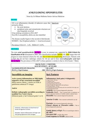
  • 1. ANKYLOSING SPONDYLITIS Notes by Col Bharat Malhotra Senior Advisor Medicine DEFINE AS is an inflammatory disorder of unknown cause that primarily affects • the axial skeleton; • peripheral joints and extraarticular structures are also frequently involved It has striking correlation to HLA-B27 Non-HLA susceptibility alleles are also identified The disease usually begins in the second or third decade. WOMEN: Has Peripheral arthritis >> Axial involvement Prevalence USA 0.9 – 1.4% INDIA 0.7- 0.9% Spectrum CRITERIA The term axial spondyloarthritis (ax-SpA) is now in common use, supported by ASAS Criteria for Classification of AS formulated in 2009. This classification includes definite AS, early stages that will progress to meet classical criteria for AS, and one or more non progressing phenotypes. Patients with ax-SpA that do not have radiologic criteria for AS are said to have non-radiographic axial SpA (nr-ax-SpA). The prevalence of HLA-B27 in these patients is similar to that in AS (Female > Male). BACK PAIN > 3 MONTHS AGE OF ONSET < 45 YEARS SACROILIITIS ON IMAGING PLUS > 1 SpA Feature OR HLA-B27 PLUS >2 SpA Features Sacroiliitis on imaging Active (acute) inflammation on MRI highly suggestive of SpA associated sacroiliitis (Bone marrow edema + osteitis on STIR or gadolinium enhanced T1 image) And/or Definite radiographic sacroiliitis according to modified New York Criteria (Bilateral grade >2 or Unilateral grade 3 or 4) SpA Features Inflammatory back pain (>4 diagnostic) (1) age of onset <40 years; (2) insidious onset; (3) improvement with exercise; (4) no improvement with rest; and (5) pain at night with improvement upon getting up; (6) morning stiffness >30 min; (7) awakening from back pain during only the second half of the night; and (8) alternating buttock pain. Arthritis -Past or present diagnosed by physician Enthesitis (heel) – Past or present pain or tenderness on exam at calcaneus insertion of Achilles tendon or plantar fascia Dactylitis- Past or present diagnosed by physician Anterior Uveitis - Past or present confirmed by eye spl Psoriasis- Past or present diagnosed by physician Chron’s disease or Ulcerative colitis - Past or present diagnosed by physician Family history of SpA- First or second-degree relatives HLAB27 Elevated CRP- exclude other causes Good response to NSAID- relief in 24-48h Enthesitis -Inflammation of attachment of tendon & ligament to bone. Dactylitis- Severe Inflammation of Finger and toes giving appearance like sausages.
  • 2. PATHOGENESIS – IT IS IMMUNE MEDIATED (Auto Inflammatory pathogenesis) HLA B27 plays direct role, Association of AS with ERAP1 which influences MHC class 1 peptide This causes B27 heavy chain to unusual tendency to misfold → process that can be proinflammatory Unlike RA synovium, citrullinated proteins and cartilage gp39 peptide-MHC are ABSENT BRIEF OVERVIEW IL-23 / IL-17 PATHWAY New bone formation – WNT signaling pathways SYNDESMOPHYTES
  • 3. PATHOLOGY Sacroiliitis Macrophages, T cells, plasma cells, and osteoclasts are prevalent. • Sacroiliitis is often an early manifestation of AS and nr-ax-SpA. • Synovitis and myxoid marrow represent the earliest changes • Followed by pannus and subchondral granulation tissue • Marrow edema, enthesitis, and chondroid differentiation found. • If the process continues to progress, eventually the eroded joint margins are gradually replaced by fibrocartilage regeneration and then by ossification. Spine • Inflammatory granulation tissue in the paravertebral connective tissue at the junction of annulus fibrosus and vertebral bone, and in some cases along the entire outer annulus. • The outer annular fibers are eroded and eventually replaced by bone, forming the beginning of a syndesmophyte. • It then grows by continued endochondral ossification, ultimately bridging the adjacent vertebral bodies. Ascending progression of this process can lead to the “bamboo spine.” • Other lesions in the spine o Diffuse osteoporosis (loss of trabecular bone despite accretion of periosteal bone), o Erosion of vertebral bodies at the disk margin, o Inflammation and destruction of the disk-bone border. Inflammatory arthritis • Inflammatory arthritis of the apophyseal (facet) joints is common, • With synovitis, inflammation at the bony attachment of the joint capsule, and subchondral bone marrow granulation tissue. • Erosion of joint cartilage by pannus is often followed by bony ankylosis. • Formation of syndesmophytes bridging the adjacent disks. • Bone mineral density is diminished in the spine and proximal femur early in the disease course. The characteristics of peripheral arthritis in AS and other forms of SpA are similar, and distinct from those of RA. Fibrocartilaginous enthesis Inflammation in the fibrocartilaginous enthesis, the region where a tendon, ligament, or joint capsule attach to bone, is a characteristic lesion in AS and other SpAs, both at axial and peripheral sites. Enthesitis is associated with prominent edema of the adjacent bone marrow and is often characterized by erosive lesions that eventually undergo ossification. Subclinical intestinal inflammation has been found in the colon or distal ileum in a majority of patients with SpA
  • 4. CLINICAL MANIFESTATIONS Age Late adolescence or early adulthood Usually < 45 years (in 5% symptoms begin after 40 years) Initial symptoms LBA • Usually dull pain, Insidious in onset • Felt deep in the lower lumbar or gluteal region, • Accompanied by low-back morning stiffness of up to a few hours’ duration that improves with activity and returns following inactivity • Within a few months, the pain usually become persistent and bilateral. • Nocturnal exacerbation of pain often forces the patient to rise and move around. Enthesitis or Osteitis Dactylitis In some patients, bony tenderness (presumably reflecting enthesitis or osteitis) may accompany back pain or stiffness. Common sites are Costo-sternal junctions, spinous processes, iliac crests, greater trochanters, ischial tuberosities, tibial tubercles and heels Dactylitis may be seen due to severe inflammation of fingers and toes. Asymmetric Oligoarthrits Arthritis of peripheral joints other than the hips and shoulders, usually asymmetric, may occur at any point in the disease course Hip & Shoulder joint Hip and shoulder (“root” joint) arthritis is considered part of axial disease. Hip arthritis occurs in 25–35% of patients. Shoulder involvement may be at least as common, but is usually less symptomatic Late – Cervical Spine Neck pain and stiffness from involvement of the cervical spine are usually relatively late manifestations, but are occasionally dominant symptoms. Juvenile onset In juvenile onset spondylarthritis, peripheral arthritis and enthesitis predominate, with axial symptoms supervening in late adolescence. It should be emphasized that in early mild, or atypical cases, the symptoms and/or physical findings may be subtle and/or nonspecific. Axial physical findings mirror the inflammatory process. Limitation of motion >> ankylosing spondylosis (Due to pain & spasm) Loss of spinal mobility imitation of anterior and lateral flexion limited extension of the lumbar spine Limited chest expansion Pain in the sacroiliac joints tenderness on posterior spinous processes symptomatic bony tenderness Enthesitis
  • 5. Modified Schober test The patient stands erect, with heels together, and marks are made on the spine at the lumbosacral junction (identified by a horizontal line between the posterosuperior iliac spines) and 10 cm above. The patient then bends forward maximally with knees fully extended, and the distance between the two marks is measured. This distance increases by ≥5 cm in the case of normal mobility and by <4 cm in the case of decreased mobility. Wall Occiput Distance Perpendicular distance between • bony prominence of the occiput • and the wall in a standing position Lateral bending Lateral bending measures the distance the patient’s middle finger travels down the leg with maximal lateral bending. Normal is >10 cm. Chest expansion Chest expansion is measured as the difference between maximal inspiration and maximal forced expiration in the fourth intercostal space in males or just below the breasts in females, with the patient’s hands resting on or just behind the head. Normal chest expansion is ≥5 cm Hips or Shoulders Limitation or pain with motion of the hips or shoulders is usually present if these joints are involved. OUTCOME MEASURES EVALUATION SCALE Schober Test Oswestry Disability Index (ODI) Neck Disability Index (NDI) Visual Analogue Scale Patient Specific Functional Scale Bath Ankylosing Spondylitis Metrology Index (BASMI) Revised Leeds Disability Questionnaire (RLDQ) European Quality of Life (EuroQoL) Bath AS Disease Activity Index (BASDAI; 0-100) AMOR criteria for Spondylarthritis BASFI index (Bath Ankylosing Spondylitis Functional Index) BAS-G index (Bath Ankylosing Spondylitis Global Index) The course of ax-SpA is extremely variable Mild stiffness and normal radiographs → totally fused spine and severe bilateral hip arthritis, accompanied by severe peripheral arthritis and extraarticular manifestations. Pain tends to be persistent early in the disease and intermittent later, with alternating exacerbations and quiescent periods. Patient’s posture undergoes characteristic changes, with obliterated lumbar lordosis, buttock atrophy, and accentuated thoracic kyphosis. There may be a forward stoop of the neck or flexion contractures at the hips, compensated by flexion at the knees. Disease progression can be estimated clinically from loss of height, limitation of chest expansion and spinal flexion, and occiput-to-wall distance.
  • 6. EXTRAARTICULAR MANIFESTATION Acute Anterior Uveitis 40% of patients Can antedate the spondylitis Attacks are typically unilateral, causing pain, photophobia, and increased lacrimation. These tend to recur, often in the opposite eye. Cataracts and secondary glaucoma may ensue. Inflammation in the Colon or Ileum 60% of patients usually asymptomatic, but frank IBD occurs in 5–10% Psoriasis 10% of patients Acne fulminans or hidradenitis suppurativa can occur There is an apparently increased risk of ischemic heart disease Aortic insufficiency occurs in a small percentage of patients. Third-degree heart block may occur alone or together with aortic insufficiency, and association with lesser degrees of heart block has been described. Cauda equina syndrome and upper pulmonary lobe fibrosis are rare late complications. Prostatitis has been reported Amyloidosis is rare COMPLICATION The most serious complication of the spinal disease is spinal fracture, which can occur with even minor trauma to the rigid, osteoporotic spine lower cervical spine Its most commonly involved. These fractures are often displaced, causing spinal cord injury. thoracolumbar spine Occasionally, fracture through a disk vertebral junction and adjacent neural arch, termed pseudoarthrosis, most common in the thoracolumbar spine, can be an unrecognized source of persistent localized pain and/or neurologic dysfunction. thoracic vertebrae Wedging of thoracic vertebrae is common and correlates with accentuated kyphosis. Mortality attributable to AS is largely the result of spinal trauma, aortic insufficiency, respiratory failure, amyloid nephropathy, or complications of therapy such as upper gastrointestinal hemorrhage. The impact of anti-TNF therapy on outcome and mortality is not yet known, except for significantly improved work productivity. LABORATORY FINDINGS No laboratory test is diagnostic of AS. Patients are rheumatoid factor (RF) negative, hence seronegative AS. HLA-B27 is present in 75–90% of patients
  • 7. In most ethnic groups, HLA-B27 is present in 75–90% of AS patients. HLA B27 is present in 5% healthy individuals 10–15% of patients with AS may be HLA B27 negative HLA-B27 positivity, in general, confers a greater risk of disease severity and also a concentration of disease in the spine and the eye. HLA-B27-negative patients are more likely to develop peripheral arthritis, skin, and nail disease, or IBD. In the absence of any preventative treatment, asymptomatic children of AS patients should not be tested. Testing is recommended only in symptomatic children. Erythrocyte sedimentation rate (ESR) and C-reactive protein (CRP) are often, but not always, elevated. Mild anemia may be present. Patients with severe disease may show an elevated alkaline phosphatase level. Elevated serum IgA levels are common. Rheumatoid factor, anti-cyclic citrullinated peptide (CCP), and antinuclear antibodies (ANAs) are largely absent unless caused by a coexistent disease, although ANAs may appear with anti-TNF therapy. Synovial fluid from peripheral joints in AS is nonspecifically inflammatory. Restricted chest wall motion causes decreased vital capacity, but ventilatory function is usually well maintained. HLA-B27 HLA-B27 is a MHC class I molecule consisting of an alpha chain encoded in the MHC region on chromosome 6 and a non-MHC encoded beta chain, β2 microglobulin. The development of ankylosing spondylitis may be promoted by increased expression of HLA-B27 on the surface of peripheral blood mononuclear cell. The arthritogenic peptide hypothesis suggests that HLA-B27 has a unique ability to bind antigens from a microorganism that trigger a CD8 T-cell response that then cross-reacts with a HLA-B27/self-peptide pair. Furthermore, it has been shown that HLA-B27 can bind peptides at the cell surface.
  • 8. RADIOGRAPHIC FINDINGS The diagnosis of AS is clinico-radiologic. Sacroiliitis is considered the radiographic hallmark of AS. Xray S I Joint Radiographically demonstrable sacroiliitis, usually symmetric. • Blurring of the cortical margins of the subchondral bone • Followed by erosions and sclerosis. • Progression of the erosions leads to “pseudo-widening” of the joint space • As fibrous and then bony ankylosis supervene, the joints may become obliterated Sacroiliitis grading – New York criteria Grade 0: normal Grade I: suspicious changes (some blurring of the joint margins) Grade II: minimum abnormality (small localized areas with erosion or sclerosis, with no alteration in the joint width) Grade III: unequivocal abnormality (moderate or advanced sacroiliitis with erosions, evidence of sclerosis, widening, narrowing, or partial ankylosis) Grade IV: severe abnormality (complete ankylosis) MRI SI Joint – with contrast or STIR image MRI is thus much more useful for the timely diagnosis of ax-SpA. Active sacroiliitis is best visualized by dynamic MRI • Either T2-weighed (STIR) image: bone marrow edema • Or T1-weighted images with contrast enhancement- synovitis & capsulitis. These techniques identify early intraarticular inflammation, cartilage changes, and underlying bone marrow edema in sacroiliitis. Active inflammation of the sacroiliac joints as determined by dynamic MRI is considered equivalent to definite radiographic sacroiliitis. X RAY Lumbosacral Spine In the lumbar spine, progression of the disease can lead to • Loss of lordosis, • Osteitis of the anterior corners of the vertebral bodies with subsequent erosion: Romanus lesions • Leading to “squaring” or even “barreling” of one or more vertebral bodies. • Linear ossification along central spine: Dagger spine • Progressive ossification leads to eventual formation of marginal syndesmophytes: Bamboo spine
  • 9. Hatchet Sign (Humeral Head) refers to the limited erosion of the lateral aspect of the humeral head that produces a hatchet- shaped deformity. This finding is typically associated with ankylosing spondylitis. Shiny Corner Sign (Spine) is a spinal finding in ankylosing spondylitis, representing reactive sclerosis secondary to inflammatory erosions at the superior and inferior endplates (corners on lateral radiograph) of the vertebral bodies which are known as Romanus lesions. Eventually, the vertebral bodies become Squared. Dagger Sign (Spine) is a radiographic feature seen in ankylosing spondylitis as a single central radio dense line on frontal radiographs related to ossification of the supraspinous and interspinous ligaments secondary to enthesitis. Bamboo spine Diffuse syndesmophytic ankylosis. DEXA SCAN Reduced bone mineral density can be detected by dual-energy x-ray absorptiometry of the femoral neck and the lumbar spine. Use of a lateral projection of the L3 vertebral body can prevent falsely elevated readings related to spinal ossification. DIAGNOSIS It is important to recognize ax-SpA before the development of irreversible deformity. This goal is challenging for several reasons: (1) only a minority of back pain patients have ax-SpA; (2) an early diagnosis often relies on clinical grounds and/or an appropriate MRI protocol requiring considerable expertise; (3) young individuals with symptoms of ax-SpA often do not seek medical care; (4) reliance on definite radiographic sacroiliitis causes early or mild cases to be missed. Low back ache > 3 months Age of onset < 45 years Defined radiological sacroiliitis Presence of spondyloarthritis features – Inflammatory back pain, asymmetric arthritis, enthesitis, dactylitis, anterior uveitis, IBD, psoriasis, family history of SpA, Elevated ESR/CRP, good response to NSAID. Confirm as AS > 4 features 2-3 Features 0-1 Features Compelling clinical picture suggest AS HLA-B27 If positive → AS If negative do MRI SI Joint HLA-B27 If positive → AS If negative do MRI SI Joint HLA-B27 If positive also do MRI SI Joint DIFFERENTIAL DIAGNOSIS • Less-common metabolic, infectious, and malignant causes of back pain must also be differentiated from AS, including infectious spondylitis, spondylodiskitis, and sacroiliitis, and primary or metastatic tumor. • Ochronosis can produce a phenotype similar to AS.
  • 10. • Calcification and ossification of paraspinous ligaments occur in diffuse idiopathic skeletal hyperostosis (DISH), which occurs in the middle-aged and elderly and is usually not symptomatic. Ligamentous calcification gives the appearance of “flowing wax” on the anterior bodies of the vertebrae. Intervertebral disk spaces are preserved, and sacroiliac and apophyseal joints appear normal, helping to differentiate DISH from spondylosis and from AS, respectively. • Both primary and secondary hyperparathyroidism can cause subchondral bone resorption around the SI joints, with bilateral widened and ill-defined joints on radiographs, but without joint space narrowing. MANAGEMENT TREATMENT PLAN EXERSIZE Exercise program to maintain posture and range of motion. NSAID Nonsteroidal anti- inflammatory drugs (NSAIDs) are the first line of pharmacologic therapy. They reduce pain and tenderness and increase mobility in many patients. Continuous high-dose NSAID therapy may slow radiographic progression, particularly in patients who are at higher risk for progression. ANTI-TNF-Α THERAPY However, many patients have continued symptoms despite NSAID therapy and are likely to benefit from anti-TNF-α therapy. Patients with AS treated with infliximab (chimeric human/mouse anti-TNF-α monoclonal antibody), etanercept (soluble p75 TNF-α receptor–IgG fusion protein), adalimumab, or golimumab (human anti-TNF-α monoclonal antibodies, or certolizumab pegol [humanized mouse anti-TNF-α monoclonal antibody]) have shown rapid, profound, and sustained reductions in all clinical and laboratory measures of disease activity. Indications of anti-TNF-α therapy Because of the expense, potentially serious side effects, and unknown long-term effects of these agents, their use should be restricted to patients with a definite diagnosis and active disease (BASDAI ≥4 out of 10 and expert opinion) that is inadequately responsive to therapy with at least two different NSAIDs. Before initiation of anti-TNF therapy, all patients should be tested for tuberculin (TB) reactivity, and reactors (≥5 mm on PPD testing or a positive quantiferon test) should be treated with anti-TB agents.
  • 11. Biologics are indicated for a disease that is active and refractory. Refractory axial disease is defined as one that has not responded to at least 2 NSAIDs tried in full doses over 4 weeks. Refractory peripheral disease is one that is unresponsive to intra-articular steroids (if indicated) and sulfasalazine (2–3 Gm/day) given for 3 months after reaching the target dose. Active AS is defined by BASDAI score >4/10 or ASDAS >2.1 Dose of anti-TNF-α therapy Infliximab IV 5mg/kg Stat, 2 weeks, 6 weeks Then 6-8 weeks interval Etanercept S/C 50mg Once weekly Adalimumab S/C 40mg Biweekly Golimumab S/C 50-100mg Every 4 weeks Certolizumab pegol S/C 400mg Every 4 weeks Side effects of anti-TNF-α therapy These potent immunosuppressive agents are relatively safe, but patients are at increased risk for serious infections, including disseminated tuberculosis. Hypersensitivity infusion or injection site reactions are not uncommon. Cases of anti-TNF-induced psoriasis have been increasingly recognized. Rare cases of systemic lupus erythematosus (SLE)–related disease have been reported, as have hematologic disorders such as pancytopenia, demyelinating disorders, exacerbation of congestive heart failure, and severe liver disease. The overall incidence of malignancy does not appear to be increased in AS patients treated with anti-TNF therapy, but isolated cases of hematologic malignancy have occurred shortly after the start of treatment. Contraindications Contraindications include active infection or high risk of infection; malignancy or premalignancy; and history of SLE, multiple sclerosis, or related autoimmunity. Pregnancy and breast-feeding are no longer considered contraindications if appropriate precautions are taken. Infants exposed to anti-TNF in utero should not be given live vaccines before age 6 months. Response Continuation beyond 12 weeks of therapy requires either a 50% reduction in BASDAI or absolute reduction of ≥2 out of 10, and favorable expert opinion. Switching to a second anti- TNF agent may be effective, especially if there was a response to the first that was lost rather than primary failure. ASDAS CUT OFF CRITERIA ASDAS IMPROVEMENT CRITERIA
  • 12. In a good response, there is significant improvement in both objective and subjective indicators of disease activity and function, including morning stiffness, pain, spinal mobility, peripheral joint swelling, CRP, ESR, and bone mineral density. MRI studies indicate substantial resolution of bone marrow edema, enthesitis, and joint effusions in the sacroiliac joints, spine, and peripheral joints. About one-half of the patients achieve a ≥50% reduction in the BASDAI. The response tends to persist over time, and partial or full remissions are common. Predictors of the best responses include younger age, shorter disease duration, higher baseline inflammatory markers, and lower baseline functional disability. Nonetheless, some patients with long-standing disease and even spinal ankylosis can obtain significant benefit. Syndesmophyte formation may continue despite the therapy, but this may apply mainly during the early years of therapy. Although less well studied, the response of patients with nr-ax-SpA to anti-TNF therapy is generally similar to that of patients with AS. HUMAN MONOCLONAL ANTIBODY TO IL-17A Secukinumab, a human monoclonal antibody to IL-17A, shows dramatic efficacy in AS, similar to that seen with TNF inhibitors, and is effective in some patients who have failed or not tolerated anti-TNF therapy. Most recommendations place the use of TNFi ahead of IL-17 inhibitors. This is primarily because of the earlier introduction and greater familiarity with these agents. In primary nonresponders, to TNFi, secukinumab or ixekizumab are preferred over treatment with a different TNFi. In secondary nonresponders, a different TNFi can also be tried. Secukinumab S/C 150mg Weekly x 4 weeks Then 4-week interval Ixekizumab Precautions regarding infection are similar to those for anti-TNF agents. An additional concern is potential exacerbation of underlying IBD, whether previously recognized or not, and careful monitoring is advised. ORAL JAK INHIBITOR, TOFACITINIB The oral Jak inhibitor, tofacitinib, showed efficacy in AS, with reduction of inflammation evident on MRI, in a 16-week phase 2 study. DMARD- SULFASALAZINE Sulfasalazine, in doses of 2–3 g/d, has modest benefit, primarily for peripheral arthritis. Methotrexate, although widely used, has not been shown to be of benefit in AS, nor has any therapeutic role for gold or oral glucocorticoids been documented.
  • 13. LOCAL STEROIDS Unlike RA, the response to systemic corticosteroids is not as good in AS. Prolonged oral corticosteroid therapy does more harm than good and should be avoided. Intra-articular corticosteroids for the persistently active joint are helpful, provided septic arthritis has been ruled out. Painful enthesopathy or refractory plantar fasciitis may also benefit from local corticosteroid injection. Direct injection into tendons is best avoided because of risk of tendon rupture. SURGERY The most common indication for surgery in patients with AS is severe hip joint arthritis, the pain and stiffness of which are usually dramatically relieved by total hip arthroplasty. Rare patients may benefit from surgical correction of extreme flexion deformities of the spine or of atlantoaxial subluxation. EXTRAARTICULAR MANIFESTATIONS TREATMENT Attacks of uveitis are usually managed effectively with local glucocorticoids and mydriatic agents, although systemic glucocorticoids, immunosuppressive drugs, or anti-TNF therapy may be required. TNF inhibitors reduce the frequency of attacks of uveitis in patients with ax- SpA, and adalimumab has recently been approved by the FDA for treating uveitis. Cases of new or recurrent uveitis after use of a TNF inhibitor have been observed, especially with etanercept. Anti-IL-17 does not appear as effective for uveitis as anti-TNF therapy. Management of axial osteoporosis is at present similar to that used for primary osteoporosis, since data specific for AS are not available.
  • 14. Notes from: Harrison’s Principle of Internal medicine 20th Ed (2018) chapter 255 Few points from • Clinical Rheumatology (2021) by Rohini Handa • Firestein Kelley’s Textbook of Rheumatology, 11th Ed (2020) • Targeting inflammatory pathways in axial spondyloarthritis. Arthritis Research & Therapy volume 21, Article number: 135 (2019) open access NOTES PREPARED BY COL BHARAT MALHOTRA SR ADV MEDICINE (Jun 2021) PATHOGENESIS CLINICAL FINDINGS TREATMENT