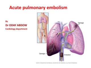
SlideShare a Scribd company logo
1 of 58
Acute pulmonary embolism
By
Dr ODAY ABDOW
Cardiology department
Introduction
- Venous thromboembolism (VTE) clinically presenting as DVT
or PE is globally the third most frequent acute cardiovascular
syndrome behind myocardial infarction and stroke.
- Annual incidince rates for PE in epidemiological studies ranges
from 39-115 per100,000 population.
- Proximal DVTs (popletial vein and above)have an estimated
risk of PE of 50% if not treated.
- Other causes: air, fat embolism, amniotic fluid .
Why is it important?
 PE remains the most common preventable cause of hospital
death.
 Diagnosis can be difficult ,and the majority of patients die
because of failure of diagnosis rather than inadequate therapy.
 Early treatment is highly effective ,in fact The mortality rate of PE
without treatment is 30% whereas it is only 2-8% with adequate
treatment.
Predesposing factors for VTE
Strong risk factors
- fracture of lower limb
- hospitalization for heart failure or atrial fibrilaation/flutter
within previous 3 months
- Hip or knee replacement
- Major trauma
- Myocardial infarction within previous 3 months
- Previous VTE
- Spinal cord injuiry
Moderate risk factors
-Arthroscopic knee surgery
- central venous lines
-Intravenous catheters and leads
-Blood transfusion
-Chemotherapy
-cancer(the highest risk is metastatic disease)
-Congestive heart failure or respiratory failure
-Hormone replacement therapy
-Oral contraceptive pills
-Postpartum period
-Paralytic stroke
-Autoimmune disease
-Inflammatory bowl disease
-Superficial vein thrombosis
-Thrombophilia
-Erythropoiesis stimulating agents
-In vitro fertilization
Weak risk factors
- Bed rest>3 days
- Immobility due to sitting(eg,prolonged car or air travel)
- Diabetes mellitus
- Hypertension
- Obesity
- Increasing age
- Pregnancy
- Varicose veins
- Laparoscopic surgery(eg,cholecystectomy)
pathophysiology
• Anatomical obstruction and hypoxic vasoconstriction in the
affected lung area lead to an, increase in PVR
Abrupt increase in PVR results in RV dilation which alters
contractile properties of the RV myocardium via frank starling
mechanism, icreasing RV pressure and wall tesion and
neurohormonal activation.
• Prolongation of RV contraction time into early diastole in LV
leads to lefward bowing of the IVS,the dysynchronization of the
ventricles may be exacerpated by development of RBBB ,as a
result LV filling is imended in early diastole and may lead to
reduction in cardiac output and sysymic hypotension and
hemodynamic instability.
Key factors contributing to haemodynamic collapse and
death in acute PE
diagnosis
Clinical presentation
The clinical signs of acute PE are nonspecific
In most cases PE is suspected in a patient with
 dyspnea
chest pain
syncope ,presyncope
haemoptysis
- Chest pain is a frequent symptom of PE and is usually caused
by pleural irritation due to distal emboli causing pulmonary
infarction.
- In central PE chest pain may have a typical angina character
possibly reflecting RV ischemia and requiring differential
diagnosis from an acute coronary syndrome or aortic
dissection.
• Dyspnea may be acute and sever in central PE,
In small peripheral PE dyspnea it is often mild and may be
transient.
• In patients with preexisting heart failure or pulmonary
disease worsening dyspnea may be the only symptom
indicative of PE.
• Syncope may occur and is associated with higher
prevelence of hemodynamic instability and RV dysfunction.
• In some cases PE may be asymptomatic and discovered
incidentally during diagnostic workup for another disease.
High risk PE
Definition of haemodynamic instability
One of the following clinical mainfestations at presentation:
1-cardiac arrest: Need for cardiopulmonary resuscitation.
2-obstructive shock: SBP< 90mmHG or vasopressors rquired to
achieve BP≥ 90mmHG despite adequate filling status and end
organ hypoperfusion (altered mental status, cold clammy skin,
oliguria/anuria, increased serum lactate).
3-persistent hypotension:SBP <90mmHG or SBPdrope≥ 40mmHG
lasting longer than 15 min and not caused by newonset
arrhythmia,hypovolemia or sepsis.
Physical examination
Common signs include:
Tachypnea
Tachycardia
Calf or thigh swelling, erythema ,edema or tenderness
Accentuated pulmonic component of S2
Jugular venous distension
Fever mimicking pneumonia
Assesment of pretest probability
 The combination of symptoms and clinical findings with the
presence of predisposing factors allows the classification of
patients with suspected PE into distinct categories of clinical
pretest probability.
 This can be done either by empirical clinical judgement or by
using prediction rules.
 The most frequently used prediction rules are GENEVA rule
and WELLS rule,both prediction rules have been simplified in an
attempt to increase their adoptioninto clinical practice.
The revised Geneva prediction rule for pulmonary embolism
ABG
Unexplained hypoxemia in the setting of normal CXR should
raise the clinical suspicion of PE and prompt further evaluation
But up to 40%of the patients have normal arterial oxygen
saturation
Troponin
As a marker of RV dysfunction cardiac troponin T or I are
elevated in 30-60-% of patients with acute PE and associated
with increased mortality
BNP and NT-pro BNP
myocardial stretch as a result of RV pressure overload increases
the release of natriuretic peptides which reflect the sevferity of
RV dysfunction
D-dimer
• Plasma D-dimer measurment preferably high sensitive assay
is recommended in outpatients/emergency department
patients with low or intermediate clinical probability or those
that PE unlikely to reduce the need for unnecessary imaging
and irradiation
• D-dimer measurment is not recommended in patients with
high clinical probability as normal results doesn’t exclude PE
even when using high sensitive assay
New recommendation in 2019 ESC guideline
• (classIIaB)As an alternative to a fixed D-dimer cut-off
anegative D-dimer test using an age adjusted cut-
off(age ×10µg/l in patients aged >50years)should be
considered for excluding PE in patients with low or
intermediate clinical probability or those that are PE
unlikely
• (classIIaB) as an alternative to fixed or age adjusted D-
dimer cut-off ,D-dimer levels adapted to clinical
probability hould be considered to exclude PE
ECG changes
 The most common findings are sinus tachycardia and
nonspecific STsegment and T wave changes
 More specific Changes indicative of RV strain are usually seen
in more sever cases of PE such as:
T wave inversion in leads V1-V4 ¹ inferior leads II,III,aVF
QR pattern in V1
S1Q3T3 pattern
 complete or incomplete RBBB
 Other findings (right axis deviation ,clockwise rotation ,atrial
fibrillation)
 Arround 18% of patients with PE will have a completely normal
ECG
Simultaneous T wave inversions in the inferior (II,III,aVF) leads and
right pericardial leads (V1-V4) is the most specific finding in favour of
PE with reported specificities up to 99% in one study
PULMONARY EMBOLISM
RBBB
Extreme right axis deviation
S1Q3T3
T wave inversion in V1-V3 and lead III
Chest radiography
- Non specific abnormalities on chest radiograph
(eg,atelectasis,effusion…) are common but nonspecific
- A normal CXR can be seen in 10-22% of patient
- It may be useful for excluding other causes of dyspnea or chest
pain
- It is also performed to detemine elegibility for V/Q scanning
- The classic signs are uncommon , including ( Hampton hump,
Westermark sign, Palla sign)
Hampton hump sign Hump- shaped opacify in the periphery of the
lung with its base against the pleural surface and hump towards the
hilum
echocardiography
 Echocardiography is not mandatory as part of routin
diagnostic workup in hemodynamically stable patients with
suspected PE ,although it may be useful in the differential
diagnosis of acute dyspnea.
 This is in contrast to suspected high risk PE in which the
absence of echocardiographic signs of RV overload or
dysfunction excludes PE as the cause of hemodynamic
instability.
 also in the latter case it may help in the differential diagnosis
of the cause of shock by detecting pericardial tamponade,
acute valvular dysfunction, sever global or regional dysfunction
,aortic dissection or hypovolemia.
 note In hemodynamically compromised patients with
suspected PE,unequevocal signs of RV pressure overload
especially with more soecific echocardiographic signs
(60/60sign, Mc connel sign ,right heart thrombi) justify emergency
reperfusion treatment for PE if immediate CT angiography is not
feasible in a patient with high clinical probability and no other
obvious causes of RV pressure overlaod.
 Note in some patients with suspected acute PE
,echocardiography may detect RV wall thickness and TV
insufficiency jet velocity beyond values compatible with acute RV
pressure overload (>3.8m/s or tricuspid valve peak systolic
gradient >60mmHG) in these cases chronic thromboembolic
pulmonary hypertension ( CTPH)or other types of PH should be
included in the differential diagnosis.
Graphic presentation of TTE parameters in the assesment of RV
dysfunction and pressure overload
Coputed tomographic pulmonary angiography
(CTPA)
Strengths:
Readily avilable in most centers
Excellent accuracy
Low rate of inconclusive results(3-5%)
May provide alternative diagnosis if PE excluded
Short acquisition time
Weaknesses/limitations:
Tendency to overuse because of easy accessibility
Radiation exposure
Contraindicated in sever renal failure
Limited use in iodin allergy hyperthyroidism
Clinical relevance of CTPA diagnosis of subsegmental PE is
unknown
CPTA
• It is recommended to accept the diagnosis of PE (without
further testing)if CPTA shows a segmental or more proximal
filling defect in a patient with intermediate or high clinical
probability (class1B).
• It is recommended to reject the diagnosis of PE(without further
testing if CPTA is normal in a patient with low or intermediate
clinical probability or who is PE-unlikely class1A.
•It should be considered to reject the diagnosis of PE without
further testing if CPTA is normal in apatient with high clinical
probability or who is PE-likely classIIaB.
V/Q scintigraphy
•It is recommended to reject the diagnosis of PE if the perfusion
lung scan is normal class IA
•It should be considered to accept the diagnosisof PE if theV/Q
scan yieldshigh probability for PE classIIaB
Lower –limb CUS (Compression ultrasonography)
•It is recommended to accept the diagnosis of VTE (and PE) if a
CUS shows a proximal DVT in a patient with clinical suspecion of
PE classIA
• if CUS shows only a distalDVT,further testing should be
considered to confirm PE classIIaB
Pulmonary angiography
- Historically the golden standard,but it is now rarely
performed as less invasive CTPA offers similar diagnostic
accuracy
- Invasive procedure
- Not readily available in all centers
- Highest radiation
MRA
It is not recommended for ruling out PE
1- suspected PE with haemodynamic instability
• In suspected high risk PE as indicated by the presense of
haemodynamic instability bedside echocardiography or emergent
CTPA (depending on availability and clinical circumstances)is
recommended for diagnosis
• IV UFH including weigh–adjusted bolus ingection is recommended
without delay in patients with suspected high risk PE
2-Suspected PE without haemodynamic instability
• Initiation of anticoagulation is recommended without delay in
patients with high or intermediate clinical probability of PE while
diagnostic workup is in progress
Assesment of PE severity
• Risk stratification of patients with acute PE is mandatory for
determining the appropriate therapeutic management
aporoach.
• Initial risk stratification is based on clinical symptoms and
signs of hemodynamic instability which indicate a high risk of
early death
• In the large remaining group of patients of PE who present
without hemodynamic instability ,further risk stratification
requires the assesment of two sets of prognostic criteria:
1- Clinical imaging and laboratory indicators of PE severity
mostly related to the RV dysfunction
2- Presence of comorbidity and any other aggravation
conditions that may affect early prognosis
Classification of PE severity and the risk of early death
Acute phase treatment of high risk PE
- UFH –including a weight djusted bolus dose is recommended
without delay classIC
- Systemic thrombolysis is recommended for high risk PE classIB
- Surgical pulmonary embolectomy is recommended for high risk
PE patients in whom thrombolysis is contraindicated or has
failed classIC
- Pecutaneous catheter directed treatment for high risk PE
patients in whom thrombolysis is contraindicated or has failed
classIIac
- Norepinephrine and or dobutamine should be considered in
patients with high risk PE classIIac
- ECMO may be considered in patients with refractory circulatory
collapse or cardiac arrest classIIbC
systemic thrombolysis
• Thrombolytic therapy leads to faster improvements in pulmonary
obstruction , PAP,and PVR in patients with PE compared with UFH
alone.
• These improvements are accompanied by a reduction in RV
dilation on echocardiography.
• The greatest benefit is observed when treatment is initiated
within 48h of symptoms onset, but thrombolysis can still be useful
in patients who have had symptoms for 6-14 days.
• Unsuccessful thrombolysis as judged by persistent clinical
instability and unchanged RV dysfunction on echocardiography
after 36h has been reported in 8% of high risk PE patients.
Thrombolytic regimens, doses in PE
Acute phase treatment of intermediate or low risk PE
Duration of treatment
Therapeutic anticoagulation for ≥ 3 months is
recommended for all patients with PE(classIA)
Regimen and duration of anticoagulation after PE in patients
without cancer
 Patients in whom discontinuation of anticoagulation after 3
months is recommended class IB
Patients with first PE/VTE secondary to major transient/reversible
risk factor discontinuation of anticoagulation is recommended after
3 months
 Patients in whom extension of anticoagulation beyond
3months is recommended classIB
• Patients presenting with recurrent VTE (at least one previous
episode of PE or DVT)not related to major transient or reversible
risk factor
• VKA for an indefinite period is recommendefor patients with
antiphospholipid syndrome
Patients in whom extension of anticoagulation beyond 3
months should be considered (claasIIa)
• Patients with first episode of PE and no identifiable risk
factors
• Patients with first episode of PE associated with persistent
risk factor other than antiphospholipid antibody syndrome
•Patients with first episode of PE associated with a minor
transient or reversible risk factor
Patients with PE and active cancer
• LMWH should be considered for first 6 months over VKA
• Edoxaban should be considered as an alternative to LMWH in
patients without gastrointestinal cancer
• Rivaroxaban should be considered as an alternative to LMWH
in patients without gastrointestinal cancer
• Anticoagulation ahould be considered beyond the first 6
months for an indefinite period or until the cancer is cured
PE and pregnancy
•LMWH based on early pregnancy body weight is the
recommended therapy for PE during pregnancy without
hemodynamic instability
•Throbbolysis or surgical embolictomy should be considered for
high risk PE during pregnancy
•NOACs are not recommended during pregnancy
•Amniotic fluid embolism should be considered in a pregnant or
postpartum woman with unexplained cardiac arrest ,sustained
hypotension,or respiratory deterioration espicially if
accompanied by DIC
references
- 2019 ESC guidelines for the diagnosis
and management of acute pulmonary
embolism
- The manual of cardiovascular
medicine 5th edition
- Up to date

More Related Content

What's hot

Pulmonary Embolism
Pulmonary Embolism	Pulmonary Embolism
Pulmonary Embolism Khalid
 
L9 pulmonary embolism
L9 pulmonary embolismL9 pulmonary embolism
L9 pulmonary embolismDr Bilal Natiq
 
Pulmonary Embolism
Pulmonary EmbolismPulmonary Embolism
Pulmonary Embolismcairo1957
 
Pulmonary embolism
Pulmonary embolismPulmonary embolism
Pulmonary embolismvkatbcd
 
Pulmonary embolism
Pulmonary embolismPulmonary embolism
Pulmonary embolismAmir Mahmoud
 
Deep Venous Thrombosis and Pulmonary Embolism : Diagnostic Approach and Curre...
Deep Venous Thrombosis and Pulmonary Embolism : Diagnostic Approach and Curre...Deep Venous Thrombosis and Pulmonary Embolism : Diagnostic Approach and Curre...
Deep Venous Thrombosis and Pulmonary Embolism : Diagnostic Approach and Curre...Bassel Ericsoussi, MD
 
Pulmonary embolism
Pulmonary embolism Pulmonary embolism
Pulmonary embolism RahulGupta1687
 
Pulmonary thromboembolism Management and prophylaxis
Pulmonary thromboembolism Management and prophylaxisPulmonary thromboembolism Management and prophylaxis
Pulmonary thromboembolism Management and prophylaxisMd Shahid Iqubal
 
Update on Pulmonary Embolism
Update on Pulmonary EmbolismUpdate on Pulmonary Embolism
Update on Pulmonary EmbolismMuhanad Majeed
 
Eisenmenger Syndrome Dr md toufiqur rahman cardiologist nicvd
Eisenmenger Syndrome Dr md toufiqur rahman cardiologist nicvdEisenmenger Syndrome Dr md toufiqur rahman cardiologist nicvd
Eisenmenger Syndrome Dr md toufiqur rahman cardiologist nicvdPROFESSOR DR. MD. TOUFIQUR RAHMAN
 
Cerebral Venous Thrombosis - Dr. KEO VEASNA
Cerebral Venous Thrombosis - Dr. KEO VEASNA Cerebral Venous Thrombosis - Dr. KEO VEASNA
Cerebral Venous Thrombosis - Dr. KEO VEASNA Keo Veasna
 
Pulmonary embolism,overview
Pulmonary embolism,overviewPulmonary embolism,overview
Pulmonary embolism,overviewEman Mahmoud
 
Heart Failure - What to expect from the Investigations?
Heart Failure - What to expect from the Investigations?Heart Failure - What to expect from the Investigations?
Heart Failure - What to expect from the Investigations?Praveen Nagula
 
PULMONARY EMBOLISM (Masteral reporting for finals)
PULMONARY EMBOLISM (Masteral reporting for finals)PULMONARY EMBOLISM (Masteral reporting for finals)
PULMONARY EMBOLISM (Masteral reporting for finals)Mary Ann Adiong
 
2019 ESC guidelines on pulmonary embolism
2019 ESC guidelines on pulmonary embolism2019 ESC guidelines on pulmonary embolism
2019 ESC guidelines on pulmonary embolismSaitej Reddy
 

What's hot (20)

Pulmonary Embolism
Pulmonary Embolism	Pulmonary Embolism
Pulmonary Embolism
 
Pulmonary embolism
Pulmonary embolismPulmonary embolism
Pulmonary embolism
 
Pulmonary Embolism
Pulmonary EmbolismPulmonary Embolism
Pulmonary Embolism
 
L9 pulmonary embolism
L9 pulmonary embolismL9 pulmonary embolism
L9 pulmonary embolism
 
Pulmonary Embolism
Pulmonary EmbolismPulmonary Embolism
Pulmonary Embolism
 
Acute pulmonary thromboembolism
Acute pulmonary thromboembolismAcute pulmonary thromboembolism
Acute pulmonary thromboembolism
 
Pulmonary embolism
Pulmonary embolismPulmonary embolism
Pulmonary embolism
 
af afl ppt, Virbhan
af afl ppt, Virbhanaf afl ppt, Virbhan
af afl ppt, Virbhan
 
Pulmonary embolism
Pulmonary embolismPulmonary embolism
Pulmonary embolism
 
Deep Venous Thrombosis and Pulmonary Embolism : Diagnostic Approach and Curre...
Deep Venous Thrombosis and Pulmonary Embolism : Diagnostic Approach and Curre...Deep Venous Thrombosis and Pulmonary Embolism : Diagnostic Approach and Curre...
Deep Venous Thrombosis and Pulmonary Embolism : Diagnostic Approach and Curre...
 
Pulmonary embolism
Pulmonary embolism Pulmonary embolism
Pulmonary embolism
 
Pulmonary thromboembolism Management and prophylaxis
Pulmonary thromboembolism Management and prophylaxisPulmonary thromboembolism Management and prophylaxis
Pulmonary thromboembolism Management and prophylaxis
 
Update on Pulmonary Embolism
Update on Pulmonary EmbolismUpdate on Pulmonary Embolism
Update on Pulmonary Embolism
 
Eisenmenger Syndrome Dr md toufiqur rahman cardiologist nicvd
Eisenmenger Syndrome Dr md toufiqur rahman cardiologist nicvdEisenmenger Syndrome Dr md toufiqur rahman cardiologist nicvd
Eisenmenger Syndrome Dr md toufiqur rahman cardiologist nicvd
 
Cerebral Venous Thrombosis - Dr. KEO VEASNA
Cerebral Venous Thrombosis - Dr. KEO VEASNA Cerebral Venous Thrombosis - Dr. KEO VEASNA
Cerebral Venous Thrombosis - Dr. KEO VEASNA
 
Pulmonary embolism,overview
Pulmonary embolism,overviewPulmonary embolism,overview
Pulmonary embolism,overview
 
Heart Failure - What to expect from the Investigations?
Heart Failure - What to expect from the Investigations?Heart Failure - What to expect from the Investigations?
Heart Failure - What to expect from the Investigations?
 
EISENMENGER SYNDROME- PAUL WOOD
EISENMENGER SYNDROME- PAUL WOODEISENMENGER SYNDROME- PAUL WOOD
EISENMENGER SYNDROME- PAUL WOOD
 
PULMONARY EMBOLISM (Masteral reporting for finals)
PULMONARY EMBOLISM (Masteral reporting for finals)PULMONARY EMBOLISM (Masteral reporting for finals)
PULMONARY EMBOLISM (Masteral reporting for finals)
 
2019 ESC guidelines on pulmonary embolism
2019 ESC guidelines on pulmonary embolism2019 ESC guidelines on pulmonary embolism
2019 ESC guidelines on pulmonary embolism
 

Similar to Pulmonary embolism

Diagnosis of Pulmonary Embolism - by Dr KD DELE
Diagnosis of Pulmonary Embolism - by Dr KD DELEDiagnosis of Pulmonary Embolism - by Dr KD DELE
Diagnosis of Pulmonary Embolism - by Dr KD DELEKemi Dele-Ijagbulu
 
Pulmonary embolism
Pulmonary embolismPulmonary embolism
Pulmonary embolismRamin Sadeghi
 
Acute Pulmonary Embolism: Introduction, Clinical presentation, Classification...
Acute Pulmonary Embolism: Introduction, Clinical presentation, Classification...Acute Pulmonary Embolism: Introduction, Clinical presentation, Classification...
Acute Pulmonary Embolism: Introduction, Clinical presentation, Classification...RichardKhoi
 
Pulmonary embolism
Pulmonary embolismPulmonary embolism
Pulmonary embolismDIPAK PATADE
 
Pulmonary Embolism2006
Pulmonary Embolism2006Pulmonary Embolism2006
Pulmonary Embolism2006Dang Thanh Tuan
 
Pulmonary Embolism2006
Pulmonary Embolism2006Pulmonary Embolism2006
Pulmonary Embolism2006Dang Thanh Tuan
 
Pulmonary Embolism2006
Pulmonary Embolism2006Pulmonary Embolism2006
Pulmonary Embolism2006Dang Thanh Tuan
 
Pulmonary Embolism2006
Pulmonary Embolism2006Pulmonary Embolism2006
Pulmonary Embolism2006Dang Thanh Tuan
 
Pulmonary Embolism2006
Pulmonary Embolism2006Pulmonary Embolism2006
Pulmonary Embolism2006Dang Thanh Tuan
 
Pulmonary Embolism2006
Pulmonary Embolism2006Pulmonary Embolism2006
Pulmonary Embolism2006Dang Thanh Tuan
 
Pulmonary Embolism
Pulmonary EmbolismPulmonary Embolism
Pulmonary EmbolismEneutron
 
Pulmonary hypertension.pdf
Pulmonary hypertension.pdfPulmonary hypertension.pdf
Pulmonary hypertension.pdfdonkumartiger
 
7-170713090357.pdf
7-170713090357.pdf7-170713090357.pdf
7-170713090357.pdfAugustusCaesar7
 
Pneumothorax.pptx
Pneumothorax.pptxPneumothorax.pptx
Pneumothorax.pptxMISSCOM1
 
Pulmonary embolism radiology imaging
Pulmonary embolism radiology imagingPulmonary embolism radiology imaging
Pulmonary embolism radiology imagingharshvardhan
 
Pulmonary embolism
Pulmonary embolismPulmonary embolism
Pulmonary embolismYouttam Laudari
 
Diagnosis & Classification of Pulmonary Hypertension
Diagnosis & Classification of Pulmonary HypertensionDiagnosis & Classification of Pulmonary Hypertension
Diagnosis & Classification of Pulmonary Hypertensionmediwaves
 
Pulmonary Embolism
Pulmonary Embolism Pulmonary Embolism
Pulmonary Embolism Asraf Hussain
 
Cardiogenic shock
Cardiogenic  shockCardiogenic  shock
Cardiogenic shockMohammad Ali
 
Pulmonary embolism notes 2021
Pulmonary embolism notes 2021Pulmonary embolism notes 2021
Pulmonary embolism notes 2021Best Doctors
 

Similar to Pulmonary embolism (20)

Diagnosis of Pulmonary Embolism - by Dr KD DELE
Diagnosis of Pulmonary Embolism - by Dr KD DELEDiagnosis of Pulmonary Embolism - by Dr KD DELE
Diagnosis of Pulmonary Embolism - by Dr KD DELE
 
Pulmonary embolism
Pulmonary embolismPulmonary embolism
Pulmonary embolism
 
Acute Pulmonary Embolism: Introduction, Clinical presentation, Classification...
Acute Pulmonary Embolism: Introduction, Clinical presentation, Classification...Acute Pulmonary Embolism: Introduction, Clinical presentation, Classification...
Acute Pulmonary Embolism: Introduction, Clinical presentation, Classification...
 
Pulmonary embolism
Pulmonary embolismPulmonary embolism
Pulmonary embolism
 
Pulmonary Embolism2006
Pulmonary Embolism2006Pulmonary Embolism2006
Pulmonary Embolism2006
 
Pulmonary Embolism2006
Pulmonary Embolism2006Pulmonary Embolism2006
Pulmonary Embolism2006
 
Pulmonary Embolism2006
Pulmonary Embolism2006Pulmonary Embolism2006
Pulmonary Embolism2006
 
Pulmonary Embolism2006
Pulmonary Embolism2006Pulmonary Embolism2006
Pulmonary Embolism2006
 
Pulmonary Embolism2006
Pulmonary Embolism2006Pulmonary Embolism2006
Pulmonary Embolism2006
 
Pulmonary Embolism2006
Pulmonary Embolism2006Pulmonary Embolism2006
Pulmonary Embolism2006
 
Pulmonary Embolism
Pulmonary EmbolismPulmonary Embolism
Pulmonary Embolism
 
Pulmonary hypertension.pdf
Pulmonary hypertension.pdfPulmonary hypertension.pdf
Pulmonary hypertension.pdf
 
7-170713090357.pdf
7-170713090357.pdf7-170713090357.pdf
7-170713090357.pdf
 
Pneumothorax.pptx
Pneumothorax.pptxPneumothorax.pptx
Pneumothorax.pptx
 
Pulmonary embolism radiology imaging
Pulmonary embolism radiology imagingPulmonary embolism radiology imaging
Pulmonary embolism radiology imaging
 
Pulmonary embolism
Pulmonary embolismPulmonary embolism
Pulmonary embolism
 
Diagnosis & Classification of Pulmonary Hypertension
Diagnosis & Classification of Pulmonary HypertensionDiagnosis & Classification of Pulmonary Hypertension
Diagnosis & Classification of Pulmonary Hypertension
 
Pulmonary Embolism
Pulmonary Embolism Pulmonary Embolism
Pulmonary Embolism
 
Cardiogenic shock
Cardiogenic  shockCardiogenic  shock
Cardiogenic shock
 
Pulmonary embolism notes 2021
Pulmonary embolism notes 2021Pulmonary embolism notes 2021
Pulmonary embolism notes 2021
 

Recently uploaded

Introduction to ArtificiaI Intelligence in Higher Education
Introduction to ArtificiaI Intelligence in Higher EducationIntroduction to ArtificiaI Intelligence in Higher Education
Introduction to ArtificiaI Intelligence in Higher Educationpboyjonauth
 
microwave assisted reaction. General introduction
microwave assisted reaction. General introductionmicrowave assisted reaction. General introduction
microwave assisted reaction. General introductionMaksud Ahmed
 
Incoming and Outgoing Shipments in 1 STEP Using Odoo 17
Incoming and Outgoing Shipments in 1 STEP Using Odoo 17Incoming and Outgoing Shipments in 1 STEP Using Odoo 17
Incoming and Outgoing Shipments in 1 STEP Using Odoo 17Celine George
 
Organic Name Reactions for the students and aspirants of Chemistry12th.pptx
Organic Name Reactions  for the students and aspirants of Chemistry12th.pptxOrganic Name Reactions  for the students and aspirants of Chemistry12th.pptx
Organic Name Reactions for the students and aspirants of Chemistry12th.pptxVS Mahajan Coaching Centre
 
Presiding Officer Training module 2024 lok sabha elections
Presiding Officer Training module 2024 lok sabha electionsPresiding Officer Training module 2024 lok sabha elections
Presiding Officer Training module 2024 lok sabha electionsanshu789521
 
call girls in Kamla Market (DELHI) 🔝 >༒9953330565🔝 genuine Escort Service 🔝✔️✔️
call girls in Kamla Market (DELHI) 🔝 >༒9953330565🔝 genuine Escort Service 🔝✔️✔️call girls in Kamla Market (DELHI) 🔝 >༒9953330565🔝 genuine Escort Service 🔝✔️✔️
call girls in Kamla Market (DELHI) 🔝 >༒9953330565🔝 genuine Escort Service 🔝✔️✔️9953056974 Low Rate Call Girls In Saket, Delhi NCR
 
Crayon Activity Handout For the Crayon A
Crayon Activity Handout For the Crayon ACrayon Activity Handout For the Crayon A
Crayon Activity Handout For the Crayon AUnboundStockton
 
Contemporary philippine arts from the regions_PPT_Module_12 [Autosaved] (1).pptx
Contemporary philippine arts from the regions_PPT_Module_12 [Autosaved] (1).pptxContemporary philippine arts from the regions_PPT_Module_12 [Autosaved] (1).pptx
Contemporary philippine arts from the regions_PPT_Module_12 [Autosaved] (1).pptxRoyAbrique
 
Software Engineering Methodologies (overview)
Software Engineering Methodologies (overview)Software Engineering Methodologies (overview)
Software Engineering Methodologies (overview)eniolaolutunde
 
Interactive Powerpoint_How to Master effective communication
Interactive Powerpoint_How to Master effective communicationInteractive Powerpoint_How to Master effective communication
Interactive Powerpoint_How to Master effective communicationnomboosow
 
Solving Puzzles Benefits Everyone (English).pptx
Solving Puzzles Benefits Everyone (English).pptxSolving Puzzles Benefits Everyone (English).pptx
Solving Puzzles Benefits Everyone (English).pptxOH TEIK BIN
 
Presentation by Andreas Schleicher Tackling the School Absenteeism Crisis 30 ...
Presentation by Andreas Schleicher Tackling the School Absenteeism Crisis 30 ...Presentation by Andreas Schleicher Tackling the School Absenteeism Crisis 30 ...
Presentation by Andreas Schleicher Tackling the School Absenteeism Crisis 30 ...EduSkills OECD
 
Separation of Lanthanides/ Lanthanides and Actinides
Separation of Lanthanides/ Lanthanides and ActinidesSeparation of Lanthanides/ Lanthanides and Actinides
Separation of Lanthanides/ Lanthanides and ActinidesFatimaKhan178732
 
Enzyme, Pharmaceutical Aids, Miscellaneous Last Part of Chapter no 5th.pdf
Enzyme, Pharmaceutical Aids, Miscellaneous Last Part of Chapter no 5th.pdfEnzyme, Pharmaceutical Aids, Miscellaneous Last Part of Chapter no 5th.pdf
Enzyme, Pharmaceutical Aids, Miscellaneous Last Part of Chapter no 5th.pdfSumit Tiwari
 
ECONOMIC CONTEXT - LONG FORM TV DRAMA - PPT
ECONOMIC CONTEXT - LONG FORM TV DRAMA - PPTECONOMIC CONTEXT - LONG FORM TV DRAMA - PPT
ECONOMIC CONTEXT - LONG FORM TV DRAMA - PPTiammrhaywood
 
“Oh GOSH! Reflecting on Hackteria's Collaborative Practices in a Global Do-It...
“Oh GOSH! Reflecting on Hackteria's Collaborative Practices in a Global Do-It...“Oh GOSH! Reflecting on Hackteria's Collaborative Practices in a Global Do-It...
“Oh GOSH! Reflecting on Hackteria's Collaborative Practices in a Global Do-It...Marc Dusseiller Dusjagr
 
KSHARA STURA .pptx---KSHARA KARMA THERAPY (CAUSTIC THERAPY)————IMP.OF KSHARA ...
KSHARA STURA .pptx---KSHARA KARMA THERAPY (CAUSTIC THERAPY)————IMP.OF KSHARA ...KSHARA STURA .pptx---KSHARA KARMA THERAPY (CAUSTIC THERAPY)————IMP.OF KSHARA ...
KSHARA STURA .pptx---KSHARA KARMA THERAPY (CAUSTIC THERAPY)————IMP.OF KSHARA ...M56BOOKSTORE PRODUCT/SERVICE
 
Call Girls in Dwarka Mor Delhi Contact Us 9654467111
Call Girls in Dwarka Mor Delhi Contact Us 9654467111Call Girls in Dwarka Mor Delhi Contact Us 9654467111
Call Girls in Dwarka Mor Delhi Contact Us 9654467111Sapana Sha
 

Recently uploaded (20)

Introduction to ArtificiaI Intelligence in Higher Education
Introduction to ArtificiaI Intelligence in Higher EducationIntroduction to ArtificiaI Intelligence in Higher Education
Introduction to ArtificiaI Intelligence in Higher Education
 
microwave assisted reaction. General introduction
microwave assisted reaction. General introductionmicrowave assisted reaction. General introduction
microwave assisted reaction. General introduction
 
Incoming and Outgoing Shipments in 1 STEP Using Odoo 17
Incoming and Outgoing Shipments in 1 STEP Using Odoo 17Incoming and Outgoing Shipments in 1 STEP Using Odoo 17
Incoming and Outgoing Shipments in 1 STEP Using Odoo 17
 
Model Call Girl in Bikash Puri Delhi reach out to us at 🔝9953056974🔝
Model Call Girl in Bikash Puri  Delhi reach out to us at 🔝9953056974🔝Model Call Girl in Bikash Puri  Delhi reach out to us at 🔝9953056974🔝
Model Call Girl in Bikash Puri Delhi reach out to us at 🔝9953056974🔝
 
Organic Name Reactions for the students and aspirants of Chemistry12th.pptx
Organic Name Reactions  for the students and aspirants of Chemistry12th.pptxOrganic Name Reactions  for the students and aspirants of Chemistry12th.pptx
Organic Name Reactions for the students and aspirants of Chemistry12th.pptx
 
Presiding Officer Training module 2024 lok sabha elections
Presiding Officer Training module 2024 lok sabha electionsPresiding Officer Training module 2024 lok sabha elections
Presiding Officer Training module 2024 lok sabha elections
 
call girls in Kamla Market (DELHI) 🔝 >༒9953330565🔝 genuine Escort Service 🔝✔️✔️
call girls in Kamla Market (DELHI) 🔝 >༒9953330565🔝 genuine Escort Service 🔝✔️✔️call girls in Kamla Market (DELHI) 🔝 >༒9953330565🔝 genuine Escort Service 🔝✔️✔️
call girls in Kamla Market (DELHI) 🔝 >༒9953330565🔝 genuine Escort Service 🔝✔️✔️
 
Crayon Activity Handout For the Crayon A
Crayon Activity Handout For the Crayon ACrayon Activity Handout For the Crayon A
Crayon Activity Handout For the Crayon A
 
Contemporary philippine arts from the regions_PPT_Module_12 [Autosaved] (1).pptx
Contemporary philippine arts from the regions_PPT_Module_12 [Autosaved] (1).pptxContemporary philippine arts from the regions_PPT_Module_12 [Autosaved] (1).pptx
Contemporary philippine arts from the regions_PPT_Module_12 [Autosaved] (1).pptx
 
Model Call Girl in Tilak Nagar Delhi reach out to us at 🔝9953056974🔝
Model Call Girl in Tilak Nagar Delhi reach out to us at 🔝9953056974🔝Model Call Girl in Tilak Nagar Delhi reach out to us at 🔝9953056974🔝
Model Call Girl in Tilak Nagar Delhi reach out to us at 🔝9953056974🔝
 
Software Engineering Methodologies (overview)
Software Engineering Methodologies (overview)Software Engineering Methodologies (overview)
Software Engineering Methodologies (overview)
 
Interactive Powerpoint_How to Master effective communication
Interactive Powerpoint_How to Master effective communicationInteractive Powerpoint_How to Master effective communication
Interactive Powerpoint_How to Master effective communication
 
Solving Puzzles Benefits Everyone (English).pptx
Solving Puzzles Benefits Everyone (English).pptxSolving Puzzles Benefits Everyone (English).pptx
Solving Puzzles Benefits Everyone (English).pptx
 
Presentation by Andreas Schleicher Tackling the School Absenteeism Crisis 30 ...
Presentation by Andreas Schleicher Tackling the School Absenteeism Crisis 30 ...Presentation by Andreas Schleicher Tackling the School Absenteeism Crisis 30 ...
Presentation by Andreas Schleicher Tackling the School Absenteeism Crisis 30 ...
 
Separation of Lanthanides/ Lanthanides and Actinides
Separation of Lanthanides/ Lanthanides and ActinidesSeparation of Lanthanides/ Lanthanides and Actinides
Separation of Lanthanides/ Lanthanides and Actinides
 
Enzyme, Pharmaceutical Aids, Miscellaneous Last Part of Chapter no 5th.pdf
Enzyme, Pharmaceutical Aids, Miscellaneous Last Part of Chapter no 5th.pdfEnzyme, Pharmaceutical Aids, Miscellaneous Last Part of Chapter no 5th.pdf
Enzyme, Pharmaceutical Aids, Miscellaneous Last Part of Chapter no 5th.pdf
 
ECONOMIC CONTEXT - LONG FORM TV DRAMA - PPT
ECONOMIC CONTEXT - LONG FORM TV DRAMA - PPTECONOMIC CONTEXT - LONG FORM TV DRAMA - PPT
ECONOMIC CONTEXT - LONG FORM TV DRAMA - PPT
 
“Oh GOSH! Reflecting on Hackteria's Collaborative Practices in a Global Do-It...
“Oh GOSH! Reflecting on Hackteria's Collaborative Practices in a Global Do-It...“Oh GOSH! Reflecting on Hackteria's Collaborative Practices in a Global Do-It...
“Oh GOSH! Reflecting on Hackteria's Collaborative Practices in a Global Do-It...
 
KSHARA STURA .pptx---KSHARA KARMA THERAPY (CAUSTIC THERAPY)————IMP.OF KSHARA ...
KSHARA STURA .pptx---KSHARA KARMA THERAPY (CAUSTIC THERAPY)————IMP.OF KSHARA ...KSHARA STURA .pptx---KSHARA KARMA THERAPY (CAUSTIC THERAPY)————IMP.OF KSHARA ...
KSHARA STURA .pptx---KSHARA KARMA THERAPY (CAUSTIC THERAPY)————IMP.OF KSHARA ...
 
Call Girls in Dwarka Mor Delhi Contact Us 9654467111
Call Girls in Dwarka Mor Delhi Contact Us 9654467111Call Girls in Dwarka Mor Delhi Contact Us 9654467111
Call Girls in Dwarka Mor Delhi Contact Us 9654467111
 

Pulmonary embolism

  • 1. Acute pulmonary embolism By Dr ODAY ABDOW Cardiology department
  • 2. Introduction - Venous thromboembolism (VTE) clinically presenting as DVT or PE is globally the third most frequent acute cardiovascular syndrome behind myocardial infarction and stroke. - Annual incidince rates for PE in epidemiological studies ranges from 39-115 per100,000 population. - Proximal DVTs (popletial vein and above)have an estimated risk of PE of 50% if not treated. - Other causes: air, fat embolism, amniotic fluid .
  • 3. Why is it important?  PE remains the most common preventable cause of hospital death.  Diagnosis can be difficult ,and the majority of patients die because of failure of diagnosis rather than inadequate therapy.  Early treatment is highly effective ,in fact The mortality rate of PE without treatment is 30% whereas it is only 2-8% with adequate treatment.
  • 4. Predesposing factors for VTE Strong risk factors - fracture of lower limb - hospitalization for heart failure or atrial fibrilaation/flutter within previous 3 months - Hip or knee replacement - Major trauma - Myocardial infarction within previous 3 months - Previous VTE - Spinal cord injuiry
  • 5. Moderate risk factors -Arthroscopic knee surgery - central venous lines -Intravenous catheters and leads -Blood transfusion -Chemotherapy -cancer(the highest risk is metastatic disease) -Congestive heart failure or respiratory failure -Hormone replacement therapy -Oral contraceptive pills -Postpartum period -Paralytic stroke -Autoimmune disease -Inflammatory bowl disease -Superficial vein thrombosis -Thrombophilia -Erythropoiesis stimulating agents -In vitro fertilization
  • 6. Weak risk factors - Bed rest>3 days - Immobility due to sitting(eg,prolonged car or air travel) - Diabetes mellitus - Hypertension - Obesity - Increasing age - Pregnancy - Varicose veins - Laparoscopic surgery(eg,cholecystectomy)
  • 7. pathophysiology • Anatomical obstruction and hypoxic vasoconstriction in the affected lung area lead to an, increase in PVR Abrupt increase in PVR results in RV dilation which alters contractile properties of the RV myocardium via frank starling mechanism, icreasing RV pressure and wall tesion and neurohormonal activation. • Prolongation of RV contraction time into early diastole in LV leads to lefward bowing of the IVS,the dysynchronization of the ventricles may be exacerpated by development of RBBB ,as a result LV filling is imended in early diastole and may lead to reduction in cardiac output and sysymic hypotension and hemodynamic instability.
  • 8. Key factors contributing to haemodynamic collapse and death in acute PE
  • 10. Clinical presentation The clinical signs of acute PE are nonspecific In most cases PE is suspected in a patient with  dyspnea chest pain syncope ,presyncope haemoptysis
  • 11. - Chest pain is a frequent symptom of PE and is usually caused by pleural irritation due to distal emboli causing pulmonary infarction. - In central PE chest pain may have a typical angina character possibly reflecting RV ischemia and requiring differential diagnosis from an acute coronary syndrome or aortic dissection.
  • 12. • Dyspnea may be acute and sever in central PE, In small peripheral PE dyspnea it is often mild and may be transient. • In patients with preexisting heart failure or pulmonary disease worsening dyspnea may be the only symptom indicative of PE. • Syncope may occur and is associated with higher prevelence of hemodynamic instability and RV dysfunction. • In some cases PE may be asymptomatic and discovered incidentally during diagnostic workup for another disease.
  • 13. High risk PE Definition of haemodynamic instability One of the following clinical mainfestations at presentation: 1-cardiac arrest: Need for cardiopulmonary resuscitation. 2-obstructive shock: SBP< 90mmHG or vasopressors rquired to achieve BP≥ 90mmHG despite adequate filling status and end organ hypoperfusion (altered mental status, cold clammy skin, oliguria/anuria, increased serum lactate). 3-persistent hypotension:SBP <90mmHG or SBPdrope≥ 40mmHG lasting longer than 15 min and not caused by newonset arrhythmia,hypovolemia or sepsis.
  • 14. Physical examination Common signs include: Tachypnea Tachycardia Calf or thigh swelling, erythema ,edema or tenderness Accentuated pulmonic component of S2 Jugular venous distension Fever mimicking pneumonia
  • 15. Assesment of pretest probability  The combination of symptoms and clinical findings with the presence of predisposing factors allows the classification of patients with suspected PE into distinct categories of clinical pretest probability.  This can be done either by empirical clinical judgement or by using prediction rules.  The most frequently used prediction rules are GENEVA rule and WELLS rule,both prediction rules have been simplified in an attempt to increase their adoptioninto clinical practice.
  • 16. The revised Geneva prediction rule for pulmonary embolism
  • 17. ABG Unexplained hypoxemia in the setting of normal CXR should raise the clinical suspicion of PE and prompt further evaluation But up to 40%of the patients have normal arterial oxygen saturation Troponin As a marker of RV dysfunction cardiac troponin T or I are elevated in 30-60-% of patients with acute PE and associated with increased mortality BNP and NT-pro BNP myocardial stretch as a result of RV pressure overload increases the release of natriuretic peptides which reflect the sevferity of RV dysfunction
  • 18. D-dimer • Plasma D-dimer measurment preferably high sensitive assay is recommended in outpatients/emergency department patients with low or intermediate clinical probability or those that PE unlikely to reduce the need for unnecessary imaging and irradiation • D-dimer measurment is not recommended in patients with high clinical probability as normal results doesn’t exclude PE even when using high sensitive assay
  • 19. New recommendation in 2019 ESC guideline • (classIIaB)As an alternative to a fixed D-dimer cut-off anegative D-dimer test using an age adjusted cut- off(age ×10Âľg/l in patients aged >50years)should be considered for excluding PE in patients with low or intermediate clinical probability or those that are PE unlikely • (classIIaB) as an alternative to fixed or age adjusted D- dimer cut-off ,D-dimer levels adapted to clinical probability hould be considered to exclude PE
  • 20. ECG changes  The most common findings are sinus tachycardia and nonspecific STsegment and T wave changes  More specific Changes indicative of RV strain are usually seen in more sever cases of PE such as: T wave inversion in leads V1-V4 Âą inferior leads II,III,aVF QR pattern in V1 S1Q3T3 pattern  complete or incomplete RBBB  Other findings (right axis deviation ,clockwise rotation ,atrial fibrillation)  Arround 18% of patients with PE will have a completely normal ECG
  • 21. Simultaneous T wave inversions in the inferior (II,III,aVF) leads and right pericardial leads (V1-V4) is the most specific finding in favour of PE with reported specificities up to 99% in one study
  • 22.
  • 23.
  • 24. PULMONARY EMBOLISM RBBB Extreme right axis deviation S1Q3T3 T wave inversion in V1-V3 and lead III
  • 25.
  • 26. Chest radiography - Non specific abnormalities on chest radiograph (eg,atelectasis,effusion…) are common but nonspecific - A normal CXR can be seen in 10-22% of patient - It may be useful for excluding other causes of dyspnea or chest pain - It is also performed to detemine elegibility for V/Q scanning - The classic signs are uncommon , including ( Hampton hump, Westermark sign, Palla sign)
  • 27. Hampton hump sign Hump- shaped opacify in the periphery of the lung with its base against the pleural surface and hump towards the hilum
  • 28.
  • 29.
  • 30.
  • 31. echocardiography  Echocardiography is not mandatory as part of routin diagnostic workup in hemodynamically stable patients with suspected PE ,although it may be useful in the differential diagnosis of acute dyspnea.  This is in contrast to suspected high risk PE in which the absence of echocardiographic signs of RV overload or dysfunction excludes PE as the cause of hemodynamic instability.  also in the latter case it may help in the differential diagnosis of the cause of shock by detecting pericardial tamponade, acute valvular dysfunction, sever global or regional dysfunction ,aortic dissection or hypovolemia.
  • 32.  note In hemodynamically compromised patients with suspected PE,unequevocal signs of RV pressure overload especially with more soecific echocardiographic signs (60/60sign, Mc connel sign ,right heart thrombi) justify emergency reperfusion treatment for PE if immediate CT angiography is not feasible in a patient with high clinical probability and no other obvious causes of RV pressure overlaod.  Note in some patients with suspected acute PE ,echocardiography may detect RV wall thickness and TV insufficiency jet velocity beyond values compatible with acute RV pressure overload (>3.8m/s or tricuspid valve peak systolic gradient >60mmHG) in these cases chronic thromboembolic pulmonary hypertension ( CTPH)or other types of PH should be included in the differential diagnosis.
  • 33. Graphic presentation of TTE parameters in the assesment of RV dysfunction and pressure overload
  • 34. Coputed tomographic pulmonary angiography (CTPA) Strengths: Readily avilable in most centers Excellent accuracy Low rate of inconclusive results(3-5%) May provide alternative diagnosis if PE excluded Short acquisition time Weaknesses/limitations: Tendency to overuse because of easy accessibility Radiation exposure Contraindicated in sever renal failure Limited use in iodin allergy hyperthyroidism Clinical relevance of CTPA diagnosis of subsegmental PE is unknown
  • 35. CPTA • It is recommended to accept the diagnosis of PE (without further testing)if CPTA shows a segmental or more proximal filling defect in a patient with intermediate or high clinical probability (class1B). • It is recommended to reject the diagnosis of PE(without further testing if CPTA is normal in a patient with low or intermediate clinical probability or who is PE-unlikely class1A. •It should be considered to reject the diagnosis of PE without further testing if CPTA is normal in apatient with high clinical probability or who is PE-likely classIIaB.
  • 36.
  • 37.
  • 38.
  • 39.
  • 40.
  • 41. V/Q scintigraphy •It is recommended to reject the diagnosis of PE if the perfusion lung scan is normal class IA •It should be considered to accept the diagnosisof PE if theV/Q scan yieldshigh probability for PE classIIaB Lower –limb CUS (Compression ultrasonography) •It is recommended to accept the diagnosis of VTE (and PE) if a CUS shows a proximal DVT in a patient with clinical suspecion of PE classIA • if CUS shows only a distalDVT,further testing should be considered to confirm PE classIIaB
  • 42. Pulmonary angiography - Historically the golden standard,but it is now rarely performed as less invasive CTPA offers similar diagnostic accuracy - Invasive procedure - Not readily available in all centers - Highest radiation MRA It is not recommended for ruling out PE
  • 43. 1- suspected PE with haemodynamic instability • In suspected high risk PE as indicated by the presense of haemodynamic instability bedside echocardiography or emergent CTPA (depending on availability and clinical circumstances)is recommended for diagnosis • IV UFH including weigh–adjusted bolus ingection is recommended without delay in patients with suspected high risk PE 2-Suspected PE without haemodynamic instability • Initiation of anticoagulation is recommended without delay in patients with high or intermediate clinical probability of PE while diagnostic workup is in progress
  • 44.
  • 45.
  • 46. Assesment of PE severity • Risk stratification of patients with acute PE is mandatory for determining the appropriate therapeutic management aporoach. • Initial risk stratification is based on clinical symptoms and signs of hemodynamic instability which indicate a high risk of early death • In the large remaining group of patients of PE who present without hemodynamic instability ,further risk stratification requires the assesment of two sets of prognostic criteria: 1- Clinical imaging and laboratory indicators of PE severity mostly related to the RV dysfunction 2- Presence of comorbidity and any other aggravation conditions that may affect early prognosis
  • 47. Classification of PE severity and the risk of early death
  • 48. Acute phase treatment of high risk PE - UFH –including a weight djusted bolus dose is recommended without delay classIC - Systemic thrombolysis is recommended for high risk PE classIB - Surgical pulmonary embolectomy is recommended for high risk PE patients in whom thrombolysis is contraindicated or has failed classIC - Pecutaneous catheter directed treatment for high risk PE patients in whom thrombolysis is contraindicated or has failed classIIac - Norepinephrine and or dobutamine should be considered in patients with high risk PE classIIac - ECMO may be considered in patients with refractory circulatory collapse or cardiac arrest classIIbC
  • 49. systemic thrombolysis • Thrombolytic therapy leads to faster improvements in pulmonary obstruction , PAP,and PVR in patients with PE compared with UFH alone. • These improvements are accompanied by a reduction in RV dilation on echocardiography. • The greatest benefit is observed when treatment is initiated within 48h of symptoms onset, but thrombolysis can still be useful in patients who have had symptoms for 6-14 days. • Unsuccessful thrombolysis as judged by persistent clinical instability and unchanged RV dysfunction on echocardiography after 36h has been reported in 8% of high risk PE patients.
  • 51. Acute phase treatment of intermediate or low risk PE
  • 52.
  • 53. Duration of treatment Therapeutic anticoagulation for ≥ 3 months is recommended for all patients with PE(classIA)
  • 54. Regimen and duration of anticoagulation after PE in patients without cancer  Patients in whom discontinuation of anticoagulation after 3 months is recommended class IB Patients with first PE/VTE secondary to major transient/reversible risk factor discontinuation of anticoagulation is recommended after 3 months  Patients in whom extension of anticoagulation beyond 3months is recommended classIB • Patients presenting with recurrent VTE (at least one previous episode of PE or DVT)not related to major transient or reversible risk factor • VKA for an indefinite period is recommendefor patients with antiphospholipid syndrome
  • 55. Patients in whom extension of anticoagulation beyond 3 months should be considered (claasIIa) • Patients with first episode of PE and no identifiable risk factors • Patients with first episode of PE associated with persistent risk factor other than antiphospholipid antibody syndrome •Patients with first episode of PE associated with a minor transient or reversible risk factor
  • 56. Patients with PE and active cancer • LMWH should be considered for first 6 months over VKA • Edoxaban should be considered as an alternative to LMWH in patients without gastrointestinal cancer • Rivaroxaban should be considered as an alternative to LMWH in patients without gastrointestinal cancer • Anticoagulation ahould be considered beyond the first 6 months for an indefinite period or until the cancer is cured
  • 57. PE and pregnancy •LMWH based on early pregnancy body weight is the recommended therapy for PE during pregnancy without hemodynamic instability •Throbbolysis or surgical embolictomy should be considered for high risk PE during pregnancy •NOACs are not recommended during pregnancy •Amniotic fluid embolism should be considered in a pregnant or postpartum woman with unexplained cardiac arrest ,sustained hypotension,or respiratory deterioration espicially if accompanied by DIC
  • 58. references - 2019 ESC guidelines for the diagnosis and management of acute pulmonary embolism - The manual of cardiovascular medicine 5th edition - Up to date