SlideShare a Scribd company logo
1 of 65
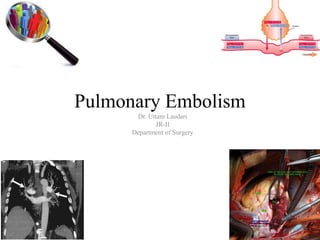
Pulmonary Embolism
Dr. Uttam Laudari
JR-II
Department of Surgery
Epidemiology
• Venous thromboembolism (VTE) encompasses deep vein thrombosis
(DVT) and pulmonary embolism (PE).
• It is the third most frequent cardiovascular disease with an overall annual
incidence of 100–200 per 100 000 inhabitants.1
• VTE may be lethal in the acute phase or lead to chronic disease and
disability, but it is also often preventable.
Heit JA. The epidemiology of venous thromboembolism in the community. Arterioscler
Thromb Vasc Biol 2008;28(3):370–372.
Disease burden
• As estimated on the basis of an epidemiological model, over 317 000
deaths were related to VTE in six countries of the European Union (with a
total population of 454.4 million) in 2004.
34%
59%
7%
percentage
sudden fatal
death
deaths from
undiagnosed
PE
deaths after
diagnosed
Cohen AT, Agnelli G, Anderson FA, Arcelus JI, Bergqvist D, Brecht JG, Greer IA, Heit JA, Hutchinson JL, Kakkar AK,
Mottier D, Oger E, Samama MM, Spannagl M. Venous thromboembolism (VTE) in Europe. The number of VTE events
and associated morbidity and mortality. Thromb Haemost 2007;98(4):756–764.
Predisposition
Patient related
PERMANENT RISK
FACTORS
UNPROVOKED
Settings related
TEMPORARY RISK
FACTORS
PROVOKED
Pulmonary Embolism
Temporary or Reversible risk factor
surgery, trauma, immobilization,
pregnancy,
oral contraceptive use or hormone
replacement therapy)
within the last 6 weeks to 3 months
before diagnosis
PROVOKED
UNPROVOKED In the absence of above
• Major trauma, surgery, lower limb fractures
and joint replacements ,and spinal cord injury,
are strong provoking factors for VTE.
• Cancer
– Haematological malignancies, lung cancer,
gastrointestinal cancer, pancreatic cancer
and brain cancer carry the highest risk
• OCPS
• Pregnancy
– risk is highest in the third trimester of pregnancy and over the 6 weeks of the postpartum
period, being up to 60 times higher compared with the risk in non-pregnant women
• post-menopausal women who receive hormone replacement therapy
• Infection
• Blood transfusion and erythropoiesis-stimulating agents
• Children's- chronic medical conditions, Central lines
• cigarette smoking, obesity, hypercholesterolaemia, hypertension and diabetes
mellitus
• Myocardial infarction and heart failure
Pathophysiology
Embolization
• When venous thrombi are dislodged from their site of formation,
• they embolize to the pulmonary arterial circulation
• or paradoxically, to the arterial circulation through a patent foramen ovale
or atrial septal defect
• About one-half of patients with pelvic vein thrombosis or proximal leg DVT
develop PE, which is often asymptomatic.
• Isolated calf vein thrombi pose a much lower risk of PE but
are the most common source of paradoxical embolism.
• These tiny thrombi can traverse a patent foramen ovale or
atrial septal defect, unlike larger, more proximal leg thrombi.
Physiology
• The most common gas exchange abnormalities are hypoxemia
(decreased arterial PO2) and an increased alveolar-arterial O2
tension gradient, which represents the inefficiency of O2
transfer across the lungs.
• Anatomic dead space
– increases because breathed gas does not enter gas exchange units of the
lung.
• Physiologic dead space
– increases because ventilation to gas exchange units exceeds venous
blood flow through the pulmonary capillaries.
Increased pulmonary
vascular resistance
pulmonary artery
obstruction
+
Serotonin ( produced by
platelets)
Impaired gaseous
exchange
•Increased dead space
•Hypoxemia – alveolar
hypoventillation
relative to perfusion in
other lung
•Right to left shunt
Increased airway
resistance
•constriction of
airways distal to the
bronchi
Decreased pulmonary compliance due to lung edema, lung
hemorrhage, or loss of surfactant
Alveolar hyperventilation due to reflex stimulation of irritant
receptors.
Right ventricular dysfunction
• pulmonary vascular resistance
increases
• Increased RV wall tension – RV
dysfunction
• interventricular septum bulges into
and compresses an intrinsically
normal left ventricle.
• Diastolic LV impairment due to
septal displacement,
• reduced LV distensibility and
impaired LV filling during diastole.
Right ventricular dysfunction
• Increased RV wall tension also compresses
the right coronary artery
diminished subendocardial perfusion
limits myocardial oxygen supply
myocardial ischemia and RV infarction
• LV Underfilling
– fall in left-ventricular
cardiac output and
systemic arterial pressure
– provoking myocardial
ischemia due to
compromised coronary
artery perfusion.
– Eventually, circulatory
collapse and death may
ensue
• Dyspnoea
– may be acute and severe in central PE
– in small peripheral PE, it is often mild and may be transient.
– In patients with pre-existing heart failure or pulmonary disease,
worsening dyspnoea may be the only symptom indicative of PE
• Chest pain
– caused by pleural irritation due to distal emboli causing pulmonary
infarction
– In central PE, chest pain may have a typical angina character, possibly
reflecting RV ischaemia and requiring differential diagnosis with
acute coronary syndrome (ACS) or aortic dissection
• ABG-
– hypoxaemia
– 40% of the patients have normal arterial oxygen saturation
– 20% a normal alveolar-arterial oxygen gradient.
– Hypocapnia is also often present.
• The chest X-ray is frequently abnormal
– its findings are usually non-specific in PE,
– it is useful for excluding other causes of dyspnoea or chest pain.
– focal oligemia (Westermark's sign),
– a peripheral wedged-shaped density above the diaphragm (Hampton's
hump)
– and an enlarged right descending pulmonary artery (Palla's sign).
Electrocardiographic
• changes indicative of RV strain,
– such as inversion of T waves in leads V1–V4,
– a QR pattern in V1,
– S1Q3T3 pattern,
– incomplete or complete right bundle-branch block
• These electrocardiographic changes are usually found in more
severe cases of PE
• milder cases, the only anomaly may be sinus tachycardia, present in
40% of patients.
• Finally, atrial arrhythmias, most frequently atrial fibrillation, may be
associated with acute PE
Assessment of clinical probability
Wells criteria
Previous PE or
DVT
1.5
Hear rate
>100bpm
1.5
Surgery or
immobilsation
in past 4 weeks
1.5
hemoptysis 1
Active cancer 1
Clinical signs of
DVT
3
Alternative
diagnosis less
likely than PE
3
Clinical
probability
score- three
level
PE
probablity
Low 0-1 10%
Intermediate 2-6 30%
high >7 65%
Clinical
probability
score- three
level
Low 0-3 (10%)
Intermediate 4-10(30%)
high >11(65%)
D- dimer
fibrin is also produced in a
wide variety of conditions
such as- cancer,
inflammation, bleeding,
trauma, surgery and
necrosis.
sensitivity of the d-dimer
is >80% for DVT (including
isolated calf DVT) and
>95% for PE.
D- dimer
• The d-dimer is less sensitive for DVT than for PE because the
DVT thrombus size is smaller.
• The d-dimer is a useful "rule out" test.
• More than 95% of patients with a normal (<500 ng/mL) d-
dimer do not have PE.
• The specificity of D-dimer in suspected PE decreases steadily with age, to
almost 10% in patients >80 years
• Recent evidence suggests using age-adjusted cut-offs to improve the
performance of D-dimer testing in the elderly
• In a recent meta-analysis, age-adjusted cut-off values (age x 10 mg/L
above 50 years) allowed increasing specificity from 34–46% while
retaining a sensitivity above 97%
CT Pulmonary angiography
Sensitivity-83%
specificity-96%
Also obtains
excellent images of
the RV and LV and
can be used for risk
stratification along
with its use as a
diagnostic tool
CT Pulmonary angiography
• It allows adequate visualization of the pulmonary arteries down to at least the
segmental level
• RV enlargement on chest CT indicates an increased likelihood of death within the
next 30 days compared with PE patients who have normal RV size on chest CT.
• imaging is continued below the chest to the knee pelvic and proximal leg DVT
also can be diagnosed by CT scanning.
• Rules out other
– pneumonia, emphysema, pulmonary fibrosis, pulmonary mass, and aortic pathology.
Lung scintigraphy
• second-line diagnostic test for PE
• used mostly for patients who cannot tolerate intravenous contrast
• Small particulate aggregates of albumin labeled with a gamma-
emitting radionuclide are injected intravenously and are trapped in
the pulmonary capillary bed
• perfusion scan defect indicates absent or decreased blood flow,
possibly due to PE.
• Ventilation scans, obtained with a radiolabeled inhaled gas such as
xenon or krypton, improve the specificity of the perfusion scan.
Lung scintigraphy
A high-probability scan for PE is defined as
one that indicates two or more segmental
perfusion defects in the presence of
normal ventilation
Lung scintigraphy
• Being a radiation- and contrast medium-sparing procedure, the V/Q scan
may preferentially be applied in outpatients with low clinical probability
and a normal chest X-ray,
– in young (particularly female) patients
– pregnancy
– history of contrast medium-induced anaphylaxis and strong allergic history,
– severe renal failure,
– myeloma and paraproteinaemia
Magnetic Resonance (MR)
• suspected VTE patients with renal insufficiency or contrast
dye allergy.
• may detect large proximal PE but is not reliable for smaller
segmental and subsegmental PE.
Echocardiography
• useful diagnostic tool for detecting conditions that may mimic
PE, such as acute myocardial infarction, pericardial
tamponade, and aortic dissection
• The best-known indirect sign of PE on transthoracic
echocardiography is McConnell's sign: hypokinesis of the RV
free wall with normal motion of the RV apex
Pulmonary angiography
• technically unsatisfactory chest CTs
• interventional procedure such as catheter-directed
thrombolysis or embolectomy is planned.
• visualization of an intraluminal filling defect in more than
one projection.
• Secondary signs of PE include abrupt occlusion ("cut-off")
of vessels, segmental oligemia or avascularity, a prolonged
arterial phase with slow filling, and tortuous, tapering
peripheral vessels.
diagnostic algorithm for patients with suspected high-risk PE, i.e.
presenting with shock or hypotension
Proposed diagnostic algorithm for patients with
suspected not high-risk pulmonary embolism
Treatment in Acute phase
• Hemodynamic and respiratory support
• Acute RV failure with resulting low systemic output is the
leading cause of death in patients with high-risk PE.
• Therefore, supportive treatment is vital in patients with PE and
RV failure
• Fluid
– aggressive volume expansion is of no benefit
– worsen RV function by causing mechanical overstretch, or
by reflex mechanisms that depress contractility
– modest (500 mL) fluid challenge may help to increase
cardiac index in patients with PE, low cardiac index, and
normal BP
Vasopressors
• often necessary, in parallel with (or while waiting for) pharmacological,
surgical, or interventional reperfusion treatment.
• Norepinephrine
– improve RV function via a direct positive inotropic effect
– improves RV coronary perfusion by peripheral vascular alpha-receptor
stimulation and the increase in systemic BP
– Its use should probably be limited to hypotensive patient
• Epinephrine combines the beneficial properties of
norepinephrine and dobutamine, without the systemic
vasodilatory effects of the latter.
• It may therefore exert beneficial effects in patients with PE and
shock.
Vasodilators
– decrease pulmonary arterial pressure and pulmonary vascular
resistance,
– but the main concern is the lack of specificity of these drugs for the
pulmonary vasculature after systemic (intravenous) administration
– Inhalation of nitric oxide may improve the haemodynamic status and
gas exchange of patients with PE
Respiratory support
• Hypoxaemia and hypocapnia are frequently encountered in
patients with PE, but they are of moderate severity in most
cases.
– oxygen
– mechanical ventilation
• Mechanical ventilation
– Careful to limit its adverse haemodynamic effects
– the positive intrathoracic pressuremay reduce venous
return and worsen RV failure in patients with massive PE
– Low tidal volumes (approximately 6 mL/kg lean body
weight) and end-inspiratory plateau pressure 30 cm H2O.
Anticoagulation
• In patients with acute PE, anticoagulation is recommended, with the
objective of preventing both early death and recurrent symptomatic or fatal
VTE
• The standard duration of anticoagulation should cover at least 3 months
• acute-phase treatment consists of administering parenteral anticoagulation
[unfractionated heparin (UFH), LMWH or fondaparinux] over the first 5–
10 days.
• Parenteral heparin should overlap with the initiation of a vitamin K
antagonist (VKA)
UFH
• Binds to antithombin III
• inhibits factor IIa ( Thrombin) and facto Xa and also F
IXa,XIa and XIIA of coagulation cascade
• Dose-
– IV bolus- 80u/kg
– Followed by continious IV drip at 18units/kg/hr
– T1/2- 45-90min (dose dependent)
– Monitoring- aPTT 6hourly
– aPTT goal- 1.5 to 2 times the control value
Complication of UFH
1.Hemorrahage
– Fatal, intracranial hemorrhage, retroperitoneal or
requiring >2 unit of packed red cell is aprox 5 % in
hospitalized patient
• Management
– Discontinue UFH
– Protamine sulphate
• 1mg protamine neutralizes 90-115 unit of heparin
• Dose not to exceed 50mg IV over any 10 min
Complication of UFH
2. HIT
• Results due to heparin associated antiplatelets antibody
complex
• Repeated heparin exposure( vascular Sx- 21%)
• Occurs m/c in 2nd week of therapy
• Platelet counts to be monitored periodically
• Dx- exposure to Heparin + platelets <100,000 and/ or
decline in 50% of platelet following exposure
Complication of UFH
3. Heparin induced osteopenia
impairment of bone formation and enhancement of
bone resorption by heparin
LMWH(enoxaparin)
• Derived from polymerization of porcine UFH
• Act more on F Xa
• Increased bioavailability
• 2-4 times longer half life
• Can be administered S.C without lab monitoring
• Img/kg 12hrly and 1.5mg/kg OD
• Partially reversible by protamine (60%)
• Patient requiring monitoring
– Severe renal impairment, pediatrics,pregnants, wt>120kg
• HIT <2%
• Established HIT- not be used
• Outpatient treatment
• Reduce hospital stay
Fondaparinux
• Synthetic petasaccharide
• Activated antithrombin and Xa inhibiion
• Recurrent VTE- 3.8-5%
• Major bleeding- 2-2.6%
• Administered – SC once daily dose
• Half life 17 hour
Direct thrombin inhibitors
• Hirudin,argatroban and bivalirudin
• Binds thrombin and inhibiting conversion of fribrinogen to
fibrin and fribrin induced thrombocytopenia
• Used for high suspicion/confirmed HIT or with history of
HIT or HAAb positive cases
• Requires aPTT adjustment
Vitamin K antagonist
• Main stay of long term antithrombotic therapy
• Warfarin and other coumarin derivatives
• Inhibits gamma carboxyaltion of Vit K dependent factors and protein C and S
• Requires several days to achieve full effect ( 4-5 days)
• Monitored by INR
• INR= (patient PT/lab normal PT)*ISI
• ISI- international senstivity index- strength of thromboplastin that is added to
activate the extrinsic coagualtion pathway
warfarin
• Therapeutic range- 2-3
• To be started on same day of starting parenteral
anticoagualation ( except with concomitant thrombolysis and
venous thrombectomy)
• Usual starting dose 50-10mg
• Smaller dose for older, malnourished, liver disease and CHF
• Variability of response
– Depends upon Liver funtion, diet, age and medicaitons used
Fibrinolysis
The only FDA-approved indication for PE
fibrinolysis is massive PE
Fibrinolysis
• rapidly reverses right heart failure and may result in a lower
rate of death and recurrent PE
– dissolving much of the anatomically obstructing pulmonary arterial
thrombus
– preventing the continued release of serotonin and other neurohumoral
factors that exacerbate pulmonary hypertension
– lysing much of the source of the thrombus in the pelvic or deep leg
veins, thereby decreasing the likelihood of recurrent PE.
• preferred fibrinolytic regimen
– 100 mg of recombinant tissue plasminogen activator (tPA)
administered as a continuous peripheral intravenous infusion over 2
hours.
– Patients appear to respond to fibrinolysis for up to 14 days after the PE
has occurred
Contraindication of Fibrinolysis
• intracranial disease
• recent surgery and trauma
• The overall major bleeding rate is about 10%, including a 1–
3% risk of intracranial hemorrhage.
Surgical Embolectomy
•high-risk PE
•selected patients with
intermediate-high-risk PE,
particularly
if thrombolysis is
contraindicated or has
failed.
Percutaneous catheter-directed
treatment
•Administer lytic agent alone
•Pharmacomechanical clot lysis
Venous filters
1. acute PE with absolute contraindications
to anticoagulant drugs
2. patients with objectively confirmed
recurrent PE despite adequate
anticoagulation treatment
Recommendations for duration of anticoagulation after
pulmonary embolism
PE secondary to transient risk factor 3 months
Unprovoked PE At least 3 months
Class IB
second episode of
unprovoked PE
Indefinite duration
Class IA
PE with Cancer LMWH should be considered for the
first 3–
6 months. IIA
Extended anticoagulation (beyond the
first 3–6 months) should be
considered for an indefinite
period or until the cancer is
Cured . IIC
2014 ESC Guidelines on the diagnosis and management of acute
pulmonary embolism
In patients who receive extended anticoagulation, the risk–benefit ratio
of continuing such treatment should be reassessed at regular intervals. IC
In patients who refuse to take or are unable to tolerate any form of oral
anticoagulants, aspirin may be considered for extended secondary VTE
prophylaxis. IIB
2014 ESC Guidelines on the diagnosis and management of acute
pulmonary embolism
Summary

More Related Content

What's hot

What's hot (20)

Pulmonary embolism
Pulmonary embolismPulmonary embolism
Pulmonary embolism
 
Xaheer shah...pulmonary embolism
Xaheer shah...pulmonary embolismXaheer shah...pulmonary embolism
Xaheer shah...pulmonary embolism
 
Pulmonary embolism
Pulmonary embolismPulmonary embolism
Pulmonary embolism
 
Pulmonary embolism
Pulmonary embolism Pulmonary embolism
Pulmonary embolism
 
Pulmonary hypertension
Pulmonary hypertensionPulmonary hypertension
Pulmonary hypertension
 
Pulmonary Embolism and CTEPH
Pulmonary Embolism and CTEPHPulmonary Embolism and CTEPH
Pulmonary Embolism and CTEPH
 
Pulmonary hypertension
Pulmonary hypertensionPulmonary hypertension
Pulmonary hypertension
 
Pulmonary nodules
Pulmonary nodules Pulmonary nodules
Pulmonary nodules
 
Pulmonary embolism
Pulmonary embolismPulmonary embolism
Pulmonary embolism
 
Pulmonary thromboembolism
Pulmonary thromboembolismPulmonary thromboembolism
Pulmonary thromboembolism
 
Pulmonary embolism - Diagnosis and management
Pulmonary embolism - Diagnosis and managementPulmonary embolism - Diagnosis and management
Pulmonary embolism - Diagnosis and management
 
Diseases of the aorta
Diseases of the aortaDiseases of the aorta
Diseases of the aorta
 
Pulmonaryembolism final
Pulmonaryembolism finalPulmonaryembolism final
Pulmonaryembolism final
 
Alveolar hemorrhage
Alveolar hemorrhageAlveolar hemorrhage
Alveolar hemorrhage
 
DIFFUSE ALVEOLAR HAEMORRHAGE
DIFFUSE ALVEOLAR HAEMORRHAGEDIFFUSE ALVEOLAR HAEMORRHAGE
DIFFUSE ALVEOLAR HAEMORRHAGE
 
Pleural effusion.pptx cme march
Pleural effusion.pptx cme marchPleural effusion.pptx cme march
Pleural effusion.pptx cme march
 
Aortic Aneurysms
Aortic AneurysmsAortic Aneurysms
Aortic Aneurysms
 
pulmonary edema gokull.pptx
pulmonary edema gokull.pptxpulmonary edema gokull.pptx
pulmonary edema gokull.pptx
 
Ischemic Heart Disease Surgery
Ischemic Heart Disease SurgeryIschemic Heart Disease Surgery
Ischemic Heart Disease Surgery
 
Pulmonary thromboembolism
Pulmonary thromboembolismPulmonary thromboembolism
Pulmonary thromboembolism
 

Similar to Pulmonary embolism

deep vein thrombosis and pe
deep vein thrombosis and pedeep vein thrombosis and pe
deep vein thrombosis and pePraveen Shukla
 
Diagnosis of Pulmonary Embolism - by Dr KD DELE
Diagnosis of Pulmonary Embolism - by Dr KD DELEDiagnosis of Pulmonary Embolism - by Dr KD DELE
Diagnosis of Pulmonary Embolism - by Dr KD DELEKemi Dele-Ijagbulu
 
PULMONARY EMBOLISM.pptx
PULMONARY EMBOLISM.pptxPULMONARY EMBOLISM.pptx
PULMONARY EMBOLISM.pptxDrbhagya3
 
Pulmonary embolism radiology imaging
Pulmonary embolism radiology imagingPulmonary embolism radiology imaging
Pulmonary embolism radiology imagingharshvardhan
 
Pulmonary embolism,overview
Pulmonary embolism,overviewPulmonary embolism,overview
Pulmonary embolism,overviewEman Mahmoud
 
4.Pe(English终)
4.Pe(English终)4.Pe(English终)
4.Pe(English终)Deep Deep
 
Pulmonary embolism radiology
Pulmonary embolism radiologyPulmonary embolism radiology
Pulmonary embolism radiologyAnish Choudhary
 
ashish pulm embolism.pptx
ashish pulm embolism.pptxashish pulm embolism.pptx
ashish pulm embolism.pptxashishnair22
 
Pulmonary embolism
Pulmonary embolismPulmonary embolism
Pulmonary embolismoday abdow
 
Venous thromboembolism, THROMBOPROPHYLAXIS and management
Venous thromboembolism, THROMBOPROPHYLAXIS and managementVenous thromboembolism, THROMBOPROPHYLAXIS and management
Venous thromboembolism, THROMBOPROPHYLAXIS and managementmauryaramgopal
 
Pulmonary and venous thromboembolism.pptx
Pulmonary and venous thromboembolism.pptxPulmonary and venous thromboembolism.pptx
Pulmonary and venous thromboembolism.pptxNidhiBhutada5
 

Similar to Pulmonary embolism (20)

deep vein thrombosis and pe
deep vein thrombosis and pedeep vein thrombosis and pe
deep vein thrombosis and pe
 
Pulmonary embolism
Pulmonary embolismPulmonary embolism
Pulmonary embolism
 
Diagnosis of Pulmonary Embolism - by Dr KD DELE
Diagnosis of Pulmonary Embolism - by Dr KD DELEDiagnosis of Pulmonary Embolism - by Dr KD DELE
Diagnosis of Pulmonary Embolism - by Dr KD DELE
 
PULMONARY EMBOLISM.pptx
PULMONARY EMBOLISM.pptxPULMONARY EMBOLISM.pptx
PULMONARY EMBOLISM.pptx
 
Pulmonary embolism radiology imaging
Pulmonary embolism radiology imagingPulmonary embolism radiology imaging
Pulmonary embolism radiology imaging
 
Pulmonary Embolism
Pulmonary Embolism Pulmonary Embolism
Pulmonary Embolism
 
Pulmonary embolism,overview
Pulmonary embolism,overviewPulmonary embolism,overview
Pulmonary embolism,overview
 
4.Pe(English终)
4.Pe(English终)4.Pe(English终)
4.Pe(English终)
 
VTE.pptx
VTE.pptxVTE.pptx
VTE.pptx
 
Dr.cazaam
Dr.cazaamDr.cazaam
Dr.cazaam
 
VTE AND PE.pptx
VTE AND PE.pptxVTE AND PE.pptx
VTE AND PE.pptx
 
Pulmonary embolism radiology
Pulmonary embolism radiologyPulmonary embolism radiology
Pulmonary embolism radiology
 
ashish pulm embolism.pptx
ashish pulm embolism.pptxashish pulm embolism.pptx
ashish pulm embolism.pptx
 
TB Pericarditis.pptx
TB Pericarditis.pptxTB Pericarditis.pptx
TB Pericarditis.pptx
 
Pulmonary embolism
Pulmonary embolismPulmonary embolism
Pulmonary embolism
 
Pulmonary embolism
Pulmonary embolismPulmonary embolism
Pulmonary embolism
 
Venous thromboembolism, THROMBOPROPHYLAXIS and management
Venous thromboembolism, THROMBOPROPHYLAXIS and managementVenous thromboembolism, THROMBOPROPHYLAXIS and management
Venous thromboembolism, THROMBOPROPHYLAXIS and management
 
Pulmonary and venous thromboembolism.pptx
Pulmonary and venous thromboembolism.pptxPulmonary and venous thromboembolism.pptx
Pulmonary and venous thromboembolism.pptx
 
Asd and vsd
Asd and vsdAsd and vsd
Asd and vsd
 
Pavm
PavmPavm
Pavm
 

More from Youttam Laudari

Diagnosis and management of Asymptomatic Neoplastic Pancreatic cyst.pptx
Diagnosis and management of Asymptomatic Neoplastic Pancreatic cyst.pptxDiagnosis and management of Asymptomatic Neoplastic Pancreatic cyst.pptx
Diagnosis and management of Asymptomatic Neoplastic Pancreatic cyst.pptxYouttam Laudari
 
Management of exocrine and endocrine insufficiency following pancreatic (1).pptx
Management of exocrine and endocrine insufficiency following pancreatic (1).pptxManagement of exocrine and endocrine insufficiency following pancreatic (1).pptx
Management of exocrine and endocrine insufficiency following pancreatic (1).pptxYouttam Laudari
 
Cinicopathological Meeting- Intestinal Ganglioneuromatosis
Cinicopathological Meeting- Intestinal GanglioneuromatosisCinicopathological Meeting- Intestinal Ganglioneuromatosis
Cinicopathological Meeting- Intestinal GanglioneuromatosisYouttam Laudari
 
Post cholecystectomy syndromes
Post cholecystectomy syndromesPost cholecystectomy syndromes
Post cholecystectomy syndromesYouttam Laudari
 
Monitoring Hypoxia and oxygen supplementation
Monitoring Hypoxia and oxygen supplementationMonitoring Hypoxia and oxygen supplementation
Monitoring Hypoxia and oxygen supplementationYouttam Laudari
 
Gastrointestinal stromal tumours
Gastrointestinal stromal tumoursGastrointestinal stromal tumours
Gastrointestinal stromal tumoursYouttam Laudari
 
Journal club-Determination of surgical priorities in appendicitis
Journal club-Determination of surgical priorities in appendicitisJournal club-Determination of surgical priorities in appendicitis
Journal club-Determination of surgical priorities in appendicitisYouttam Laudari
 
Dr. deepak raj singhs principles of surgey
Dr. deepak raj singhs  principles of surgeyDr. deepak raj singhs  principles of surgey
Dr. deepak raj singhs principles of surgeyYouttam Laudari
 
Standard versus tubeless mini percutaneous nephrolithotomy
Standard versus tubeless mini percutaneous nephrolithotomyStandard versus tubeless mini percutaneous nephrolithotomy
Standard versus tubeless mini percutaneous nephrolithotomyYouttam Laudari
 
Multiloculated thymic cyst poster presenation
Multiloculated thymic cyst poster presenationMultiloculated thymic cyst poster presenation
Multiloculated thymic cyst poster presenationYouttam Laudari
 
Lap versus open du perfo
Lap versus open du perfoLap versus open du perfo
Lap versus open du perfoYouttam Laudari
 

More from Youttam Laudari (20)

GE junction tumor-pptx
GE junction tumor-pptxGE junction tumor-pptx
GE junction tumor-pptx
 
Diagnosis and management of Asymptomatic Neoplastic Pancreatic cyst.pptx
Diagnosis and management of Asymptomatic Neoplastic Pancreatic cyst.pptxDiagnosis and management of Asymptomatic Neoplastic Pancreatic cyst.pptx
Diagnosis and management of Asymptomatic Neoplastic Pancreatic cyst.pptx
 
Management of exocrine and endocrine insufficiency following pancreatic (1).pptx
Management of exocrine and endocrine insufficiency following pancreatic (1).pptxManagement of exocrine and endocrine insufficiency following pancreatic (1).pptx
Management of exocrine and endocrine insufficiency following pancreatic (1).pptx
 
Intravenous Urography
Intravenous UrographyIntravenous Urography
Intravenous Urography
 
Malignancy audit 2072
Malignancy audit  2072  Malignancy audit  2072
Malignancy audit 2072
 
Evidence based practice
Evidence based practiceEvidence based practice
Evidence based practice
 
Cinicopathological Meeting- Intestinal Ganglioneuromatosis
Cinicopathological Meeting- Intestinal GanglioneuromatosisCinicopathological Meeting- Intestinal Ganglioneuromatosis
Cinicopathological Meeting- Intestinal Ganglioneuromatosis
 
Surviving sepsis
Surviving sepsisSurviving sepsis
Surviving sepsis
 
Liver hemangiona
Liver hemangionaLiver hemangiona
Liver hemangiona
 
Renal trauma
Renal traumaRenal trauma
Renal trauma
 
Renal radilogy
Renal radilogyRenal radilogy
Renal radilogy
 
Post cholecystectomy syndromes
Post cholecystectomy syndromesPost cholecystectomy syndromes
Post cholecystectomy syndromes
 
Monitoring Hypoxia and oxygen supplementation
Monitoring Hypoxia and oxygen supplementationMonitoring Hypoxia and oxygen supplementation
Monitoring Hypoxia and oxygen supplementation
 
Gastrointestinal stromal tumours
Gastrointestinal stromal tumoursGastrointestinal stromal tumours
Gastrointestinal stromal tumours
 
Journal club-Determination of surgical priorities in appendicitis
Journal club-Determination of surgical priorities in appendicitisJournal club-Determination of surgical priorities in appendicitis
Journal club-Determination of surgical priorities in appendicitis
 
Deep vein thrombosis
Deep vein thrombosis   Deep vein thrombosis
Deep vein thrombosis
 
Dr. deepak raj singhs principles of surgey
Dr. deepak raj singhs  principles of surgeyDr. deepak raj singhs  principles of surgey
Dr. deepak raj singhs principles of surgey
 
Standard versus tubeless mini percutaneous nephrolithotomy
Standard versus tubeless mini percutaneous nephrolithotomyStandard versus tubeless mini percutaneous nephrolithotomy
Standard versus tubeless mini percutaneous nephrolithotomy
 
Multiloculated thymic cyst poster presenation
Multiloculated thymic cyst poster presenationMultiloculated thymic cyst poster presenation
Multiloculated thymic cyst poster presenation
 
Lap versus open du perfo
Lap versus open du perfoLap versus open du perfo
Lap versus open du perfo
 

Recently uploaded

Call 8250092165 Patna Call Girls ₹4.5k Cash Payment With Room Delivery
Call 8250092165 Patna Call Girls ₹4.5k Cash Payment With Room DeliveryCall 8250092165 Patna Call Girls ₹4.5k Cash Payment With Room Delivery
Call 8250092165 Patna Call Girls ₹4.5k Cash Payment With Room DeliveryJyoti singh
 
Call Girls Service Jaipur {9521753030 } ❤️VVIP BHAWNA Call Girl in Jaipur Raj...
Call Girls Service Jaipur {9521753030 } ❤️VVIP BHAWNA Call Girl in Jaipur Raj...Call Girls Service Jaipur {9521753030 } ❤️VVIP BHAWNA Call Girl in Jaipur Raj...
Call Girls Service Jaipur {9521753030 } ❤️VVIP BHAWNA Call Girl in Jaipur Raj...Janvi Singh
 
Chennai ❣️ Call Girl 6378878445 Call Girls in Chennai Escort service book now
Chennai ❣️ Call Girl 6378878445 Call Girls in Chennai Escort service book nowChennai ❣️ Call Girl 6378878445 Call Girls in Chennai Escort service book now
Chennai ❣️ Call Girl 6378878445 Call Girls in Chennai Escort service book nowtanudubay92
 
Bhawanipatna Call Girls 📞9332606886 Call Girls in Bhawanipatna Escorts servic...
Bhawanipatna Call Girls 📞9332606886 Call Girls in Bhawanipatna Escorts servic...Bhawanipatna Call Girls 📞9332606886 Call Girls in Bhawanipatna Escorts servic...
Bhawanipatna Call Girls 📞9332606886 Call Girls in Bhawanipatna Escorts servic...Dipal Arora
 
Difference Between Skeletal Smooth and Cardiac Muscles
Difference Between Skeletal Smooth and Cardiac MusclesDifference Between Skeletal Smooth and Cardiac Muscles
Difference Between Skeletal Smooth and Cardiac MusclesMedicoseAcademics
 
7 steps How to prevent Thalassemia : Dr Sharda Jain & Vandana Gupta
7 steps How to prevent Thalassemia : Dr Sharda Jain & Vandana Gupta7 steps How to prevent Thalassemia : Dr Sharda Jain & Vandana Gupta
7 steps How to prevent Thalassemia : Dr Sharda Jain & Vandana GuptaLifecare Centre
 
Cardiac Output, Venous Return, and Their Regulation
Cardiac Output, Venous Return, and Their RegulationCardiac Output, Venous Return, and Their Regulation
Cardiac Output, Venous Return, and Their RegulationMedicoseAcademics
 
Call Girls Kathua Just Call 8250077686 Top Class Call Girl Service Available
Call Girls Kathua Just Call 8250077686 Top Class Call Girl Service AvailableCall Girls Kathua Just Call 8250077686 Top Class Call Girl Service Available
Call Girls Kathua Just Call 8250077686 Top Class Call Girl Service AvailableDipal Arora
 
ANATOMY AND PHYSIOLOGY OF REPRODUCTIVE SYSTEM.pptx
ANATOMY AND PHYSIOLOGY OF REPRODUCTIVE SYSTEM.pptxANATOMY AND PHYSIOLOGY OF REPRODUCTIVE SYSTEM.pptx
ANATOMY AND PHYSIOLOGY OF REPRODUCTIVE SYSTEM.pptxSwetaba Besh
 
Race Course Road } Book Call Girls in Bangalore | Whatsapp No 6378878445 VIP ...
Race Course Road } Book Call Girls in Bangalore | Whatsapp No 6378878445 VIP ...Race Course Road } Book Call Girls in Bangalore | Whatsapp No 6378878445 VIP ...
Race Course Road } Book Call Girls in Bangalore | Whatsapp No 6378878445 VIP ...dishamehta3332
 
Call Girls in Lucknow Just Call 👉👉 8875999948 Top Class Call Girl Service Ava...
Call Girls in Lucknow Just Call 👉👉 8875999948 Top Class Call Girl Service Ava...Call Girls in Lucknow Just Call 👉👉 8875999948 Top Class Call Girl Service Ava...
Call Girls in Lucknow Just Call 👉👉 8875999948 Top Class Call Girl Service Ava...Janvi Singh
 
Lucknow Call Girls Service { 9984666624 } ❤️VVIP ROCKY Call Girl in Lucknow U...
Lucknow Call Girls Service { 9984666624 } ❤️VVIP ROCKY Call Girl in Lucknow U...Lucknow Call Girls Service { 9984666624 } ❤️VVIP ROCKY Call Girl in Lucknow U...
Lucknow Call Girls Service { 9984666624 } ❤️VVIP ROCKY Call Girl in Lucknow U...Janvi Singh
 
💰Call Girl In Bangalore☎️63788-78445💰 Call Girl service in Bangalore☎️Bangalo...
💰Call Girl In Bangalore☎️63788-78445💰 Call Girl service in Bangalore☎️Bangalo...💰Call Girl In Bangalore☎️63788-78445💰 Call Girl service in Bangalore☎️Bangalo...
💰Call Girl In Bangalore☎️63788-78445💰 Call Girl service in Bangalore☎️Bangalo...gragneelam30
 
Call Girls Wayanad Just Call 8250077686 Top Class Call Girl Service Available
Call Girls Wayanad Just Call 8250077686 Top Class Call Girl Service AvailableCall Girls Wayanad Just Call 8250077686 Top Class Call Girl Service Available
Call Girls Wayanad Just Call 8250077686 Top Class Call Girl Service AvailableDipal Arora
 
ANATOMY AND PHYSIOLOGY OF RESPIRATORY SYSTEM.pptx
ANATOMY AND PHYSIOLOGY OF RESPIRATORY SYSTEM.pptxANATOMY AND PHYSIOLOGY OF RESPIRATORY SYSTEM.pptx
ANATOMY AND PHYSIOLOGY OF RESPIRATORY SYSTEM.pptxSwetaba Besh
 
💞 Safe And Secure Call Girls Coimbatore🧿 6378878445 🧿 High Class Coimbatore C...
💞 Safe And Secure Call Girls Coimbatore🧿 6378878445 🧿 High Class Coimbatore C...💞 Safe And Secure Call Girls Coimbatore🧿 6378878445 🧿 High Class Coimbatore C...
💞 Safe And Secure Call Girls Coimbatore🧿 6378878445 🧿 High Class Coimbatore C...dilbirsingh0889
 
Call Girl in Chennai | Whatsapp No 📞 7427069034 📞 VIP Escorts Service Availab...
Call Girl in Chennai | Whatsapp No 📞 7427069034 📞 VIP Escorts Service Availab...Call Girl in Chennai | Whatsapp No 📞 7427069034 📞 VIP Escorts Service Availab...
Call Girl in Chennai | Whatsapp No 📞 7427069034 📞 VIP Escorts Service Availab...amritaverma53
 
Call girls Service Phullen / 9332606886 Genuine Call girls with real Photos a...
Call girls Service Phullen / 9332606886 Genuine Call girls with real Photos a...Call girls Service Phullen / 9332606886 Genuine Call girls with real Photos a...
Call girls Service Phullen / 9332606886 Genuine Call girls with real Photos a...call girls hydrabad
 
Call Girls Mussoorie Just Call 8854095900 Top Class Call Girl Service Available
Call Girls Mussoorie Just Call 8854095900 Top Class Call Girl Service AvailableCall Girls Mussoorie Just Call 8854095900 Top Class Call Girl Service Available
Call Girls Mussoorie Just Call 8854095900 Top Class Call Girl Service AvailableJanvi Singh
 
(RIYA)🎄Airhostess Call Girl Jaipur Call Now 8445551418 Premium Collection Of ...
(RIYA)🎄Airhostess Call Girl Jaipur Call Now 8445551418 Premium Collection Of ...(RIYA)🎄Airhostess Call Girl Jaipur Call Now 8445551418 Premium Collection Of ...
(RIYA)🎄Airhostess Call Girl Jaipur Call Now 8445551418 Premium Collection Of ...TanyaAhuja34
 

Recently uploaded (20)

Call 8250092165 Patna Call Girls ₹4.5k Cash Payment With Room Delivery
Call 8250092165 Patna Call Girls ₹4.5k Cash Payment With Room DeliveryCall 8250092165 Patna Call Girls ₹4.5k Cash Payment With Room Delivery
Call 8250092165 Patna Call Girls ₹4.5k Cash Payment With Room Delivery
 
Call Girls Service Jaipur {9521753030 } ❤️VVIP BHAWNA Call Girl in Jaipur Raj...
Call Girls Service Jaipur {9521753030 } ❤️VVIP BHAWNA Call Girl in Jaipur Raj...Call Girls Service Jaipur {9521753030 } ❤️VVIP BHAWNA Call Girl in Jaipur Raj...
Call Girls Service Jaipur {9521753030 } ❤️VVIP BHAWNA Call Girl in Jaipur Raj...
 
Chennai ❣️ Call Girl 6378878445 Call Girls in Chennai Escort service book now
Chennai ❣️ Call Girl 6378878445 Call Girls in Chennai Escort service book nowChennai ❣️ Call Girl 6378878445 Call Girls in Chennai Escort service book now
Chennai ❣️ Call Girl 6378878445 Call Girls in Chennai Escort service book now
 
Bhawanipatna Call Girls 📞9332606886 Call Girls in Bhawanipatna Escorts servic...
Bhawanipatna Call Girls 📞9332606886 Call Girls in Bhawanipatna Escorts servic...Bhawanipatna Call Girls 📞9332606886 Call Girls in Bhawanipatna Escorts servic...
Bhawanipatna Call Girls 📞9332606886 Call Girls in Bhawanipatna Escorts servic...
 
Difference Between Skeletal Smooth and Cardiac Muscles
Difference Between Skeletal Smooth and Cardiac MusclesDifference Between Skeletal Smooth and Cardiac Muscles
Difference Between Skeletal Smooth and Cardiac Muscles
 
7 steps How to prevent Thalassemia : Dr Sharda Jain & Vandana Gupta
7 steps How to prevent Thalassemia : Dr Sharda Jain & Vandana Gupta7 steps How to prevent Thalassemia : Dr Sharda Jain & Vandana Gupta
7 steps How to prevent Thalassemia : Dr Sharda Jain & Vandana Gupta
 
Cardiac Output, Venous Return, and Their Regulation
Cardiac Output, Venous Return, and Their RegulationCardiac Output, Venous Return, and Their Regulation
Cardiac Output, Venous Return, and Their Regulation
 
Call Girls Kathua Just Call 8250077686 Top Class Call Girl Service Available
Call Girls Kathua Just Call 8250077686 Top Class Call Girl Service AvailableCall Girls Kathua Just Call 8250077686 Top Class Call Girl Service Available
Call Girls Kathua Just Call 8250077686 Top Class Call Girl Service Available
 
ANATOMY AND PHYSIOLOGY OF REPRODUCTIVE SYSTEM.pptx
ANATOMY AND PHYSIOLOGY OF REPRODUCTIVE SYSTEM.pptxANATOMY AND PHYSIOLOGY OF REPRODUCTIVE SYSTEM.pptx
ANATOMY AND PHYSIOLOGY OF REPRODUCTIVE SYSTEM.pptx
 
Race Course Road } Book Call Girls in Bangalore | Whatsapp No 6378878445 VIP ...
Race Course Road } Book Call Girls in Bangalore | Whatsapp No 6378878445 VIP ...Race Course Road } Book Call Girls in Bangalore | Whatsapp No 6378878445 VIP ...
Race Course Road } Book Call Girls in Bangalore | Whatsapp No 6378878445 VIP ...
 
Call Girls in Lucknow Just Call 👉👉 8875999948 Top Class Call Girl Service Ava...
Call Girls in Lucknow Just Call 👉👉 8875999948 Top Class Call Girl Service Ava...Call Girls in Lucknow Just Call 👉👉 8875999948 Top Class Call Girl Service Ava...
Call Girls in Lucknow Just Call 👉👉 8875999948 Top Class Call Girl Service Ava...
 
Lucknow Call Girls Service { 9984666624 } ❤️VVIP ROCKY Call Girl in Lucknow U...
Lucknow Call Girls Service { 9984666624 } ❤️VVIP ROCKY Call Girl in Lucknow U...Lucknow Call Girls Service { 9984666624 } ❤️VVIP ROCKY Call Girl in Lucknow U...
Lucknow Call Girls Service { 9984666624 } ❤️VVIP ROCKY Call Girl in Lucknow U...
 
💰Call Girl In Bangalore☎️63788-78445💰 Call Girl service in Bangalore☎️Bangalo...
💰Call Girl In Bangalore☎️63788-78445💰 Call Girl service in Bangalore☎️Bangalo...💰Call Girl In Bangalore☎️63788-78445💰 Call Girl service in Bangalore☎️Bangalo...
💰Call Girl In Bangalore☎️63788-78445💰 Call Girl service in Bangalore☎️Bangalo...
 
Call Girls Wayanad Just Call 8250077686 Top Class Call Girl Service Available
Call Girls Wayanad Just Call 8250077686 Top Class Call Girl Service AvailableCall Girls Wayanad Just Call 8250077686 Top Class Call Girl Service Available
Call Girls Wayanad Just Call 8250077686 Top Class Call Girl Service Available
 
ANATOMY AND PHYSIOLOGY OF RESPIRATORY SYSTEM.pptx
ANATOMY AND PHYSIOLOGY OF RESPIRATORY SYSTEM.pptxANATOMY AND PHYSIOLOGY OF RESPIRATORY SYSTEM.pptx
ANATOMY AND PHYSIOLOGY OF RESPIRATORY SYSTEM.pptx
 
💞 Safe And Secure Call Girls Coimbatore🧿 6378878445 🧿 High Class Coimbatore C...
💞 Safe And Secure Call Girls Coimbatore🧿 6378878445 🧿 High Class Coimbatore C...💞 Safe And Secure Call Girls Coimbatore🧿 6378878445 🧿 High Class Coimbatore C...
💞 Safe And Secure Call Girls Coimbatore🧿 6378878445 🧿 High Class Coimbatore C...
 
Call Girl in Chennai | Whatsapp No 📞 7427069034 📞 VIP Escorts Service Availab...
Call Girl in Chennai | Whatsapp No 📞 7427069034 📞 VIP Escorts Service Availab...Call Girl in Chennai | Whatsapp No 📞 7427069034 📞 VIP Escorts Service Availab...
Call Girl in Chennai | Whatsapp No 📞 7427069034 📞 VIP Escorts Service Availab...
 
Call girls Service Phullen / 9332606886 Genuine Call girls with real Photos a...
Call girls Service Phullen / 9332606886 Genuine Call girls with real Photos a...Call girls Service Phullen / 9332606886 Genuine Call girls with real Photos a...
Call girls Service Phullen / 9332606886 Genuine Call girls with real Photos a...
 
Call Girls Mussoorie Just Call 8854095900 Top Class Call Girl Service Available
Call Girls Mussoorie Just Call 8854095900 Top Class Call Girl Service AvailableCall Girls Mussoorie Just Call 8854095900 Top Class Call Girl Service Available
Call Girls Mussoorie Just Call 8854095900 Top Class Call Girl Service Available
 
(RIYA)🎄Airhostess Call Girl Jaipur Call Now 8445551418 Premium Collection Of ...
(RIYA)🎄Airhostess Call Girl Jaipur Call Now 8445551418 Premium Collection Of ...(RIYA)🎄Airhostess Call Girl Jaipur Call Now 8445551418 Premium Collection Of ...
(RIYA)🎄Airhostess Call Girl Jaipur Call Now 8445551418 Premium Collection Of ...
 

Pulmonary embolism

  • 1. Pulmonary Embolism Dr. Uttam Laudari JR-II Department of Surgery
  • 2. Epidemiology • Venous thromboembolism (VTE) encompasses deep vein thrombosis (DVT) and pulmonary embolism (PE). • It is the third most frequent cardiovascular disease with an overall annual incidence of 100–200 per 100 000 inhabitants.1 • VTE may be lethal in the acute phase or lead to chronic disease and disability, but it is also often preventable. Heit JA. The epidemiology of venous thromboembolism in the community. Arterioscler Thromb Vasc Biol 2008;28(3):370–372.
  • 3. Disease burden • As estimated on the basis of an epidemiological model, over 317 000 deaths were related to VTE in six countries of the European Union (with a total population of 454.4 million) in 2004. 34% 59% 7% percentage sudden fatal death deaths from undiagnosed PE deaths after diagnosed Cohen AT, Agnelli G, Anderson FA, Arcelus JI, Bergqvist D, Brecht JG, Greer IA, Heit JA, Hutchinson JL, Kakkar AK, Mottier D, Oger E, Samama MM, Spannagl M. Venous thromboembolism (VTE) in Europe. The number of VTE events and associated morbidity and mortality. Thromb Haemost 2007;98(4):756–764.
  • 4.
  • 5. Predisposition Patient related PERMANENT RISK FACTORS UNPROVOKED Settings related TEMPORARY RISK FACTORS PROVOKED Pulmonary Embolism
  • 6. Temporary or Reversible risk factor surgery, trauma, immobilization, pregnancy, oral contraceptive use or hormone replacement therapy) within the last 6 weeks to 3 months before diagnosis PROVOKED UNPROVOKED In the absence of above
  • 7. • Major trauma, surgery, lower limb fractures and joint replacements ,and spinal cord injury, are strong provoking factors for VTE. • Cancer – Haematological malignancies, lung cancer, gastrointestinal cancer, pancreatic cancer and brain cancer carry the highest risk
  • 8. • OCPS • Pregnancy – risk is highest in the third trimester of pregnancy and over the 6 weeks of the postpartum period, being up to 60 times higher compared with the risk in non-pregnant women • post-menopausal women who receive hormone replacement therapy • Infection • Blood transfusion and erythropoiesis-stimulating agents • Children's- chronic medical conditions, Central lines • cigarette smoking, obesity, hypercholesterolaemia, hypertension and diabetes mellitus • Myocardial infarction and heart failure
  • 9. Pathophysiology Embolization • When venous thrombi are dislodged from their site of formation, • they embolize to the pulmonary arterial circulation • or paradoxically, to the arterial circulation through a patent foramen ovale or atrial septal defect
  • 10. • About one-half of patients with pelvic vein thrombosis or proximal leg DVT develop PE, which is often asymptomatic. • Isolated calf vein thrombi pose a much lower risk of PE but are the most common source of paradoxical embolism. • These tiny thrombi can traverse a patent foramen ovale or atrial septal defect, unlike larger, more proximal leg thrombi.
  • 11. Physiology • The most common gas exchange abnormalities are hypoxemia (decreased arterial PO2) and an increased alveolar-arterial O2 tension gradient, which represents the inefficiency of O2 transfer across the lungs.
  • 12. • Anatomic dead space – increases because breathed gas does not enter gas exchange units of the lung. • Physiologic dead space – increases because ventilation to gas exchange units exceeds venous blood flow through the pulmonary capillaries.
  • 13. Increased pulmonary vascular resistance pulmonary artery obstruction + Serotonin ( produced by platelets) Impaired gaseous exchange •Increased dead space •Hypoxemia – alveolar hypoventillation relative to perfusion in other lung •Right to left shunt Increased airway resistance •constriction of airways distal to the bronchi
  • 14. Decreased pulmonary compliance due to lung edema, lung hemorrhage, or loss of surfactant Alveolar hyperventilation due to reflex stimulation of irritant receptors.
  • 15. Right ventricular dysfunction • pulmonary vascular resistance increases • Increased RV wall tension – RV dysfunction • interventricular septum bulges into and compresses an intrinsically normal left ventricle. • Diastolic LV impairment due to septal displacement, • reduced LV distensibility and impaired LV filling during diastole.
  • 16. Right ventricular dysfunction • Increased RV wall tension also compresses the right coronary artery diminished subendocardial perfusion limits myocardial oxygen supply myocardial ischemia and RV infarction
  • 17. • LV Underfilling – fall in left-ventricular cardiac output and systemic arterial pressure – provoking myocardial ischemia due to compromised coronary artery perfusion. – Eventually, circulatory collapse and death may ensue
  • 18.
  • 19. • Dyspnoea – may be acute and severe in central PE – in small peripheral PE, it is often mild and may be transient. – In patients with pre-existing heart failure or pulmonary disease, worsening dyspnoea may be the only symptom indicative of PE • Chest pain – caused by pleural irritation due to distal emboli causing pulmonary infarction – In central PE, chest pain may have a typical angina character, possibly reflecting RV ischaemia and requiring differential diagnosis with acute coronary syndrome (ACS) or aortic dissection
  • 20. • ABG- – hypoxaemia – 40% of the patients have normal arterial oxygen saturation – 20% a normal alveolar-arterial oxygen gradient. – Hypocapnia is also often present. • The chest X-ray is frequently abnormal – its findings are usually non-specific in PE, – it is useful for excluding other causes of dyspnoea or chest pain. – focal oligemia (Westermark's sign), – a peripheral wedged-shaped density above the diaphragm (Hampton's hump) – and an enlarged right descending pulmonary artery (Palla's sign).
  • 21. Electrocardiographic • changes indicative of RV strain, – such as inversion of T waves in leads V1–V4, – a QR pattern in V1, – S1Q3T3 pattern, – incomplete or complete right bundle-branch block • These electrocardiographic changes are usually found in more severe cases of PE • milder cases, the only anomaly may be sinus tachycardia, present in 40% of patients. • Finally, atrial arrhythmias, most frequently atrial fibrillation, may be associated with acute PE
  • 22.
  • 23. Assessment of clinical probability Wells criteria Previous PE or DVT 1.5 Hear rate >100bpm 1.5 Surgery or immobilsation in past 4 weeks 1.5 hemoptysis 1 Active cancer 1 Clinical signs of DVT 3 Alternative diagnosis less likely than PE 3 Clinical probability score- three level PE probablity Low 0-1 10% Intermediate 2-6 30% high >7 65%
  • 24. Clinical probability score- three level Low 0-3 (10%) Intermediate 4-10(30%) high >11(65%)
  • 25. D- dimer fibrin is also produced in a wide variety of conditions such as- cancer, inflammation, bleeding, trauma, surgery and necrosis. sensitivity of the d-dimer is >80% for DVT (including isolated calf DVT) and >95% for PE.
  • 26. D- dimer • The d-dimer is less sensitive for DVT than for PE because the DVT thrombus size is smaller. • The d-dimer is a useful "rule out" test. • More than 95% of patients with a normal (<500 ng/mL) d- dimer do not have PE.
  • 27. • The specificity of D-dimer in suspected PE decreases steadily with age, to almost 10% in patients >80 years • Recent evidence suggests using age-adjusted cut-offs to improve the performance of D-dimer testing in the elderly • In a recent meta-analysis, age-adjusted cut-off values (age x 10 mg/L above 50 years) allowed increasing specificity from 34–46% while retaining a sensitivity above 97%
  • 28. CT Pulmonary angiography Sensitivity-83% specificity-96% Also obtains excellent images of the RV and LV and can be used for risk stratification along with its use as a diagnostic tool
  • 29. CT Pulmonary angiography • It allows adequate visualization of the pulmonary arteries down to at least the segmental level • RV enlargement on chest CT indicates an increased likelihood of death within the next 30 days compared with PE patients who have normal RV size on chest CT. • imaging is continued below the chest to the knee pelvic and proximal leg DVT also can be diagnosed by CT scanning. • Rules out other – pneumonia, emphysema, pulmonary fibrosis, pulmonary mass, and aortic pathology.
  • 30. Lung scintigraphy • second-line diagnostic test for PE • used mostly for patients who cannot tolerate intravenous contrast • Small particulate aggregates of albumin labeled with a gamma- emitting radionuclide are injected intravenously and are trapped in the pulmonary capillary bed • perfusion scan defect indicates absent or decreased blood flow, possibly due to PE. • Ventilation scans, obtained with a radiolabeled inhaled gas such as xenon or krypton, improve the specificity of the perfusion scan.
  • 31. Lung scintigraphy A high-probability scan for PE is defined as one that indicates two or more segmental perfusion defects in the presence of normal ventilation
  • 32. Lung scintigraphy • Being a radiation- and contrast medium-sparing procedure, the V/Q scan may preferentially be applied in outpatients with low clinical probability and a normal chest X-ray, – in young (particularly female) patients – pregnancy – history of contrast medium-induced anaphylaxis and strong allergic history, – severe renal failure, – myeloma and paraproteinaemia
  • 33. Magnetic Resonance (MR) • suspected VTE patients with renal insufficiency or contrast dye allergy. • may detect large proximal PE but is not reliable for smaller segmental and subsegmental PE.
  • 34. Echocardiography • useful diagnostic tool for detecting conditions that may mimic PE, such as acute myocardial infarction, pericardial tamponade, and aortic dissection • The best-known indirect sign of PE on transthoracic echocardiography is McConnell's sign: hypokinesis of the RV free wall with normal motion of the RV apex
  • 35. Pulmonary angiography • technically unsatisfactory chest CTs • interventional procedure such as catheter-directed thrombolysis or embolectomy is planned. • visualization of an intraluminal filling defect in more than one projection. • Secondary signs of PE include abrupt occlusion ("cut-off") of vessels, segmental oligemia or avascularity, a prolonged arterial phase with slow filling, and tortuous, tapering peripheral vessels.
  • 36. diagnostic algorithm for patients with suspected high-risk PE, i.e. presenting with shock or hypotension
  • 37. Proposed diagnostic algorithm for patients with suspected not high-risk pulmonary embolism
  • 38. Treatment in Acute phase • Hemodynamic and respiratory support • Acute RV failure with resulting low systemic output is the leading cause of death in patients with high-risk PE. • Therefore, supportive treatment is vital in patients with PE and RV failure
  • 39. • Fluid – aggressive volume expansion is of no benefit – worsen RV function by causing mechanical overstretch, or by reflex mechanisms that depress contractility – modest (500 mL) fluid challenge may help to increase cardiac index in patients with PE, low cardiac index, and normal BP
  • 40. Vasopressors • often necessary, in parallel with (or while waiting for) pharmacological, surgical, or interventional reperfusion treatment. • Norepinephrine – improve RV function via a direct positive inotropic effect – improves RV coronary perfusion by peripheral vascular alpha-receptor stimulation and the increase in systemic BP – Its use should probably be limited to hypotensive patient
  • 41. • Epinephrine combines the beneficial properties of norepinephrine and dobutamine, without the systemic vasodilatory effects of the latter. • It may therefore exert beneficial effects in patients with PE and shock.
  • 42. Vasodilators – decrease pulmonary arterial pressure and pulmonary vascular resistance, – but the main concern is the lack of specificity of these drugs for the pulmonary vasculature after systemic (intravenous) administration – Inhalation of nitric oxide may improve the haemodynamic status and gas exchange of patients with PE
  • 43. Respiratory support • Hypoxaemia and hypocapnia are frequently encountered in patients with PE, but they are of moderate severity in most cases. – oxygen – mechanical ventilation
  • 44. • Mechanical ventilation – Careful to limit its adverse haemodynamic effects – the positive intrathoracic pressuremay reduce venous return and worsen RV failure in patients with massive PE – Low tidal volumes (approximately 6 mL/kg lean body weight) and end-inspiratory plateau pressure 30 cm H2O.
  • 45. Anticoagulation • In patients with acute PE, anticoagulation is recommended, with the objective of preventing both early death and recurrent symptomatic or fatal VTE • The standard duration of anticoagulation should cover at least 3 months • acute-phase treatment consists of administering parenteral anticoagulation [unfractionated heparin (UFH), LMWH or fondaparinux] over the first 5– 10 days. • Parenteral heparin should overlap with the initiation of a vitamin K antagonist (VKA)
  • 46. UFH • Binds to antithombin III • inhibits factor IIa ( Thrombin) and facto Xa and also F IXa,XIa and XIIA of coagulation cascade • Dose- – IV bolus- 80u/kg – Followed by continious IV drip at 18units/kg/hr – T1/2- 45-90min (dose dependent) – Monitoring- aPTT 6hourly – aPTT goal- 1.5 to 2 times the control value
  • 47. Complication of UFH 1.Hemorrahage – Fatal, intracranial hemorrhage, retroperitoneal or requiring >2 unit of packed red cell is aprox 5 % in hospitalized patient • Management – Discontinue UFH – Protamine sulphate • 1mg protamine neutralizes 90-115 unit of heparin • Dose not to exceed 50mg IV over any 10 min
  • 48. Complication of UFH 2. HIT • Results due to heparin associated antiplatelets antibody complex • Repeated heparin exposure( vascular Sx- 21%) • Occurs m/c in 2nd week of therapy • Platelet counts to be monitored periodically • Dx- exposure to Heparin + platelets <100,000 and/ or decline in 50% of platelet following exposure
  • 49. Complication of UFH 3. Heparin induced osteopenia impairment of bone formation and enhancement of bone resorption by heparin
  • 50. LMWH(enoxaparin) • Derived from polymerization of porcine UFH • Act more on F Xa • Increased bioavailability • 2-4 times longer half life • Can be administered S.C without lab monitoring • Img/kg 12hrly and 1.5mg/kg OD • Partially reversible by protamine (60%) • Patient requiring monitoring – Severe renal impairment, pediatrics,pregnants, wt>120kg • HIT <2% • Established HIT- not be used • Outpatient treatment • Reduce hospital stay
  • 51. Fondaparinux • Synthetic petasaccharide • Activated antithrombin and Xa inhibiion • Recurrent VTE- 3.8-5% • Major bleeding- 2-2.6% • Administered – SC once daily dose • Half life 17 hour
  • 52. Direct thrombin inhibitors • Hirudin,argatroban and bivalirudin • Binds thrombin and inhibiting conversion of fribrinogen to fibrin and fribrin induced thrombocytopenia • Used for high suspicion/confirmed HIT or with history of HIT or HAAb positive cases • Requires aPTT adjustment
  • 53. Vitamin K antagonist • Main stay of long term antithrombotic therapy • Warfarin and other coumarin derivatives • Inhibits gamma carboxyaltion of Vit K dependent factors and protein C and S • Requires several days to achieve full effect ( 4-5 days) • Monitored by INR • INR= (patient PT/lab normal PT)*ISI • ISI- international senstivity index- strength of thromboplastin that is added to activate the extrinsic coagualtion pathway
  • 54. warfarin • Therapeutic range- 2-3 • To be started on same day of starting parenteral anticoagualation ( except with concomitant thrombolysis and venous thrombectomy) • Usual starting dose 50-10mg • Smaller dose for older, malnourished, liver disease and CHF • Variability of response – Depends upon Liver funtion, diet, age and medicaitons used
  • 55. Fibrinolysis The only FDA-approved indication for PE fibrinolysis is massive PE
  • 56. Fibrinolysis • rapidly reverses right heart failure and may result in a lower rate of death and recurrent PE – dissolving much of the anatomically obstructing pulmonary arterial thrombus – preventing the continued release of serotonin and other neurohumoral factors that exacerbate pulmonary hypertension – lysing much of the source of the thrombus in the pelvic or deep leg veins, thereby decreasing the likelihood of recurrent PE.
  • 57. • preferred fibrinolytic regimen – 100 mg of recombinant tissue plasminogen activator (tPA) administered as a continuous peripheral intravenous infusion over 2 hours. – Patients appear to respond to fibrinolysis for up to 14 days after the PE has occurred
  • 58. Contraindication of Fibrinolysis • intracranial disease • recent surgery and trauma • The overall major bleeding rate is about 10%, including a 1– 3% risk of intracranial hemorrhage.
  • 59. Surgical Embolectomy •high-risk PE •selected patients with intermediate-high-risk PE, particularly if thrombolysis is contraindicated or has failed.
  • 60. Percutaneous catheter-directed treatment •Administer lytic agent alone •Pharmacomechanical clot lysis
  • 61. Venous filters 1. acute PE with absolute contraindications to anticoagulant drugs 2. patients with objectively confirmed recurrent PE despite adequate anticoagulation treatment
  • 62. Recommendations for duration of anticoagulation after pulmonary embolism PE secondary to transient risk factor 3 months Unprovoked PE At least 3 months Class IB second episode of unprovoked PE Indefinite duration Class IA PE with Cancer LMWH should be considered for the first 3– 6 months. IIA Extended anticoagulation (beyond the first 3–6 months) should be considered for an indefinite period or until the cancer is Cured . IIC 2014 ESC Guidelines on the diagnosis and management of acute pulmonary embolism
  • 63. In patients who receive extended anticoagulation, the risk–benefit ratio of continuing such treatment should be reassessed at regular intervals. IC In patients who refuse to take or are unable to tolerate any form of oral anticoagulants, aspirin may be considered for extended secondary VTE prophylaxis. IIB 2014 ESC Guidelines on the diagnosis and management of acute pulmonary embolism
  • 64.