The document summarizes the mechanisms of human ventilation and breathing. It describes how inspiration and expiration occurs through contractions of the diaphragm and intercostal muscles, which increase and decrease the thoracic cavity volume. This causes changes in lung pressure that draw air in and push it out. Other topics covered include lung capacities such as tidal volume and vital capacity, and how spirometry can measure these.
Ventilation in Humansto understand the mechanism of ventilation to understand vital capacity and tidal volume Aims
2.
Ventilation Ventilation is the process of breathing: Inspiration or inhalation – air moves into the lungs Expiration or exhalation – air forced out of the lungs. The movement of air in and out of the lung is a result of differences in pressure, which is brought about by the changes in the volume of the thorax. For example, if air is to enter the lungs the pressure inside them must be lower that the atmospheric pressure.
3.
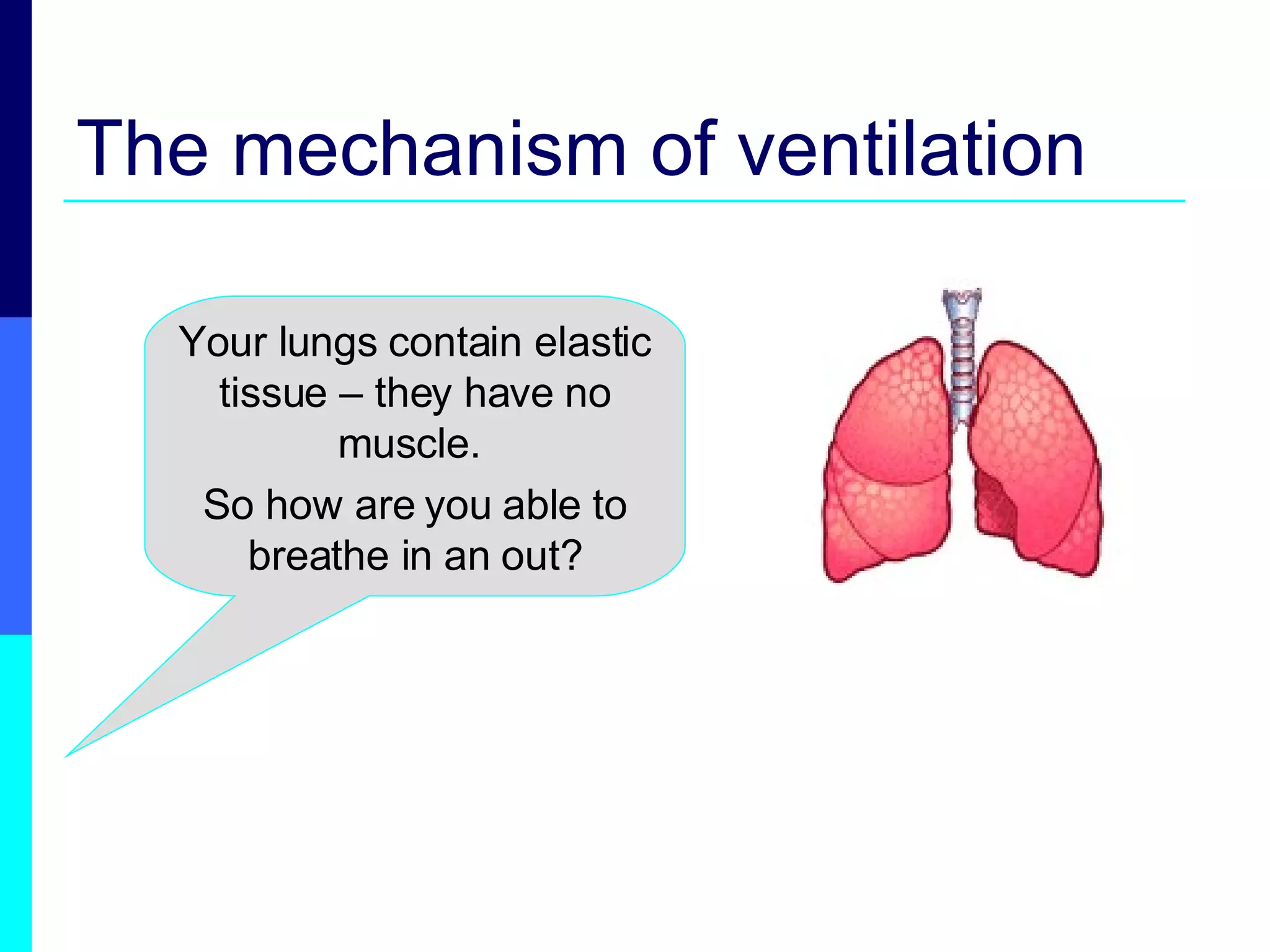
The mechanism ofventilation Your lungs contain elastic tissue – they have no muscle. So how are you able to breathe in an out?
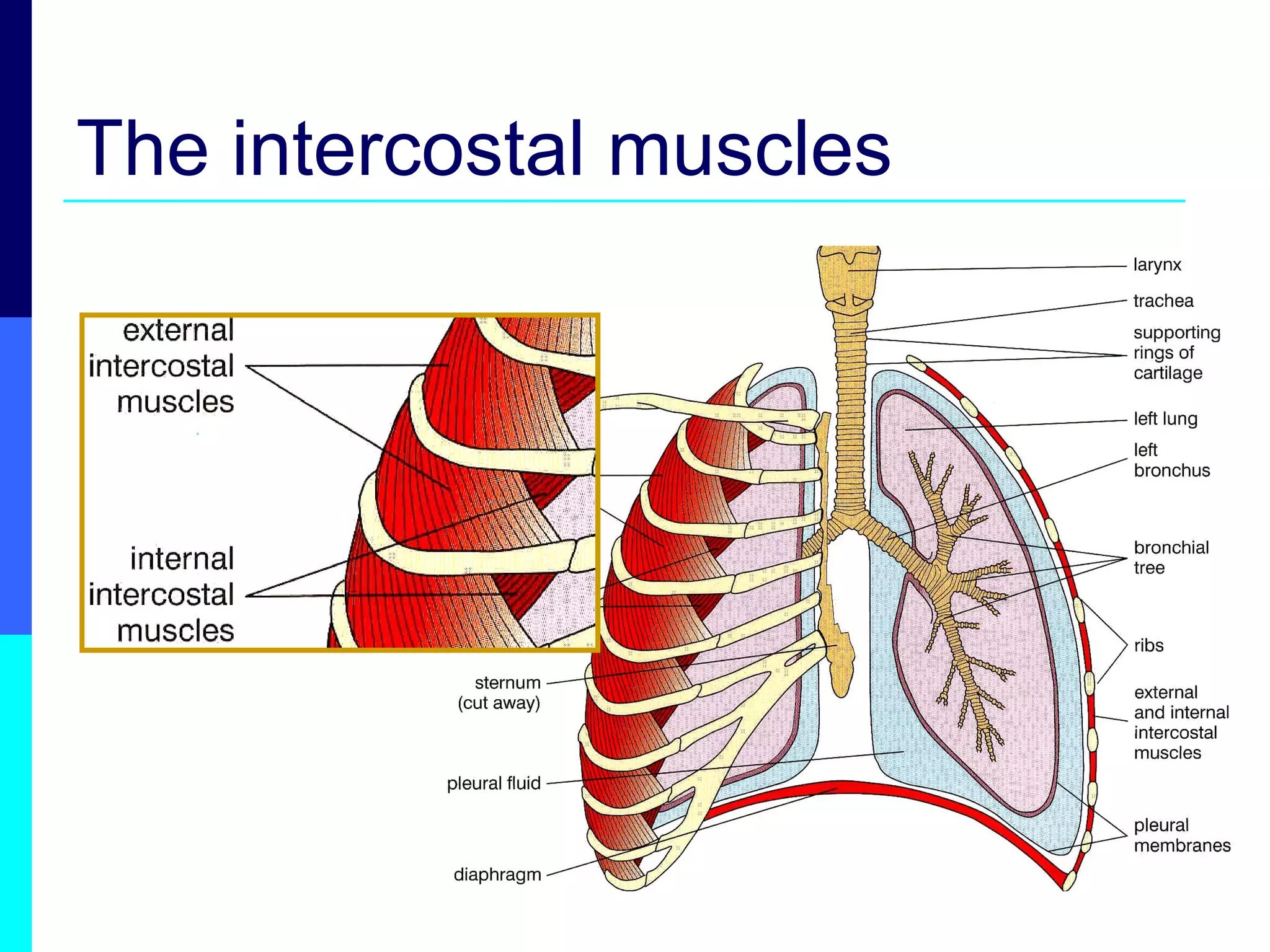
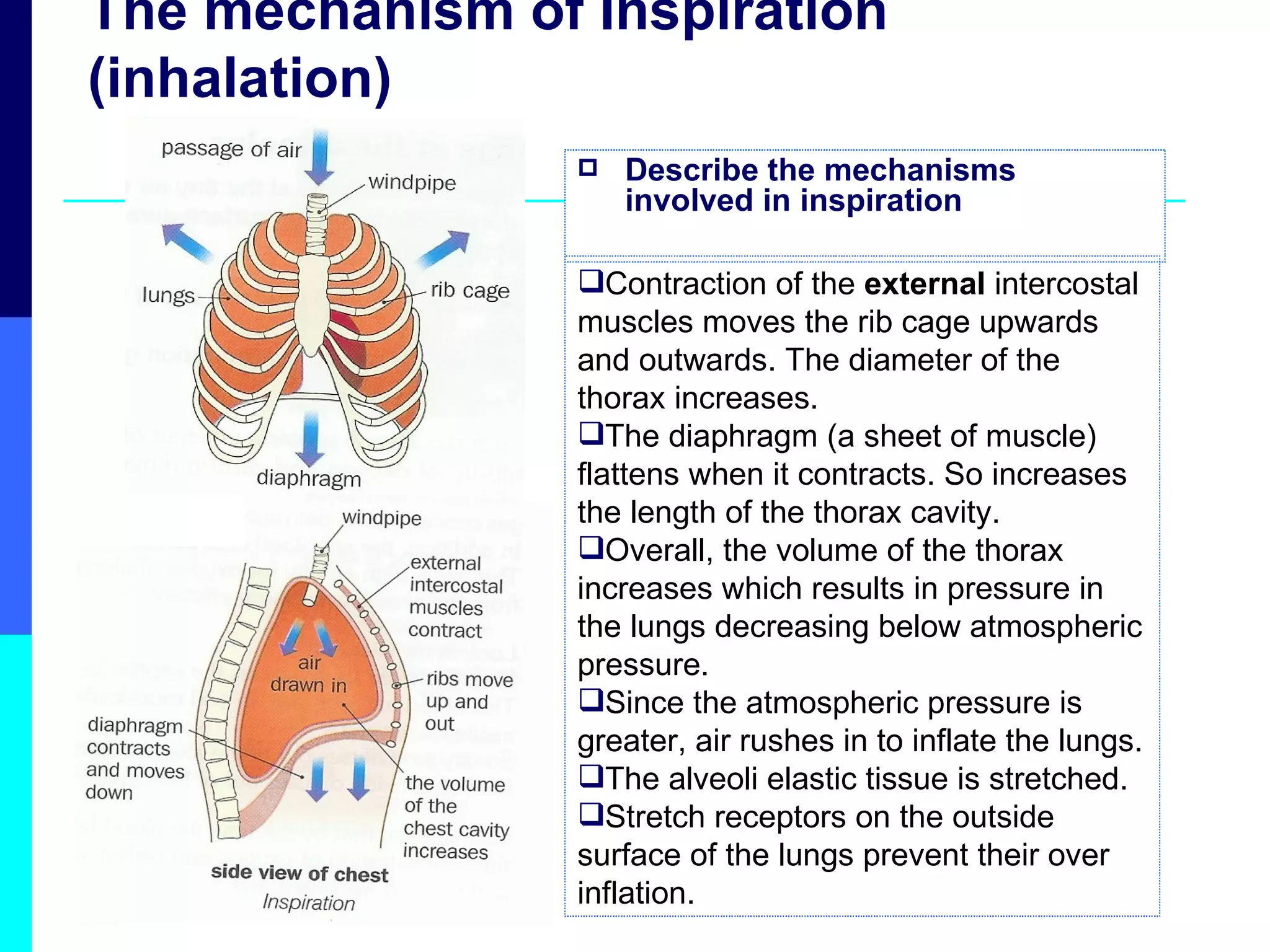
The mechanism ofInspiration (inhalation) Describe the mechanisms involved in inspiration Contraction of the external intercostal muscles moves the rib cage upwards and outwards. The diameter of the thorax increases. The diaphragm (a sheet of muscle) flattens when it contracts. So increases the length of the thorax cavity. Overall, the volume of the thorax increases which results in pressure in the lungs decreasing below atmospheric pressure. Since the atmospheric pressure is greater, air rushes in to inflate the lungs. The alveoli elastic tissue is stretched. Stretch receptors on the outside surface of the lungs prevent their over inflation.
6.
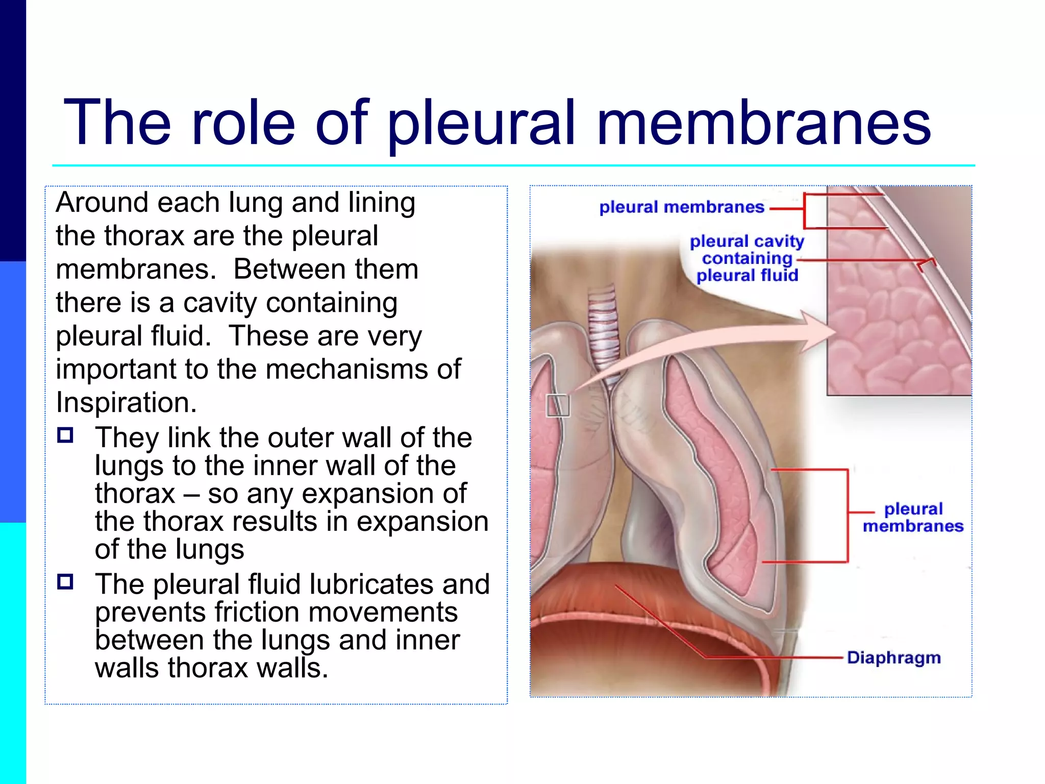
The role ofpleural membranes Around each lung and lining the thorax are the pleural membranes. Between them there is a cavity containing pleural fluid. These are very important to the mechanisms of Inspiration. They link the outer wall of the lungs to the inner wall of the thorax – so any expansion of the thorax results in expansion of the lungs The pleural fluid lubricates and prevents friction movements between the lungs and inner walls thorax walls.
7.
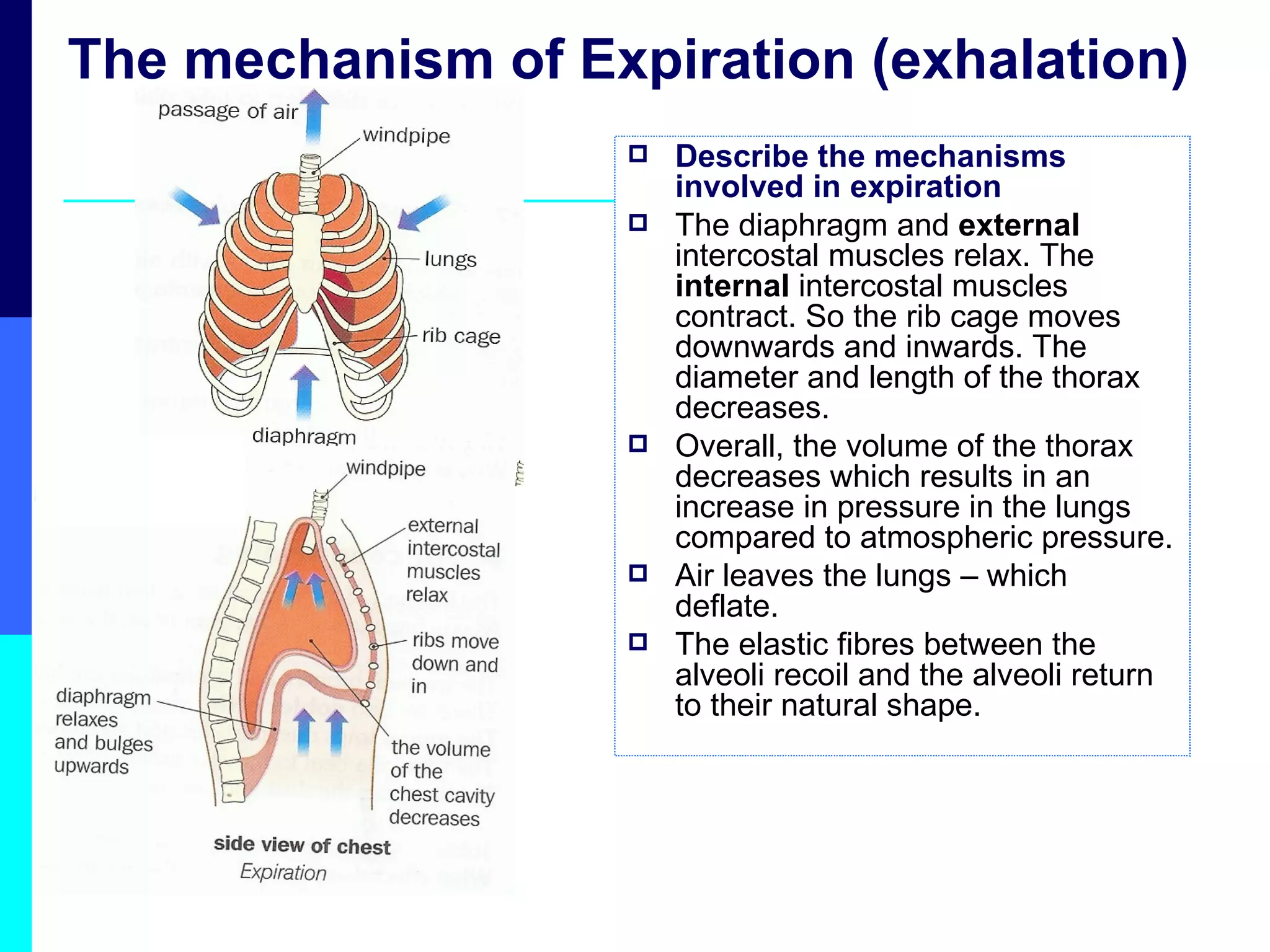
The mechanism ofExpiration (exhalation) Describe the mechanisms involved in expiration The diaphragm and external intercostal muscles relax. The internal intercostal muscles contract. So the rib cage moves downwards and inwards. The diameter and length of the thorax decreases. Overall, the volume of the thorax decreases which results in an increase in pressure in the lungs compared to atmospheric pressure. Air leaves the lungs – which deflate. The elastic fibres between the alveoli recoil and the alveoli return to their natural shape.
8.
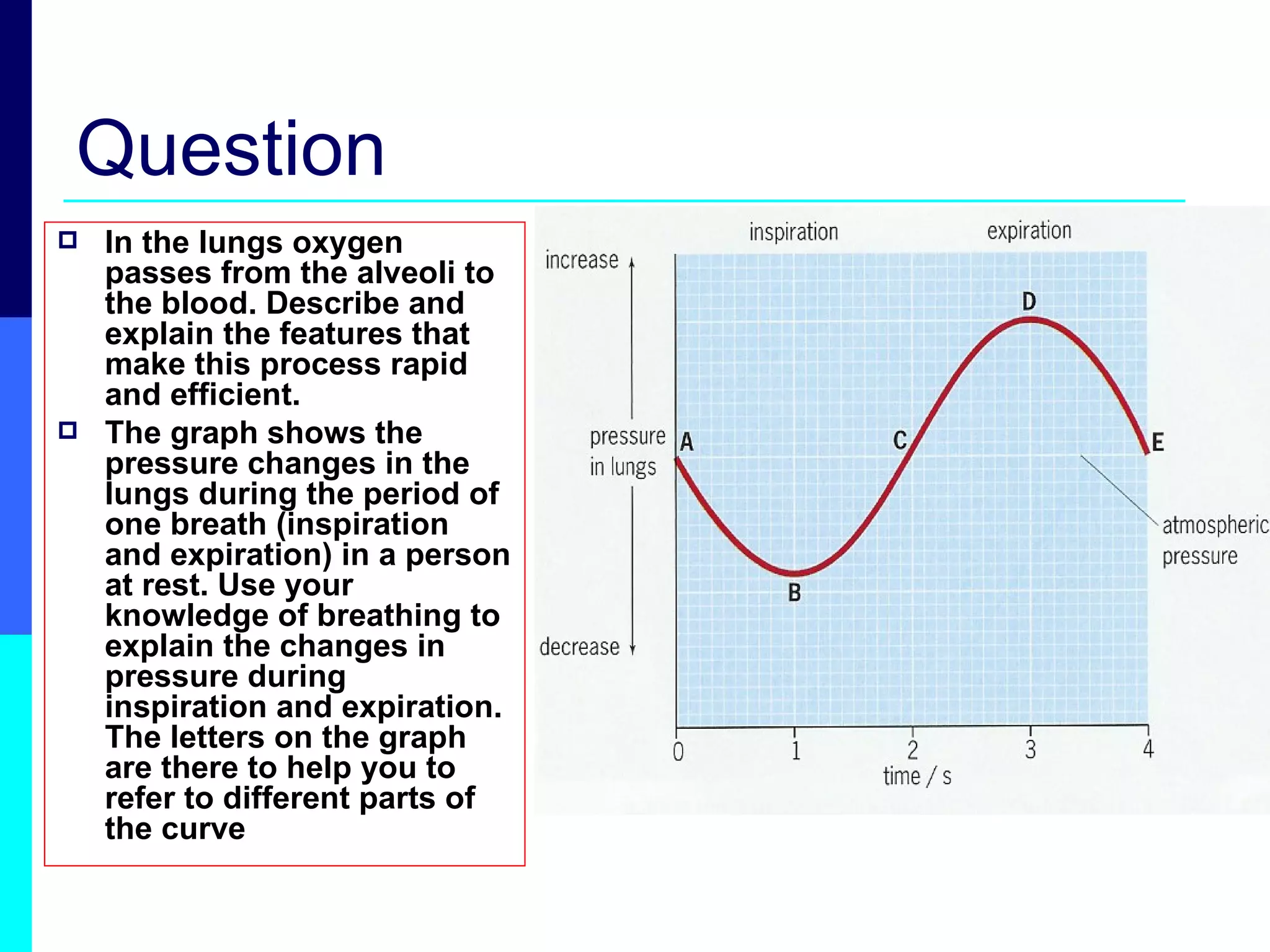
Question In thelungs oxygen passes from the alveoli to the blood. Describe and explain the features that make this process rapid and efficient. The graph shows the pressure changes in the lungs during the period of one breath (inspiration and expiration) in a person at rest. Use your knowledge of breathing to explain the changes in pressure during inspiration and expiration. The letters on the graph are there to help you to refer to different parts of the curve
9.
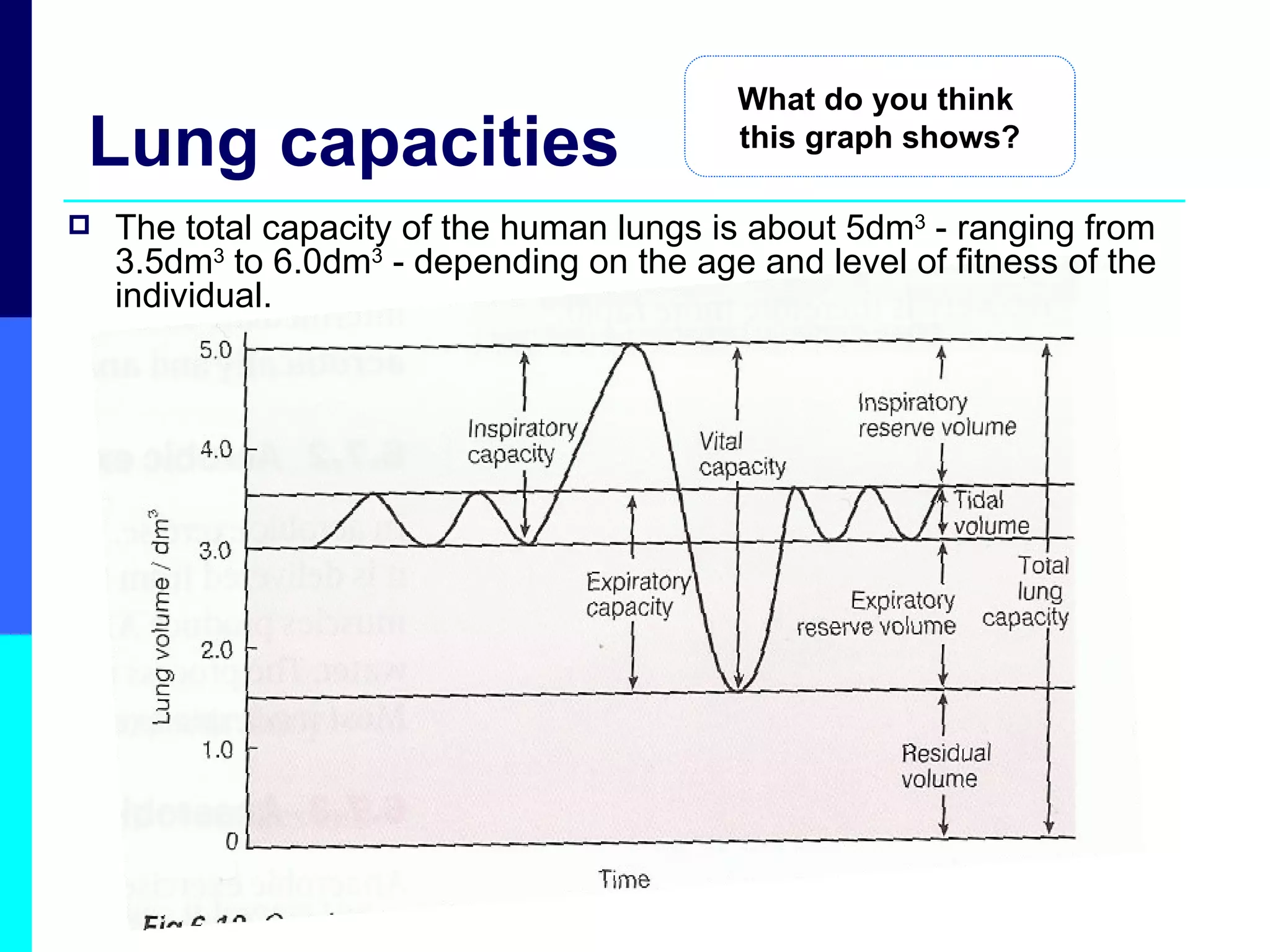
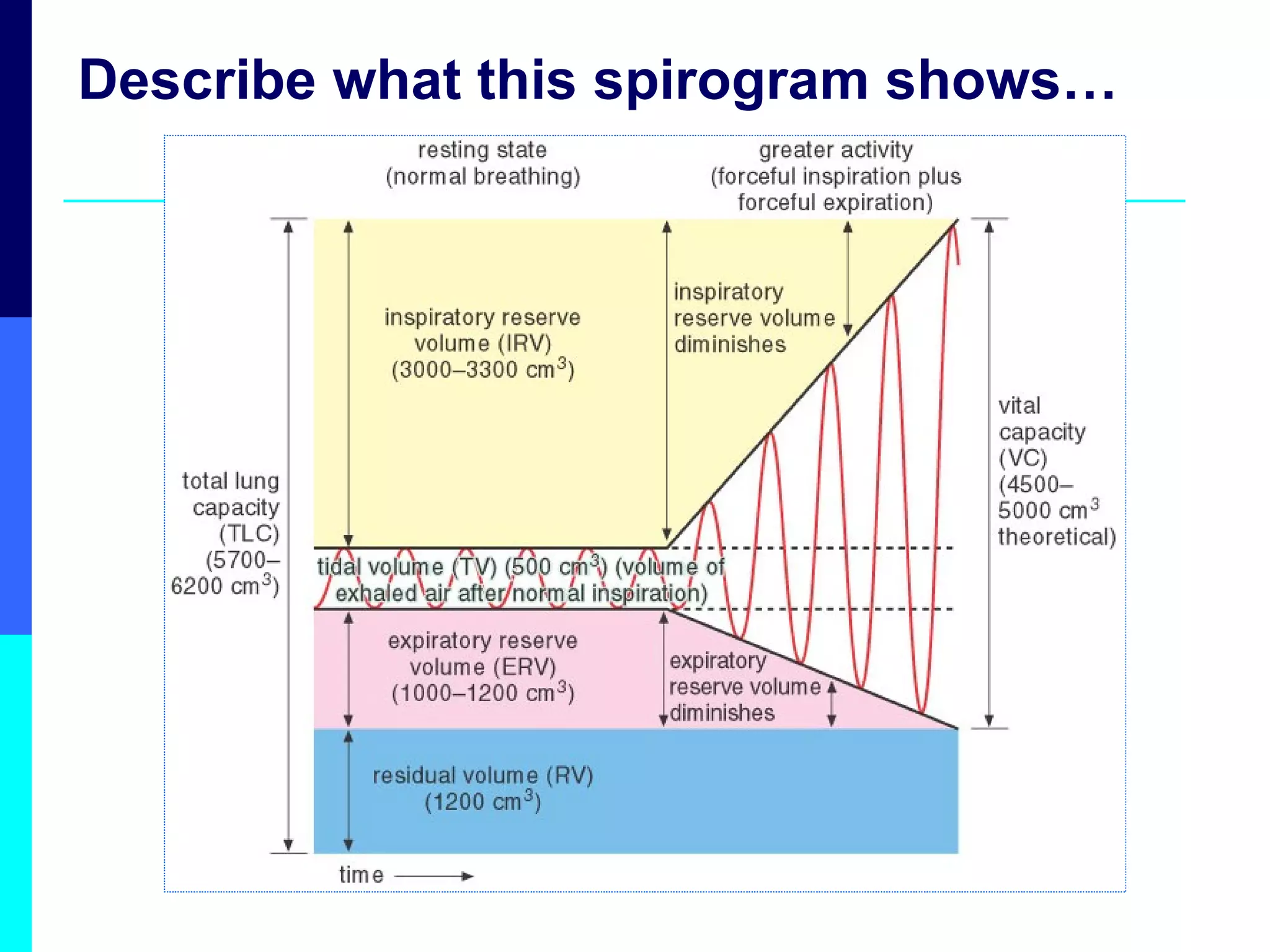
Lung capacities Thetotal capacity of the human lungs is about 5dm 3 - ranging from 3.5dm 3 to 6.0dm 3 - depending on the age and level of fitness of the individual. What do you think this graph shows?
10.
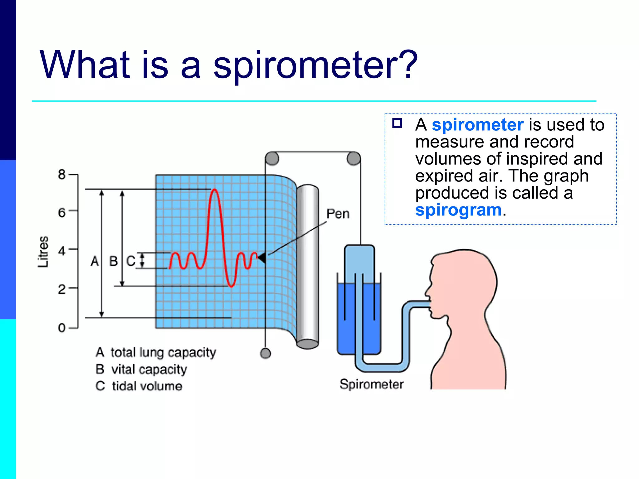
What is aspirometer? A spirometer is used to measure and record volumes of inspired and expired air. The graph produced is called a spirogram .
11.
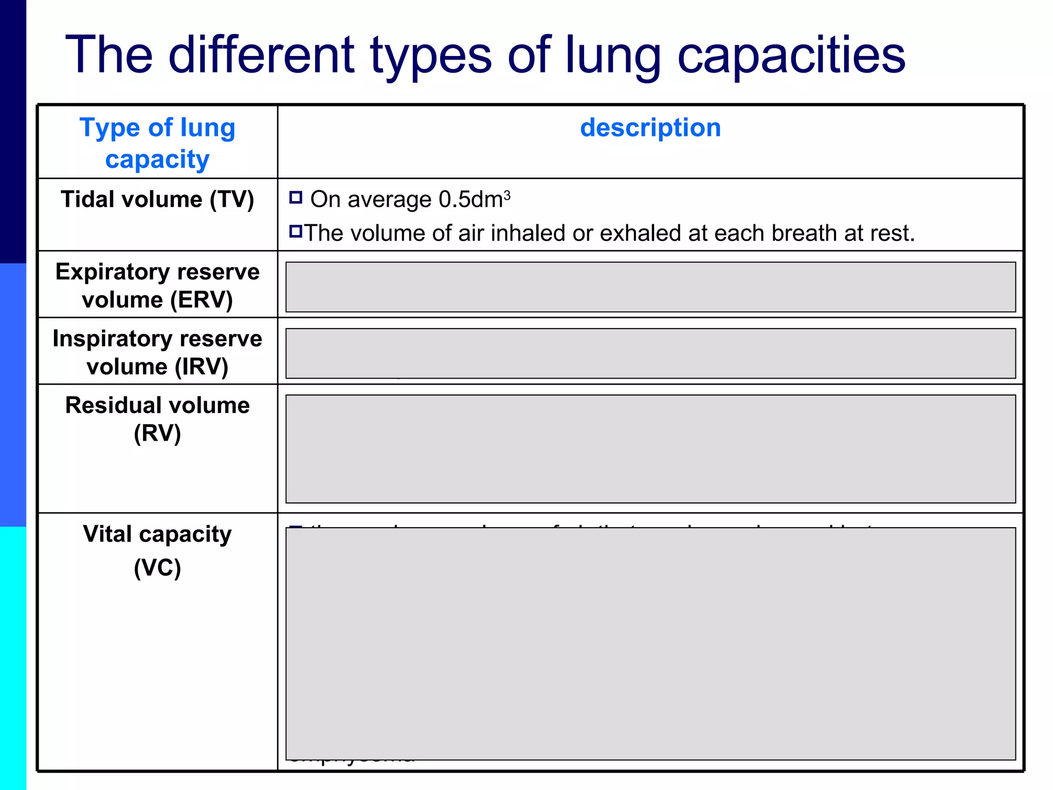
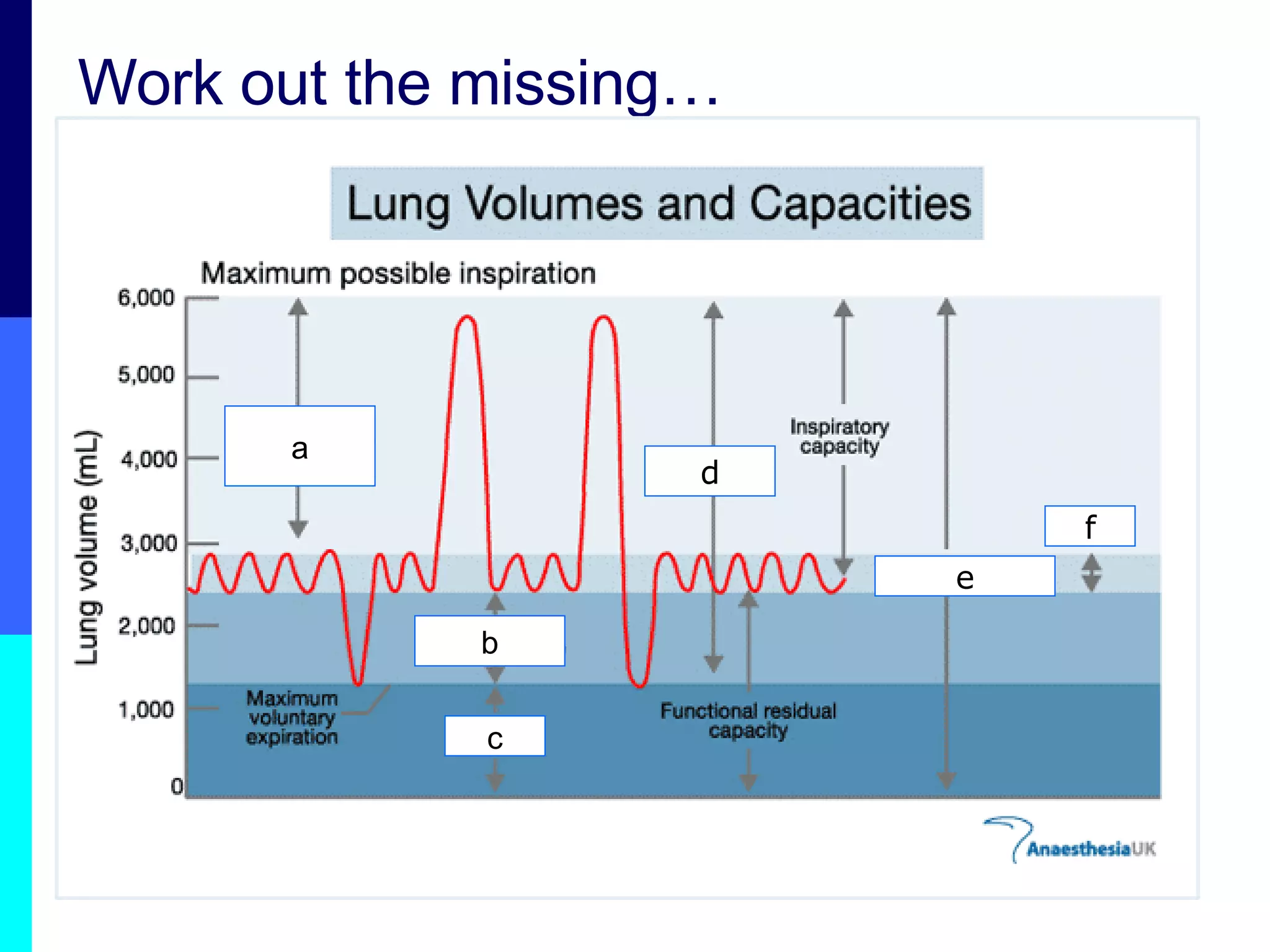
The different typesof lung capacities the maximum volume of air that can be exchanged between maximum inspiration and maximum expiration it’s the sum of IRV + TV + ERV is related to body size and on average is 2.6dm 3 for males and 2.1dm 3 for females. its higher in swimmers and divers Its lower in older people and people who have lung disease such as emphysema Vital capacity (VC) volume of air that cannot be removed and remains in the lungs at the end of a maximum expiration this air keeps the alveoli partly inflated and enables gas exchange to continue between breaths Residual volume (RV) volume breathed in by a maximum inspiration at the end of a normal inspiration Inspiratory reserve volume (IRV) volume breathed out by a maximum effort at the end of a normal expiration Expiratory reserve volume (ERV) On average 0.5dm 3 The volume of air inhaled or exhaled at each breath at rest. Tidal volume (TV) description Type of lung capacity
12.
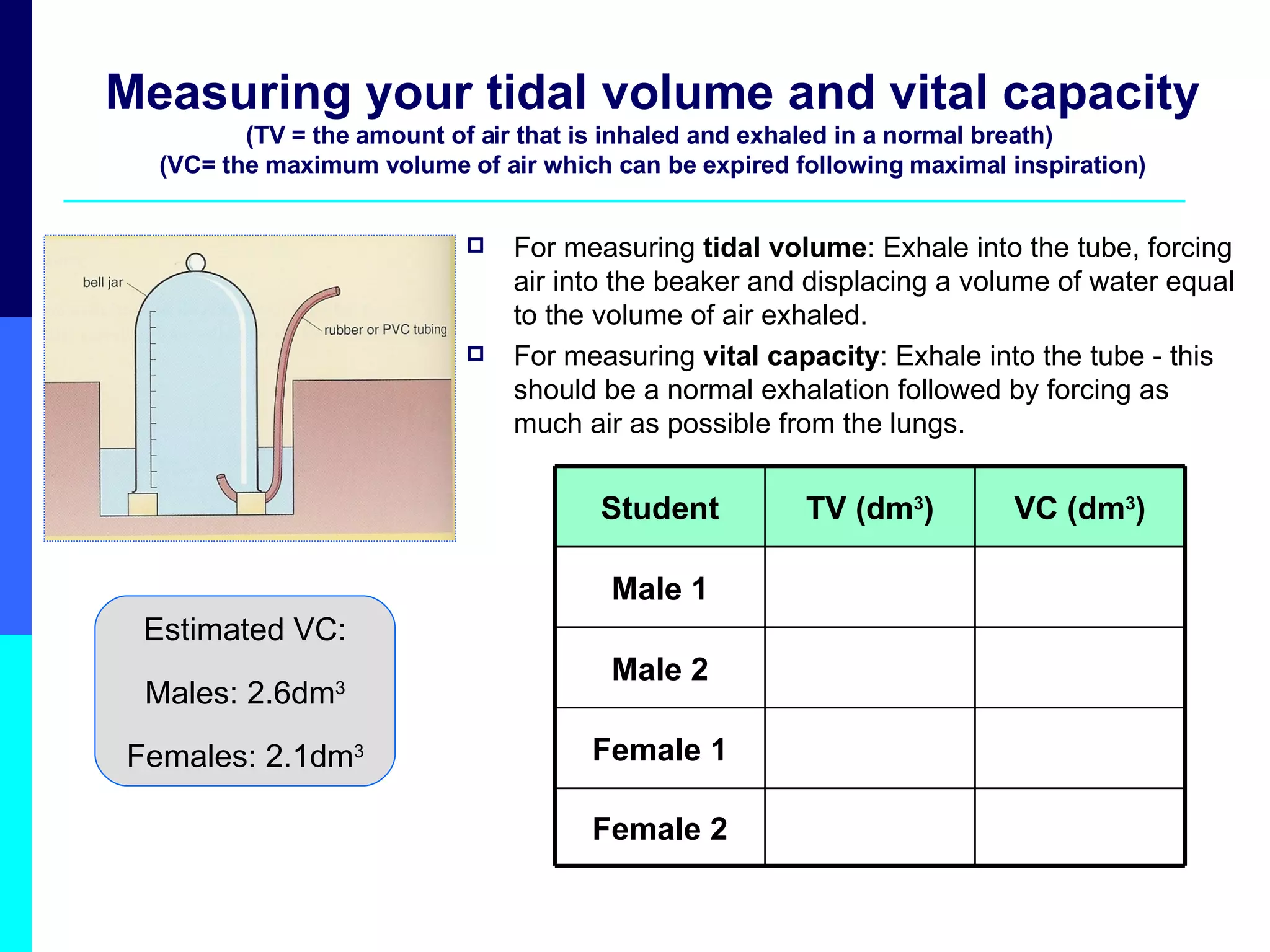
Measuring your tidalvolume and vital capacity (TV = the amount of air that is inhaled and exhaled in a normal breath) (VC= the maximum volume of air which can be expired following maximal inspiration) For measuring tidal volume : Exhale into the tube, forcing air into the beaker and displacing a volume of water equal to the volume of air exhaled. For measuring vital capacity : Exhale into the tube - this should be a normal exhalation followed by forcing as much air as possible from the lungs. . Estimated VC: Males: 2.6dm 3 Females: 2.1dm 3 Female 2 Female 1 Male 2 Male 1 VC (dm 3 ) TV (dm 3 ) Student
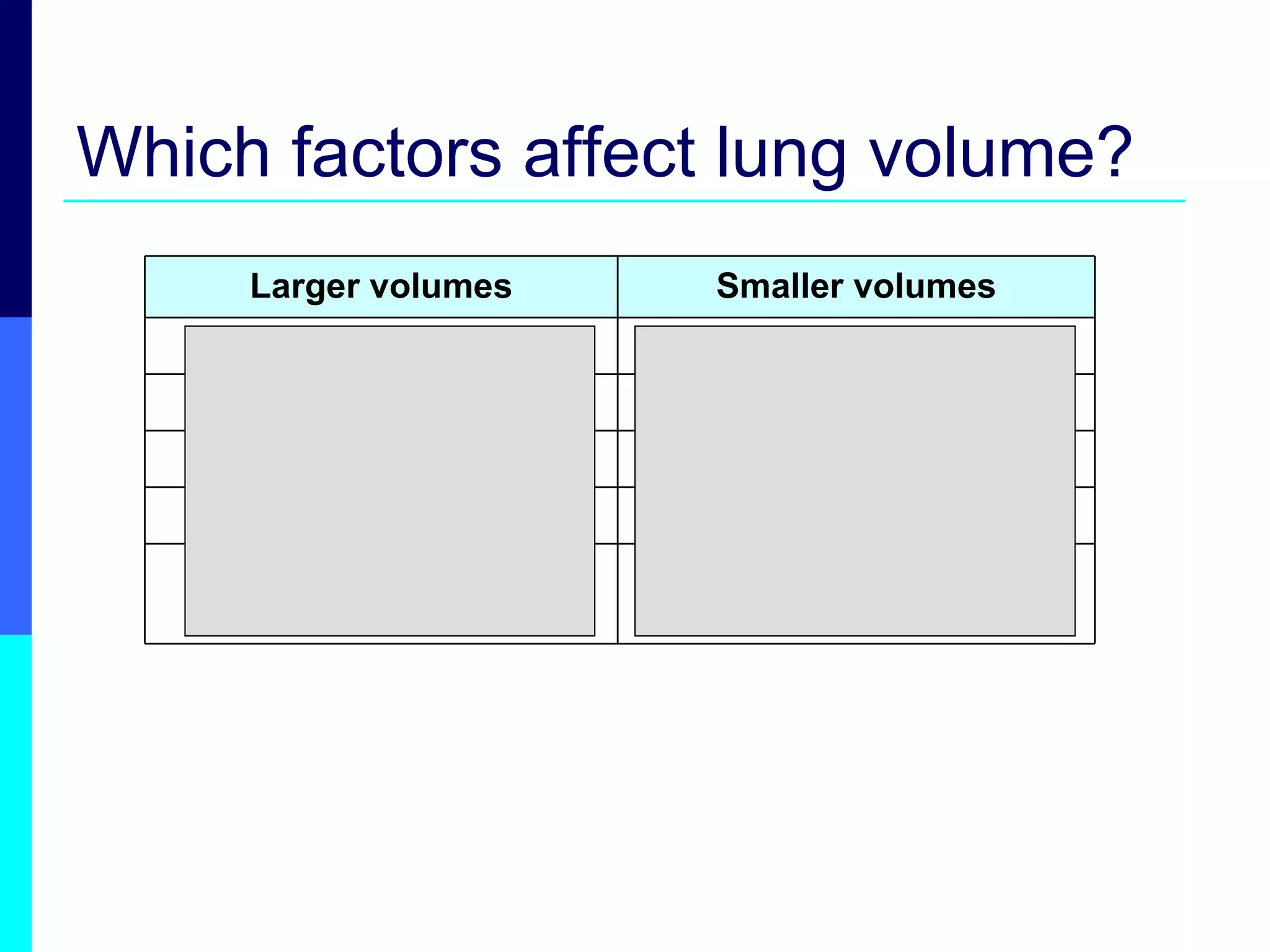
Which factors affectlung volume? people living at low altitudes people living at high altitudes non-athletes professional athletes heavy smokers Non - smokers shorter people tall people females males Smaller volumes Larger volumes
16.
Working out lungcapacities Find spirograms from different types of people
Questions 2. Duringhyperventilation, breathing is very rapid with deep inhalations and exhalations. How would a graph of hyperventilation compare to normal breathing? 3. Emphysema reduces the flexibility of alveolar walls in the lungs so that they are unable to recoil normally from expiration. This results in an increased residual volume. How would a spirogram of a person with emphysema compare to that of a healthy person, assuming the same total lung capacity? How would emphysema affect the vital capacity?
19.
Simple respirometers Q16.NAS p114: What is a simple respirometer? Diagram – labelled and explained What is the purpose of it? How does it work?