call girls in Kamla Market (DELHI) 🔝 >༒9953330565🔝 genuine Escort Service 🔝✔️✔️
Urinary system
1. URINARY SYSTEM
Assoc. Prof Dr. Karim Al-Jshamy
IMS/MSU 2010
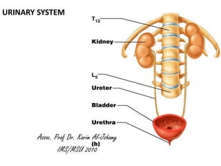
2. • The kidneys, ureters, urinary bladder and urethra are the main components
of the urinary system.
• A function of the urinary system that immediately comes to mind is the
excretion of waste products from the body. This is only one of many functions
of the system. Others are
• elimination of foreign substances
• regulation of the amount of water in the body
• control of the concentration of most compounds in the extracellular fluid
• Most of these tasks are performed in the kidneys. Functionally the processes
can be divided into two steps, each of which have their anatomical correlate:
• filtration - glomeruli of the kidney
• selective resorption and excretion - tubular system of the kidney
• In addition, the kidney also functions as an endocrine organ. Fibrocytes in the
cortex release the hormone erythropoietin, which stimulates the formation of
red blood cells.
• Modified fibrocytes of the medulla secrete prostaglandins which are able to
decrease blood pressure.
3. Overall Organization of the Kidney
Stroma
Capsule
Dense FECT
Myofibroblast layer
Interstitial stroma (loose FECT)
Parenchyma
Nephrons
Collecting ducts
Vascular components
Organized into
cortex and medulla
4.
5.
6. Kidney the tubular system
Glomeruli and
are both part of the basic functional
unit of the kidney, the nephron.
The Glomerulus (or renal corpuscle)
The glomerulus is the round (~0.2
mm in diameter) blind beginning of
the nephron. It is invaginated by a
tuft of capillaries at the vascular
pole of the glomerulus.
The tuft of capillaries and other
cells in contact with them form the
anatomical glomerulus. Glomerulus.
The anatomical glomerulus is enclosed by two layers of epithelium, Bowman's
capsule. Cells of the outer or parietal layer of Bowman's capsule form a simple
squamous epithelium.
Cells of the inner layer, podocytes in the visceral layer, are extremely complex in
shape. Small foot-like processes, pedicles, of their cytoplasm form a fenestrated
epithelium around the fenestrated capillaries of the glomerulus.
7. • The openings between the pedicles are
called filtration slits. They are spanned
by a thin membrane, the filtration slit
membrane.
• Between the podocytes and the
endothelial cells of the capillaries we
find a comparatively thick basal lamina,
which can be subdivided into an outer
lamina rara externa, a middle lamina
densa and an inner lamina rara interna.
The basal lamina and the slit
membranes form the glomerular
filtration barrier,
• Mesangial cells in the glomerulus form
the connective tissue that gives
structural support to podocytes and
vessels.
Blood pressure is the driving force in the formation of about 125 ml of
glomerular filtrate per minute.
About 124 ml of the glomerular filtrate is reabsorbed in the tubules of the
nephron.
8. Kidney
Capsule as thin membrane of
connective tissue, cortex of the kidney
and scan over the tissue, presence of
glomeruli (convoluted parts of
proximal and distal tubuli).
• to identify the vascular pole of a good
glomerulus by the attachment of the
capillary tuft to the wall of the
glomerulus.
• The nuclei are located side by side or
may even overlap.
• Proximal tubules are characterised by
their eosinophilic (pink) low columnar
cells and by large amounts of fuzzy
material, which may fill the entire
lumen of the tubulus.
9. The anatomical glomerulus,
the parietal blade of
Bowman's capsule
(squamous cells),
podocytes (fairly large and
light nuclei ),
endothelial cells (smaller and
darker nuclei), vascular pole.
10.
11. Tubules of the Nephron
• The tubular system can be divided into
proximal and distal tubules, which in turn
have convoluted and straight portions.
• Intermediate tubules connect the proximal
and distal tubules. Running from the cortex
of the kidney towards the medulla
(descending), then turning and running back
towards the cortex (ascending), the tubules
form the loop of Henle.
• The proximal tubule is the longest section of
the nephron (about 14 mm).
• The convoluted part of the proximal tubules
coils close to the glomerulus in the cortex.
• The proximal tubules are formed by a low
columnar epithelium. The eosinophilic cells
of the epithelium have a wide brush border
(long microvilli) and are active in
endocytosis.
12. • They almost completely resorb substances
of nutritional value from the glomerular
filtrate (glucose, amino acids, protein,
vitamins etc.)
• In the proximal tubules the volume of the
glomerular filtrate is reduced by about 75%.
Sodium ions are actively resorbed from the
glomerular filtrate.
• hey are followed by passively diffusing
chloride ions and the osmotic absorption of
water. The straight portion of the proximal
tubule descends towards the medulla.
• The straight portion of the proximal tubule
merges with the intermediate tubule (thin
segment of the loop of Henle).
• A flattened, only ~1-2 µm high epithelium
forms the intermediate tubule, which is only
~15 µm wide. Descending parts of the
straight proximal and intermediate tubules
are permeable to water but not to solutes.
13. Kidney
The medulla of the kidney, there
is a collecting ducts (cuboidal to
columnar cells, well-defined
boundaries between cells,
cytoplasm only weakly stained or
unstained, large ducts)
An intermediate (very flat
epithelium, nuclei bulge into the
lumen of the tubulus, diameter of
the duct is small) and distal tubule
(cuboidal epithelium, cells stain
weakly pink).
14. The Juxtaglomerular Apparatus
• The distal tubule contacts the
glomerulus forming a
specialized section of tubular
epithelium, the macula densa.
• At the point of contact with the
glomerulus, the distal tubule is
always in close contact with the
efferent and afferent arterioles
of the glomerulus.
• The juxtaglomerular (JG)
apparatus are extraglomerular
mesangial cells and the
juxtaglomerular cells
surrounding the afferent
arteriole (modified smooth
muscle cells), which produce
and secrete renin.
15. • URETER
• The urine flows through these
structures to the ureter and is
channelled to the bladder.
• The mucosa is lined with a transitional
epithelium , which occurs exclusively
in the urinary system.
• The lamina propria consists mainly of
dense connective tissue, with many
bundles of coarse collagenous fibres.
• The muscularis usually consists of an
inner longitudinal and outer circular
layer of smooth muscle cells .
• In lower parts of the ureter and the
bladder an additional outer
longitudinal layer of muscles is added
to the first two.
21. BLADDER
Histology of Bladder: mucosa of transitional epithelium, Submucosa,
and thick muscular layer know as the detrusor muscle
These are retroperitoneal structures. They enter the bladder at an oblique angle which
helps to prevent backflow of urine. Smooth muscle in the wall of the ureters rhythmically
contracts (peristalsis) to move urine into the bladder
24. The Urethra
• Initially, the urethra is lined by a
transitional epithelium in males and
females.
• In males, it is replaced by a
pseudostratified or stratified columnar
epithelium below the openings of the
ejaculatory ducts into the urethra.
• The distal parts of the female urethra
and the distal end of the male urethra
are lined by a stratified squamous
epithelium.
• The lamina propria contains loose
connective tissue. Smooth muscle cells
in the muscularis are mainly oriented
longitudinally.
• They are surrounded, in the middle
part of the urethra (below the prostate
in males), by striated muscle cells of
the sphincter urethrae.