Downloaded 587 times
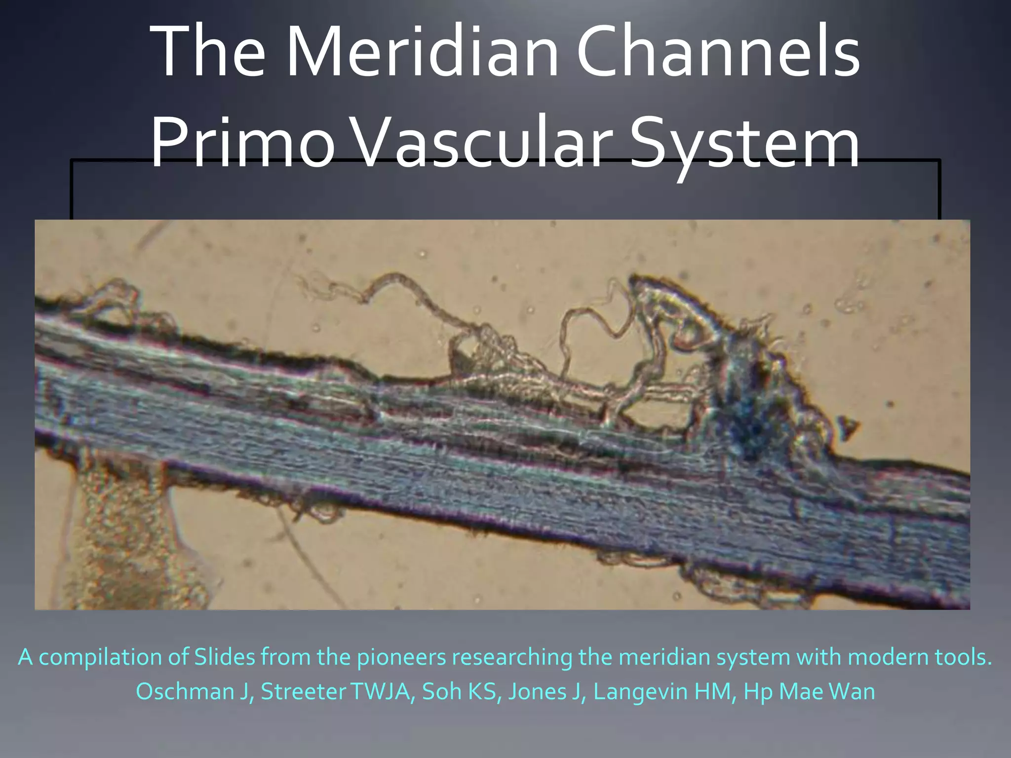
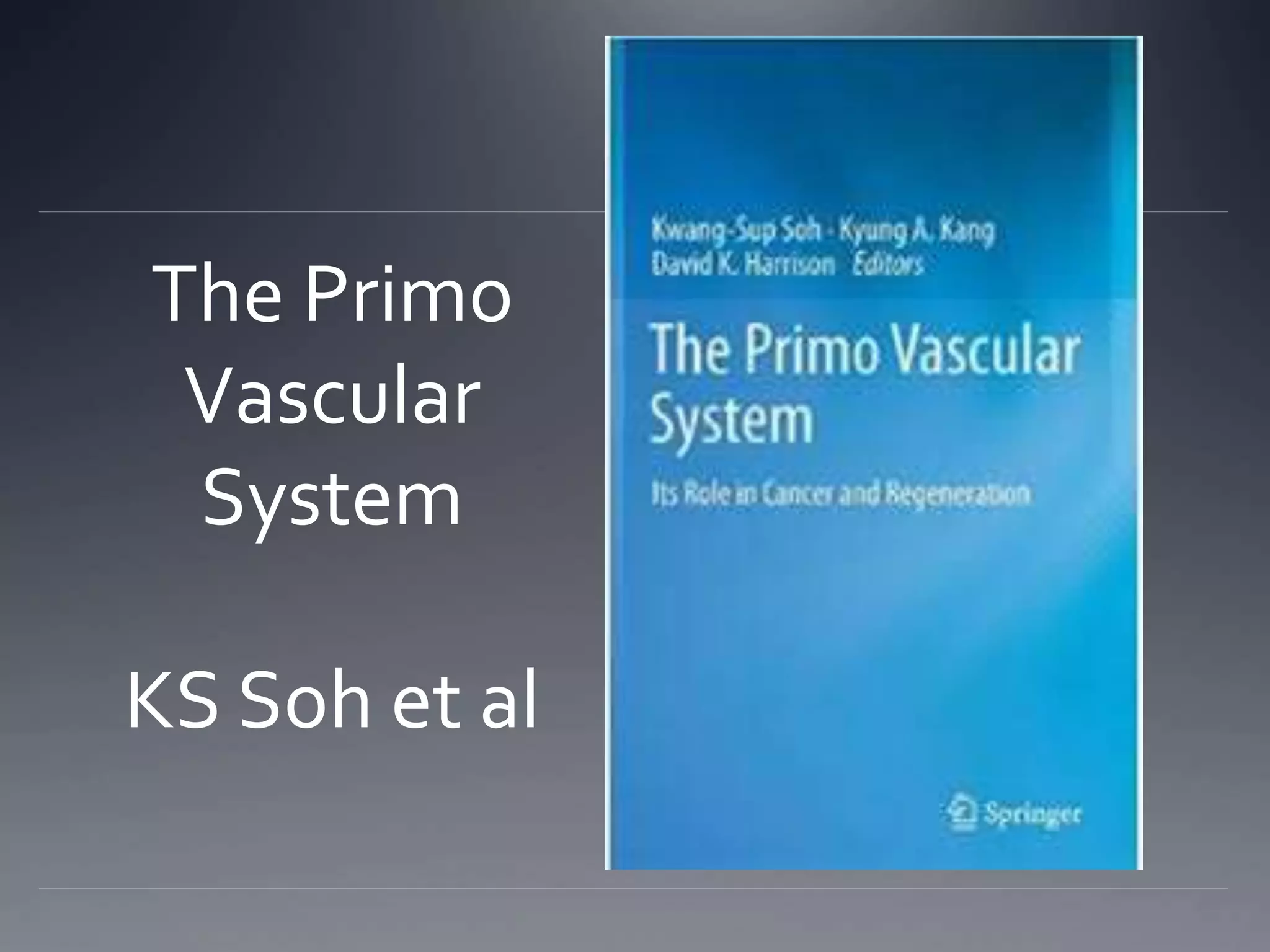
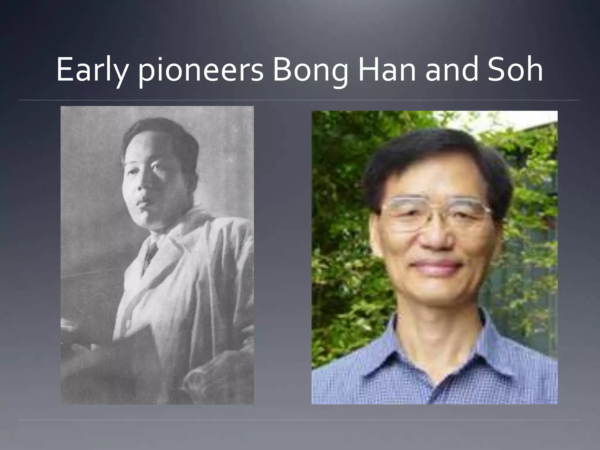
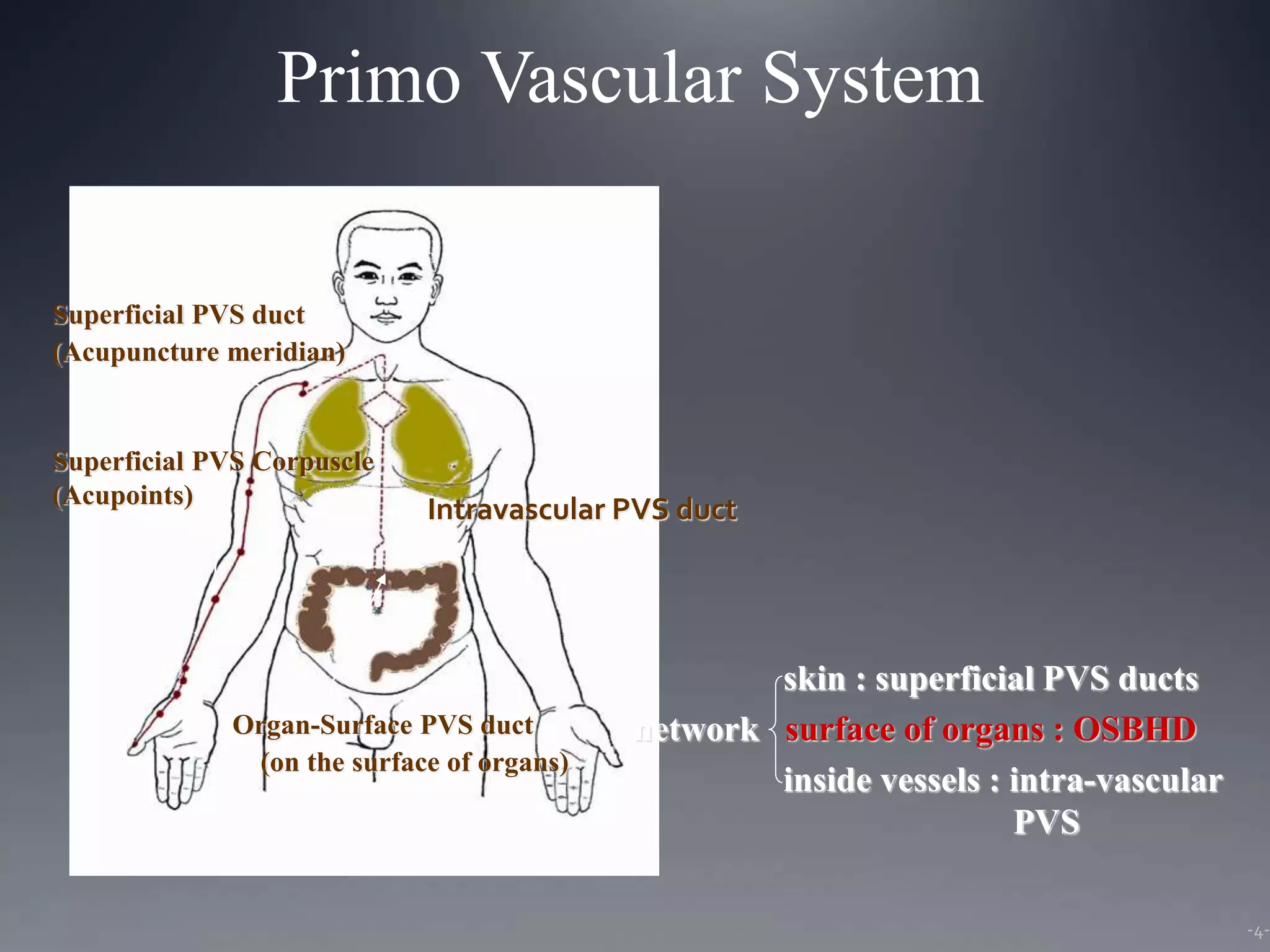
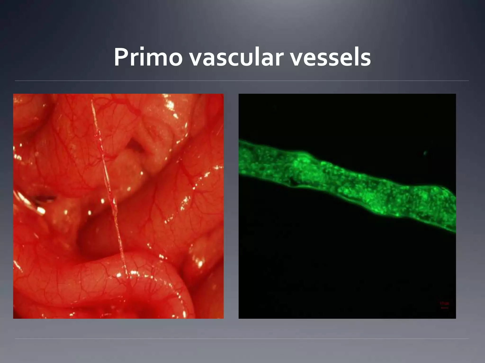
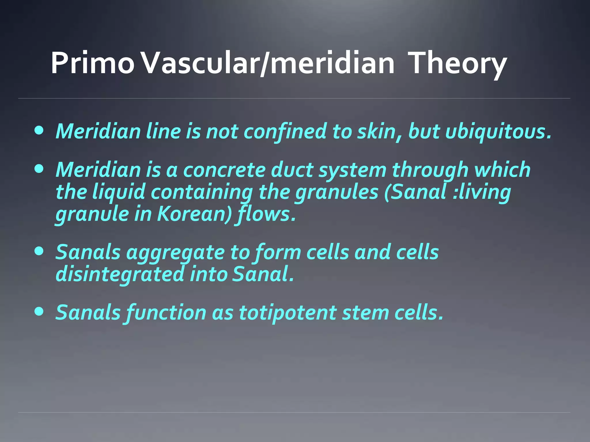
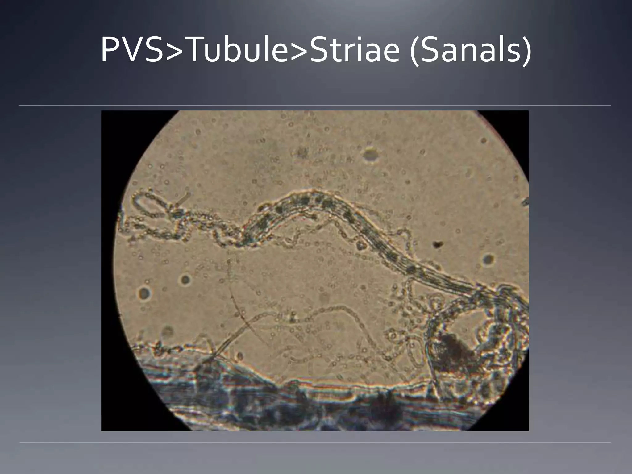
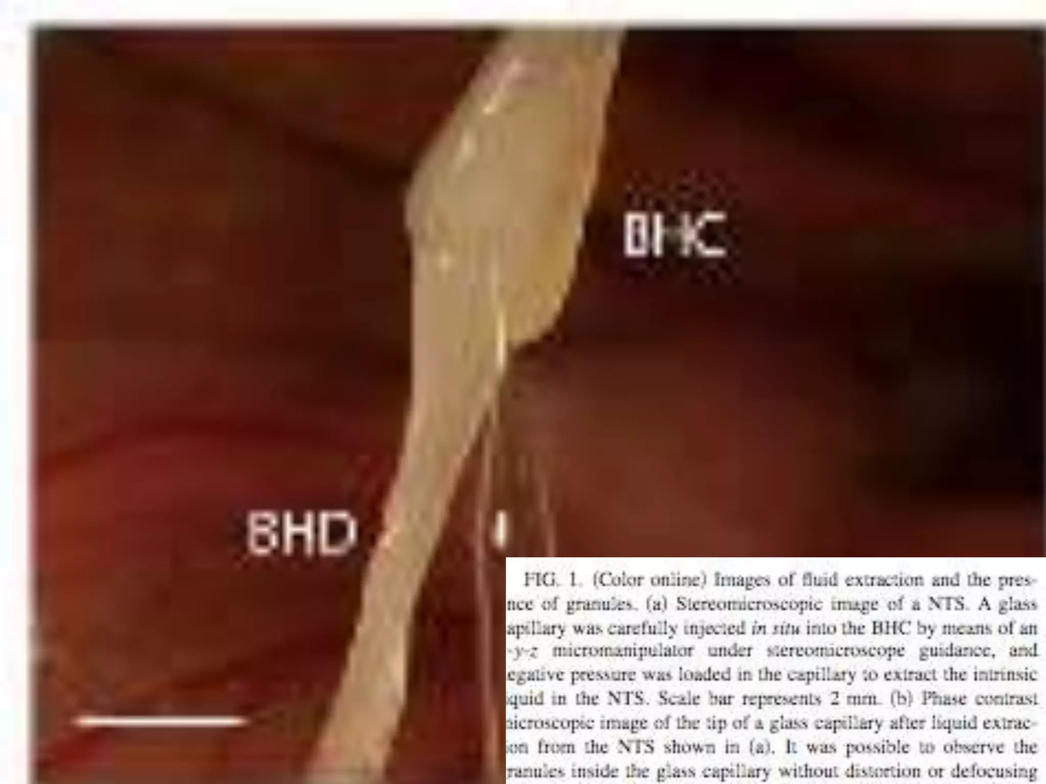
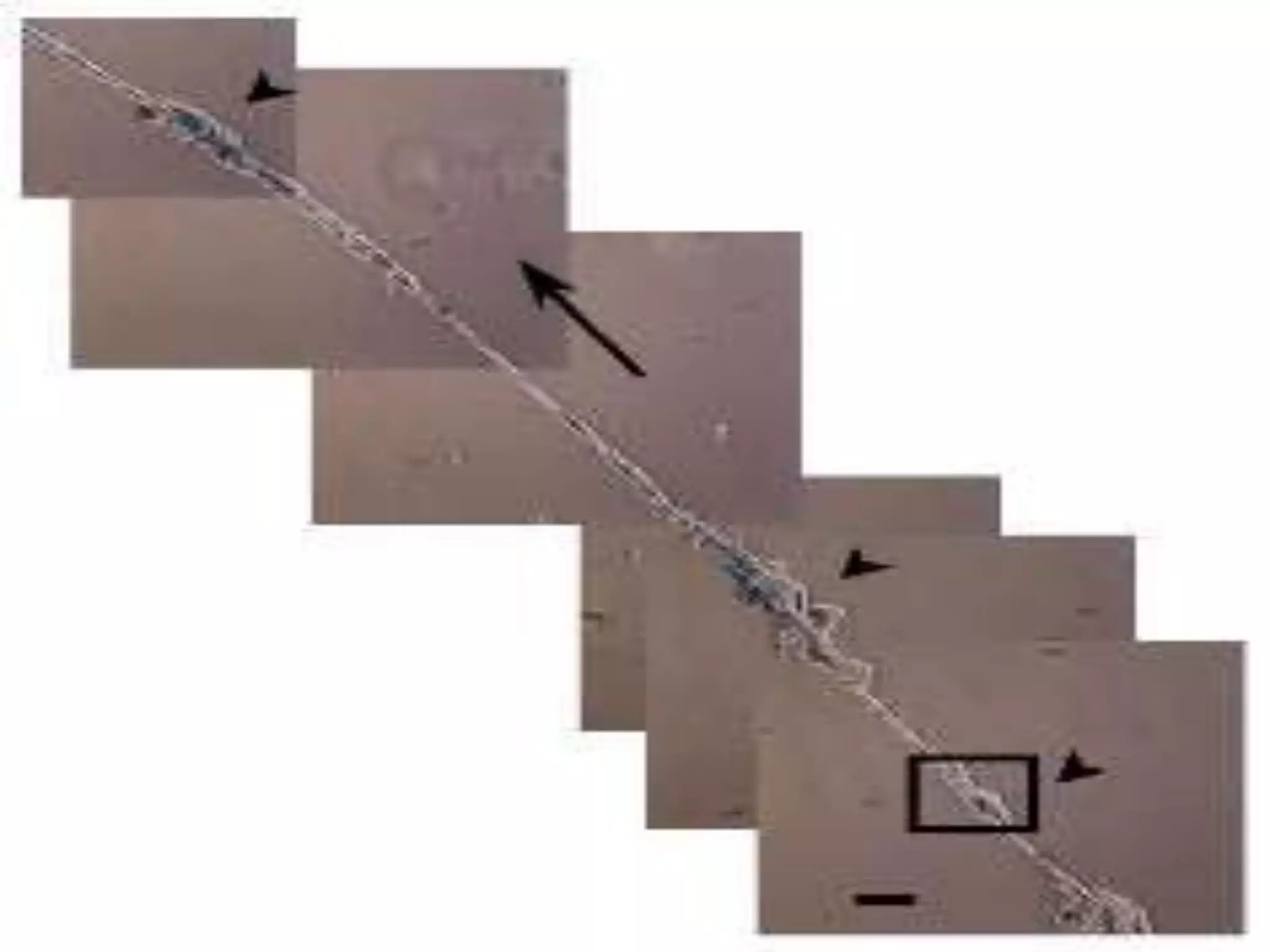
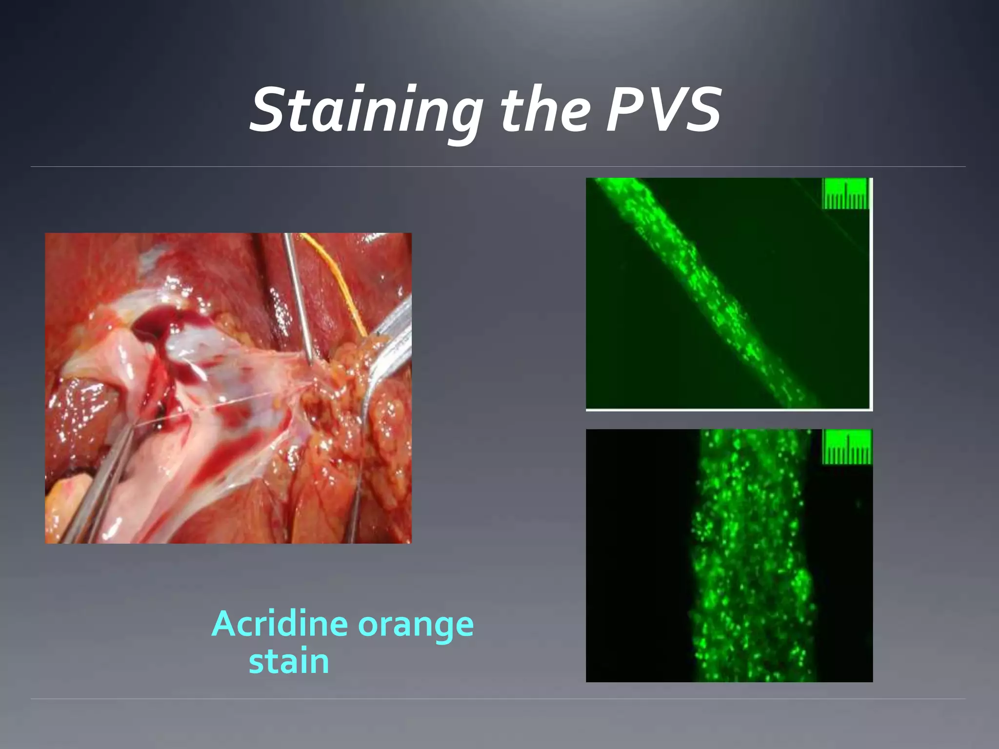
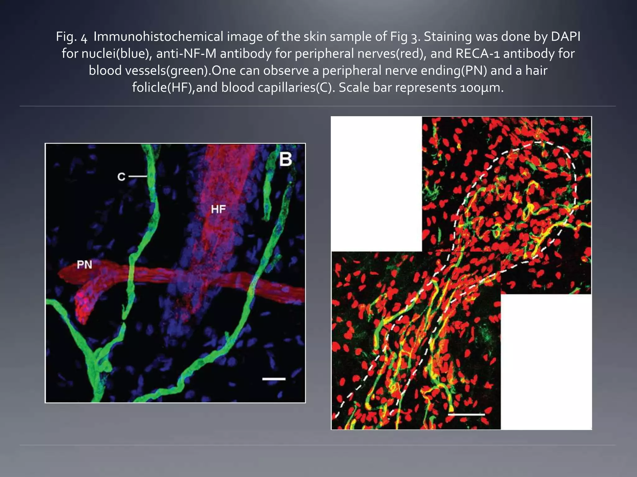
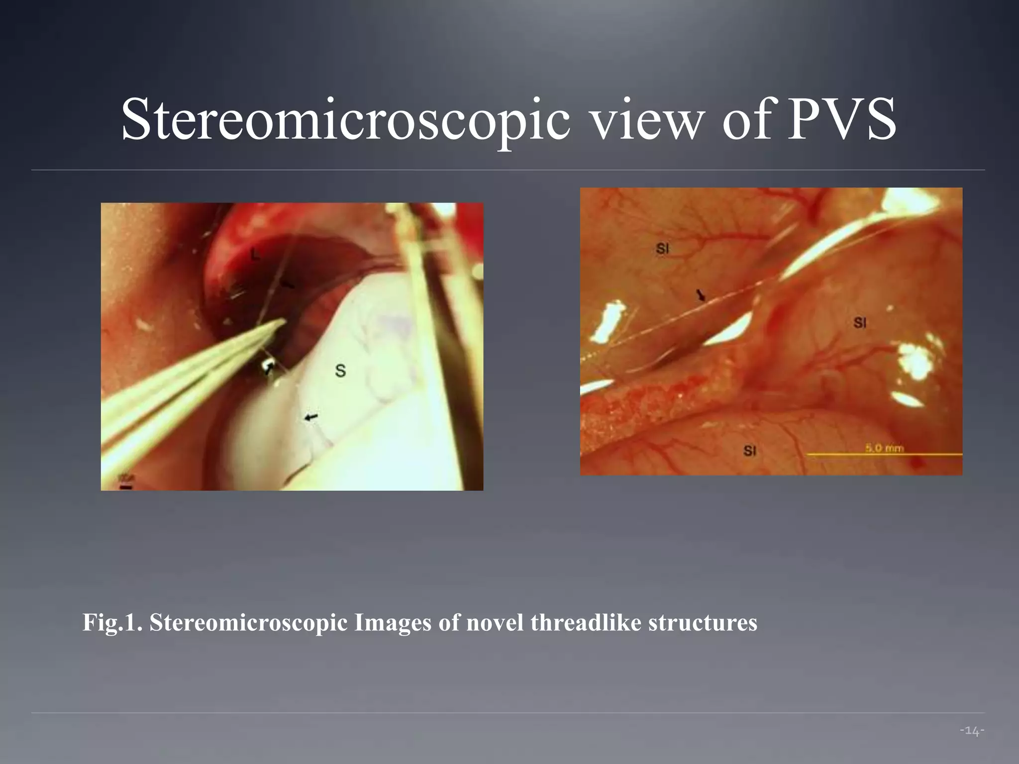
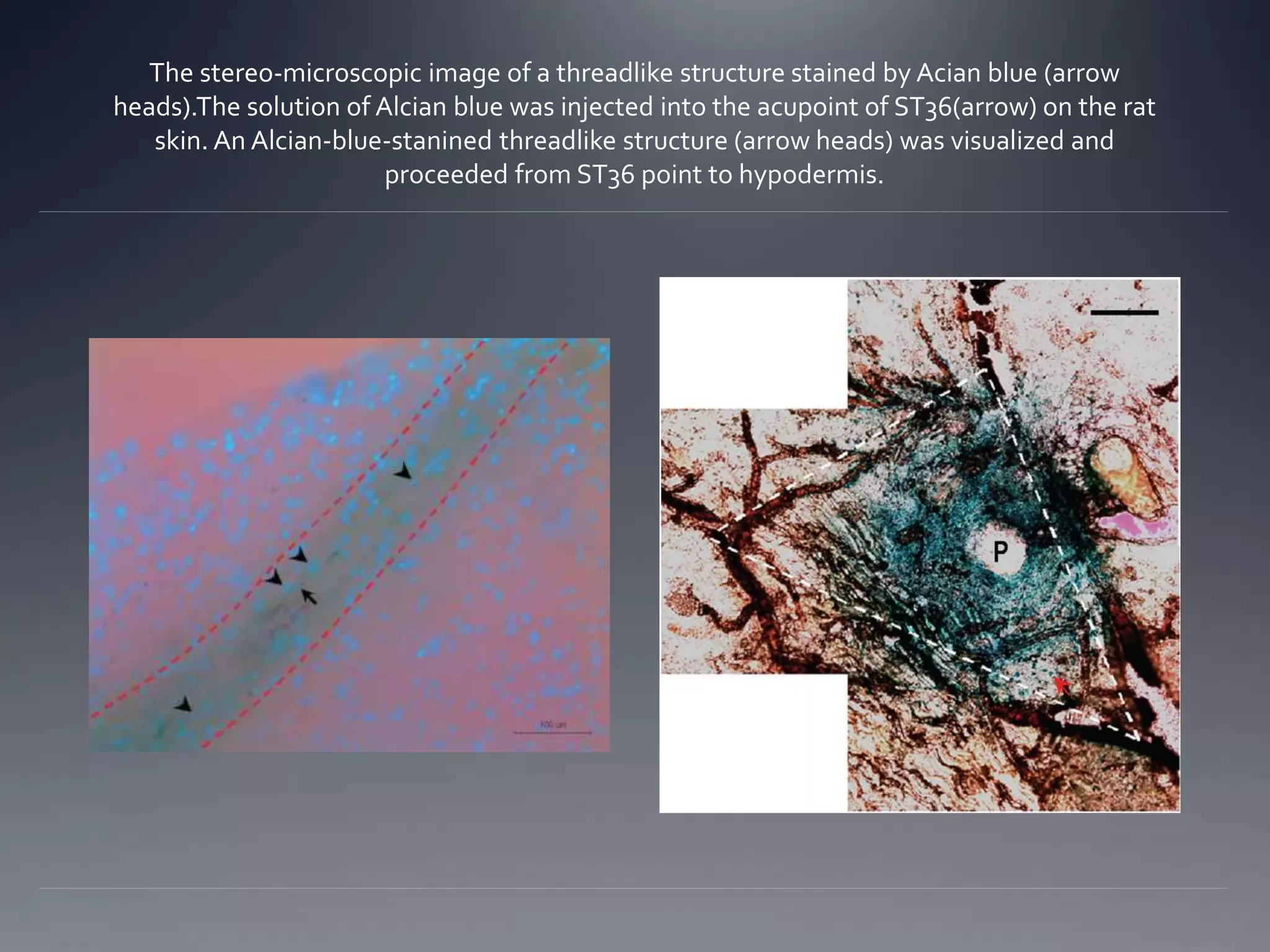
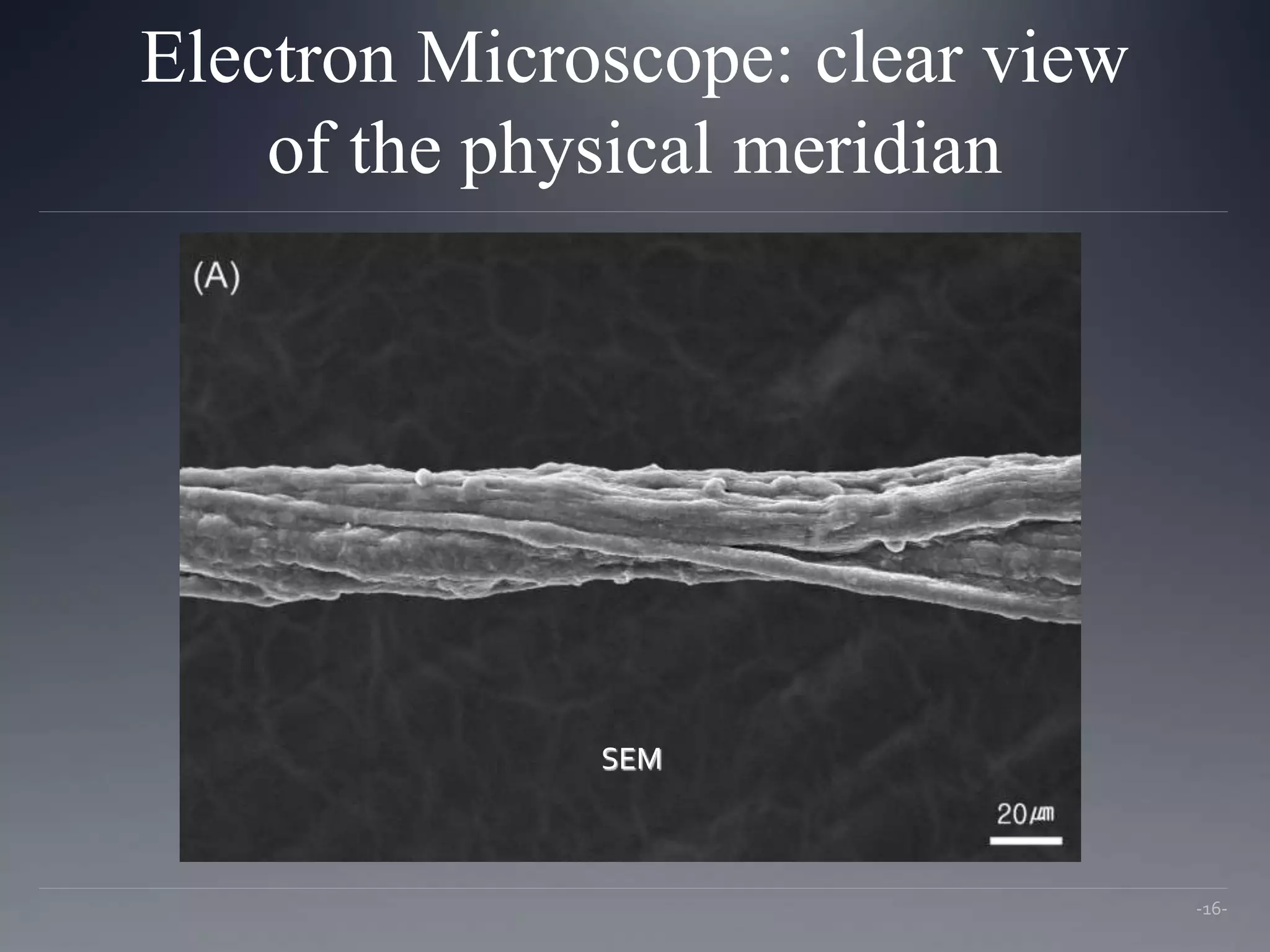
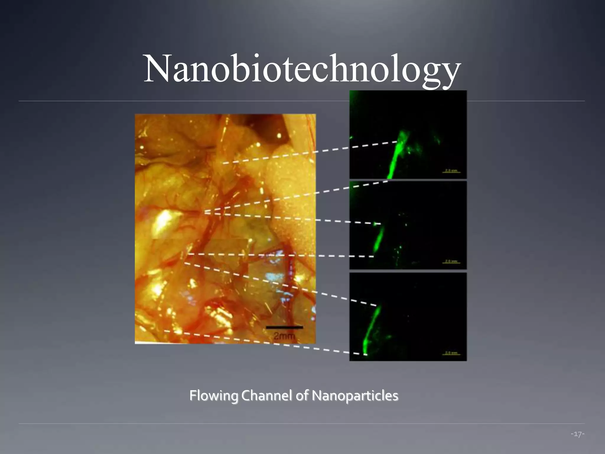
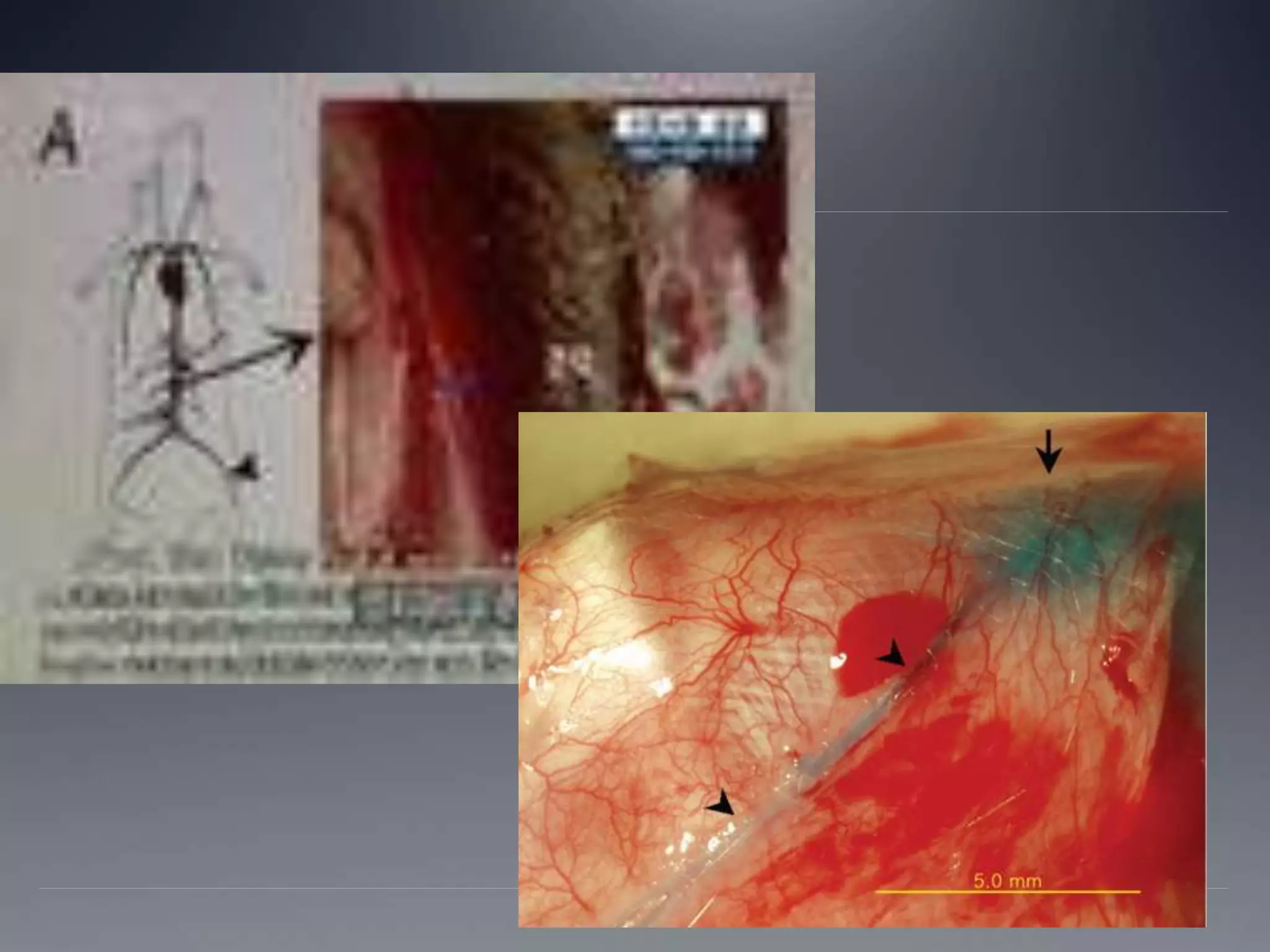


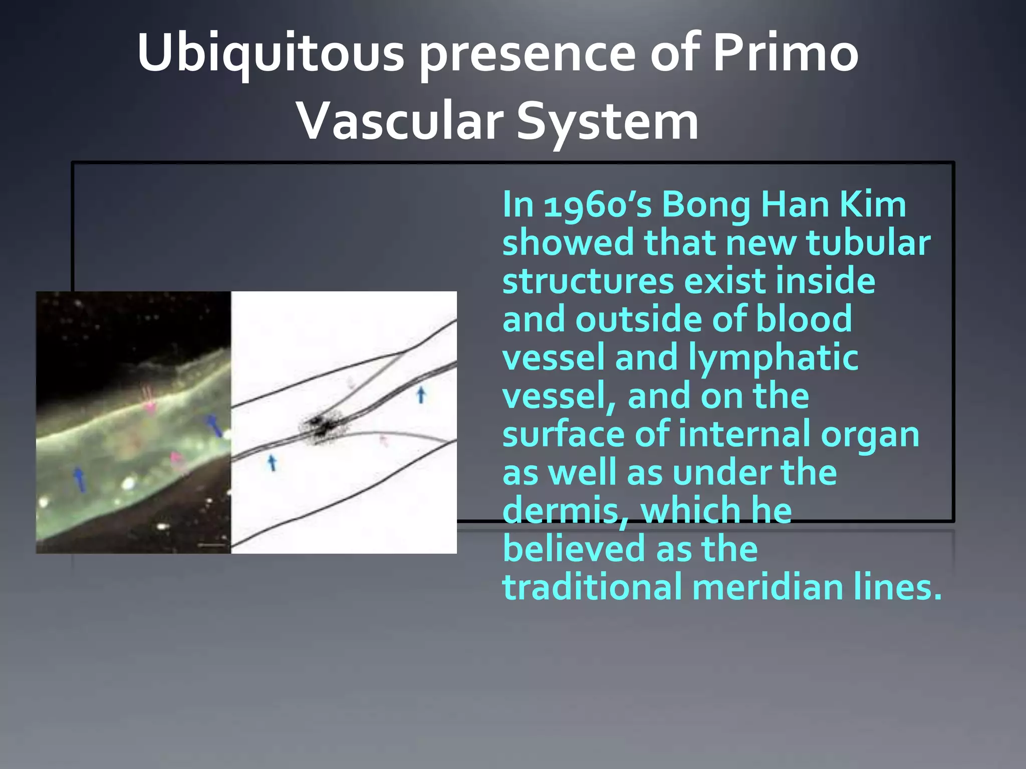
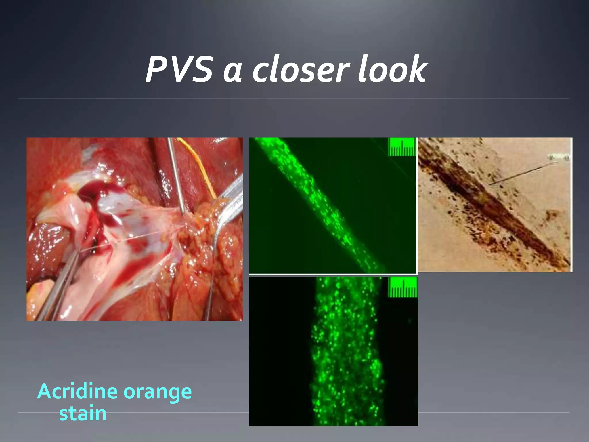
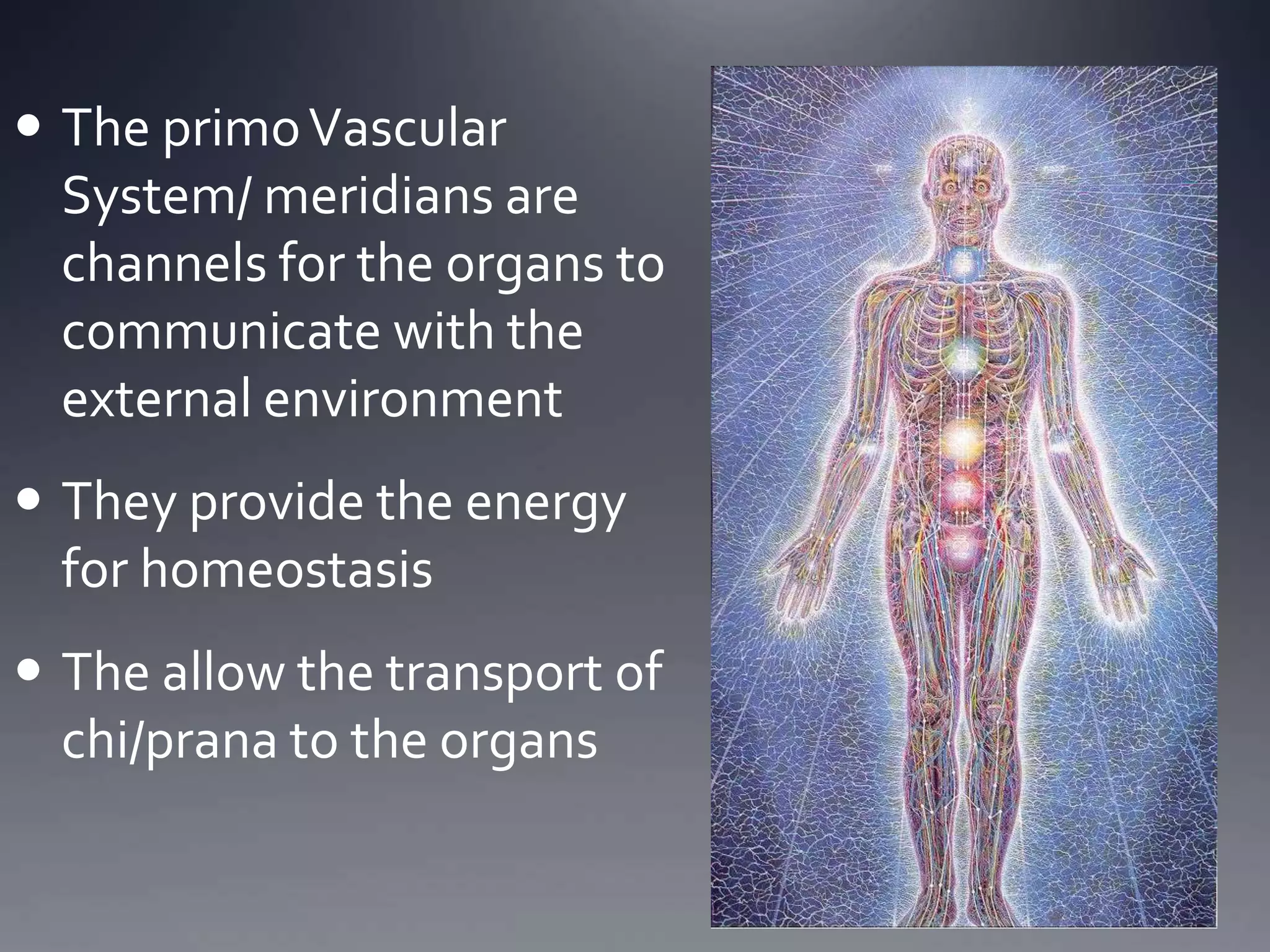
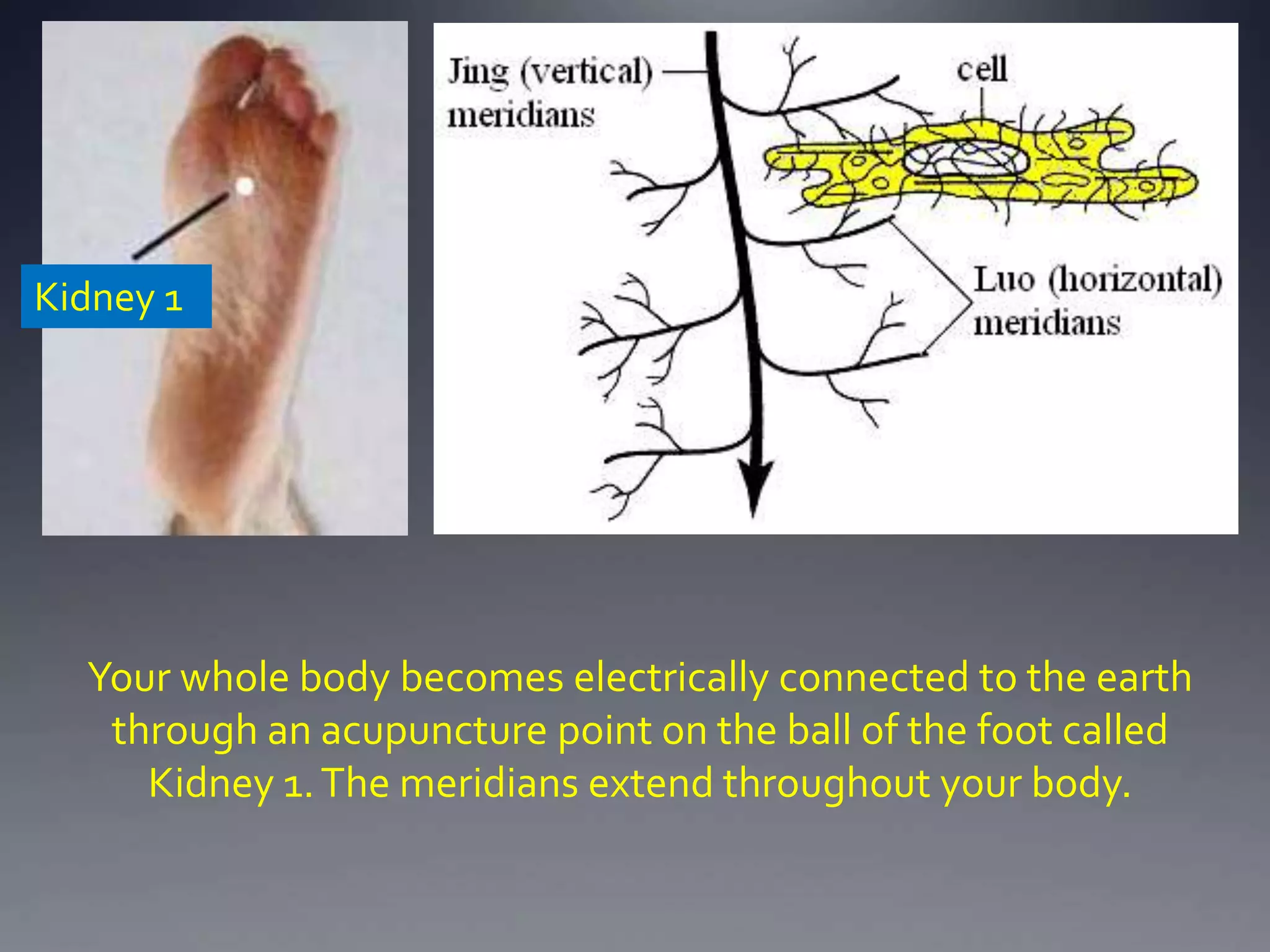
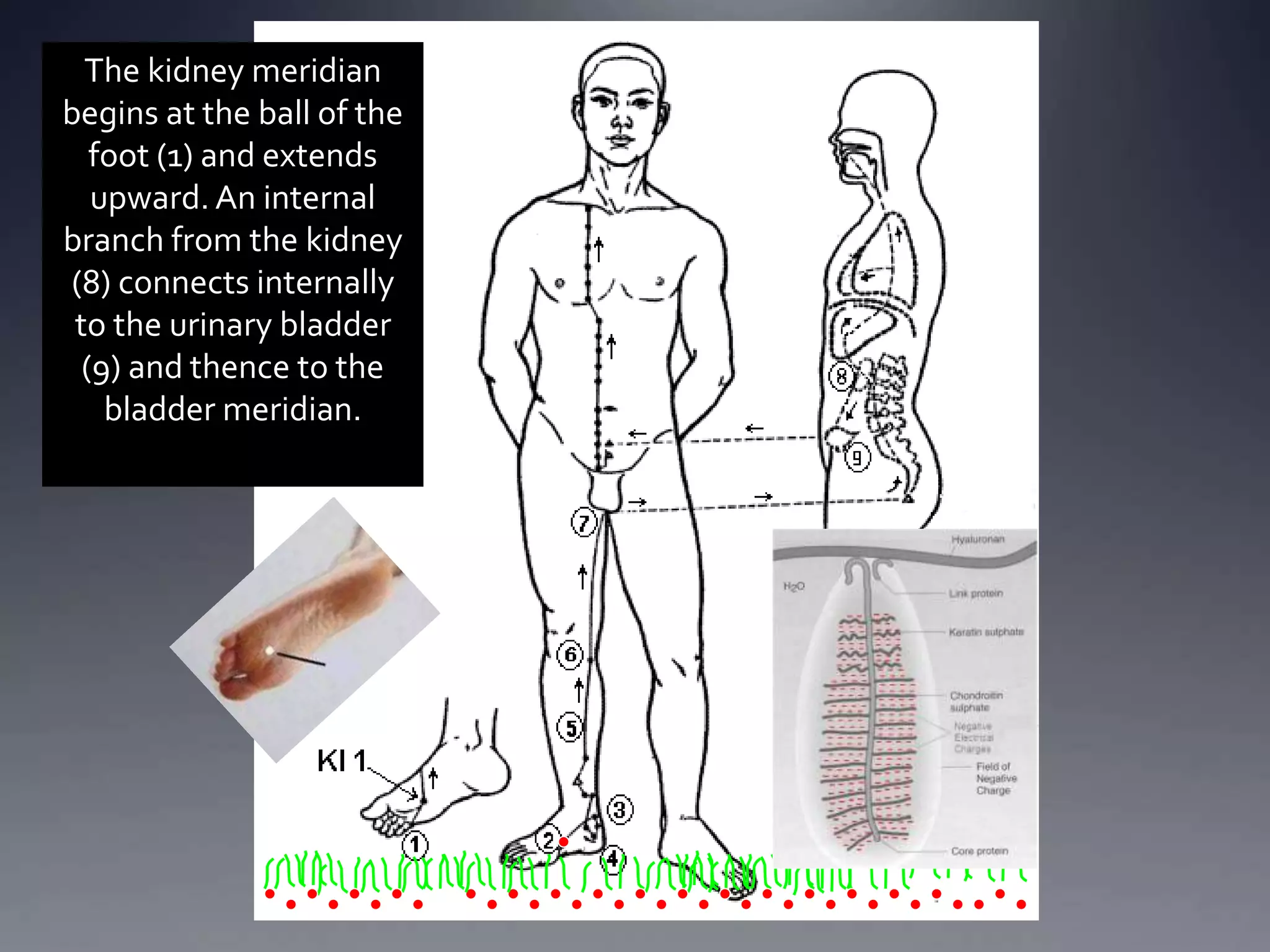
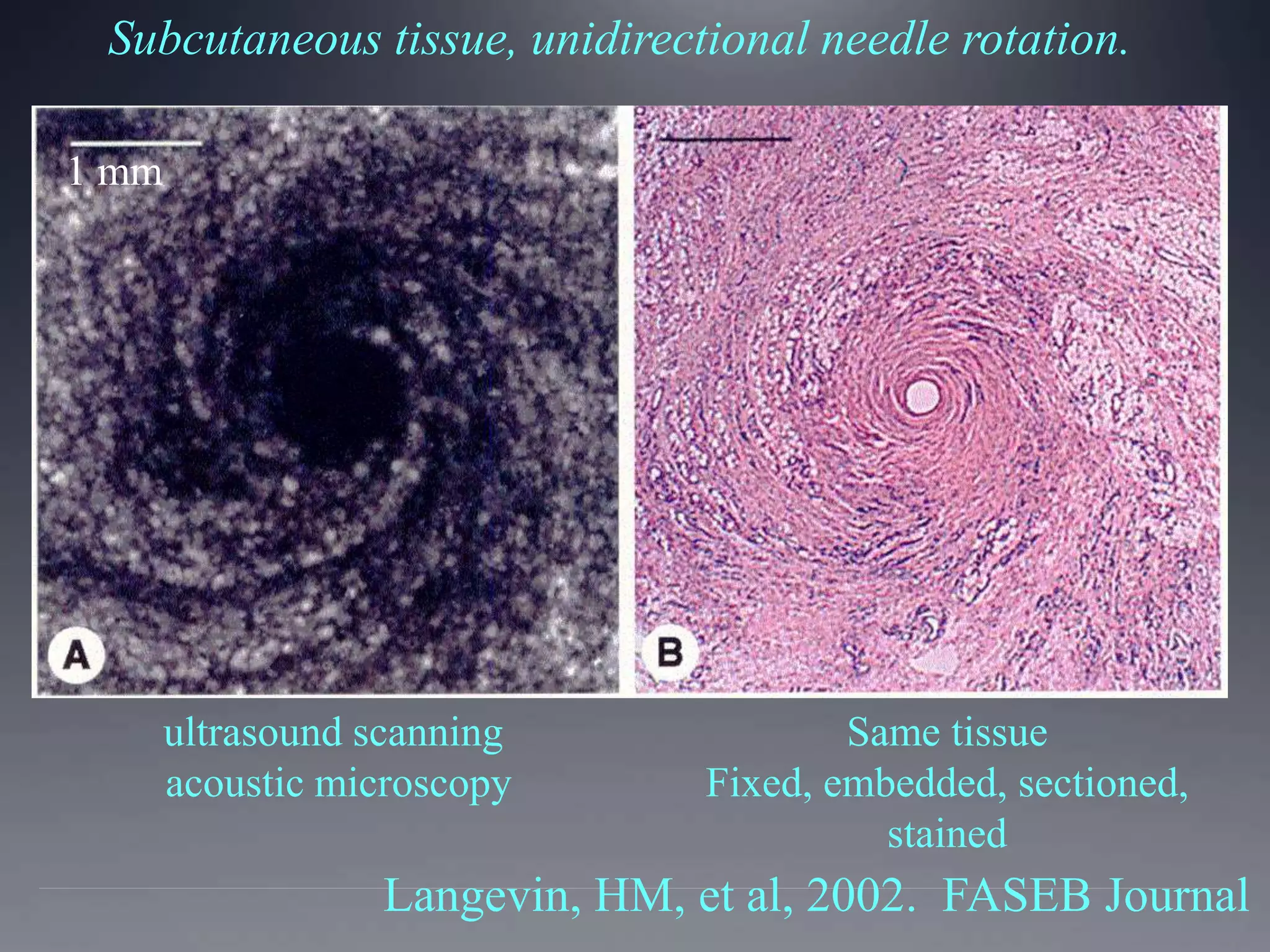
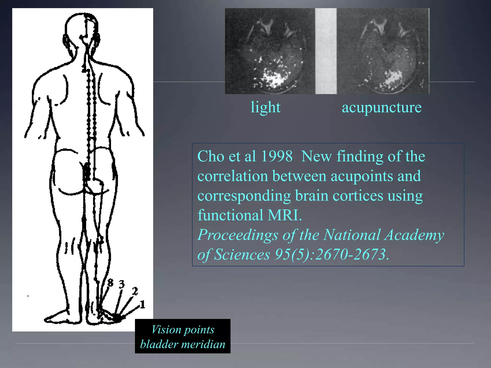
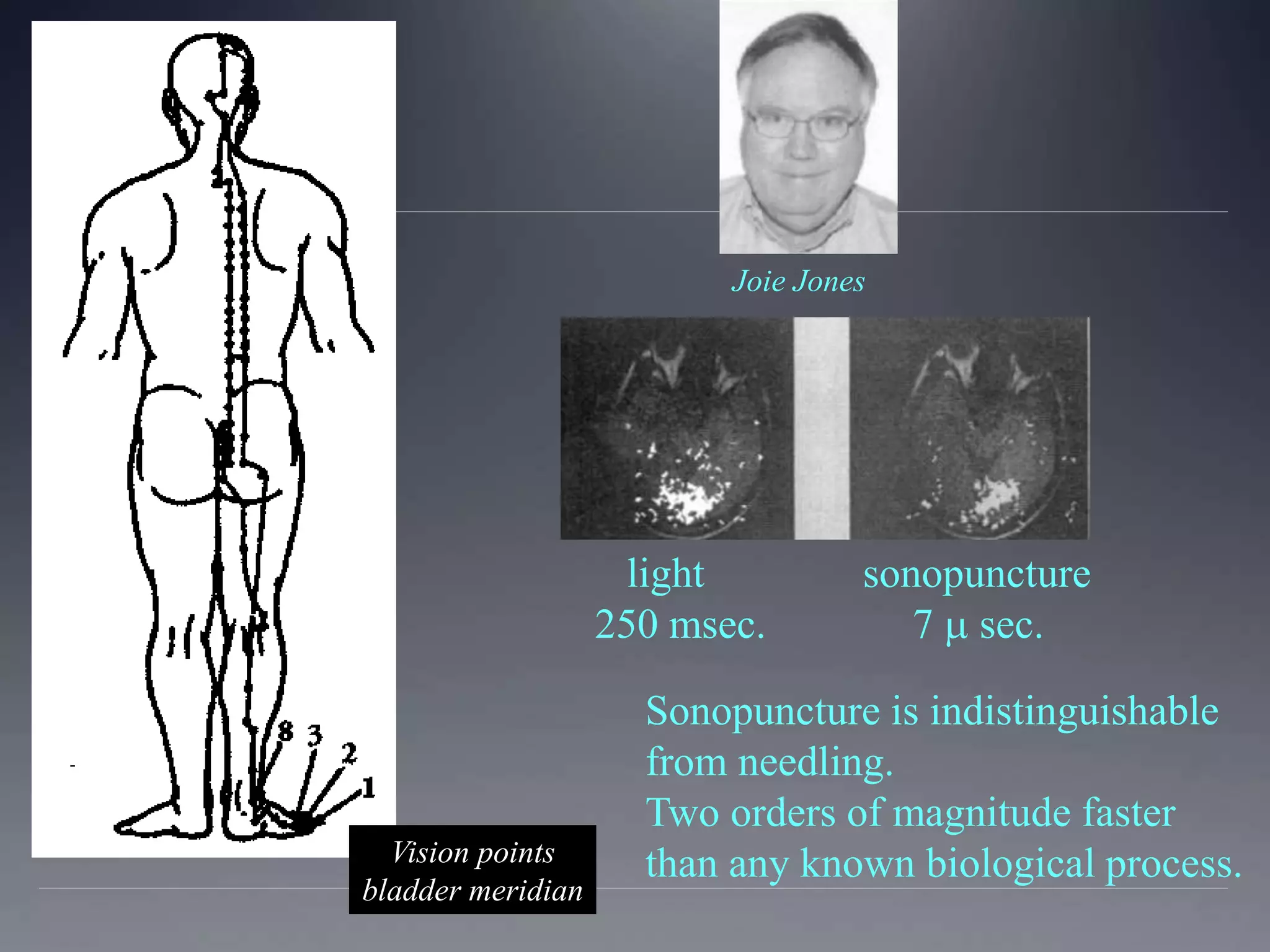
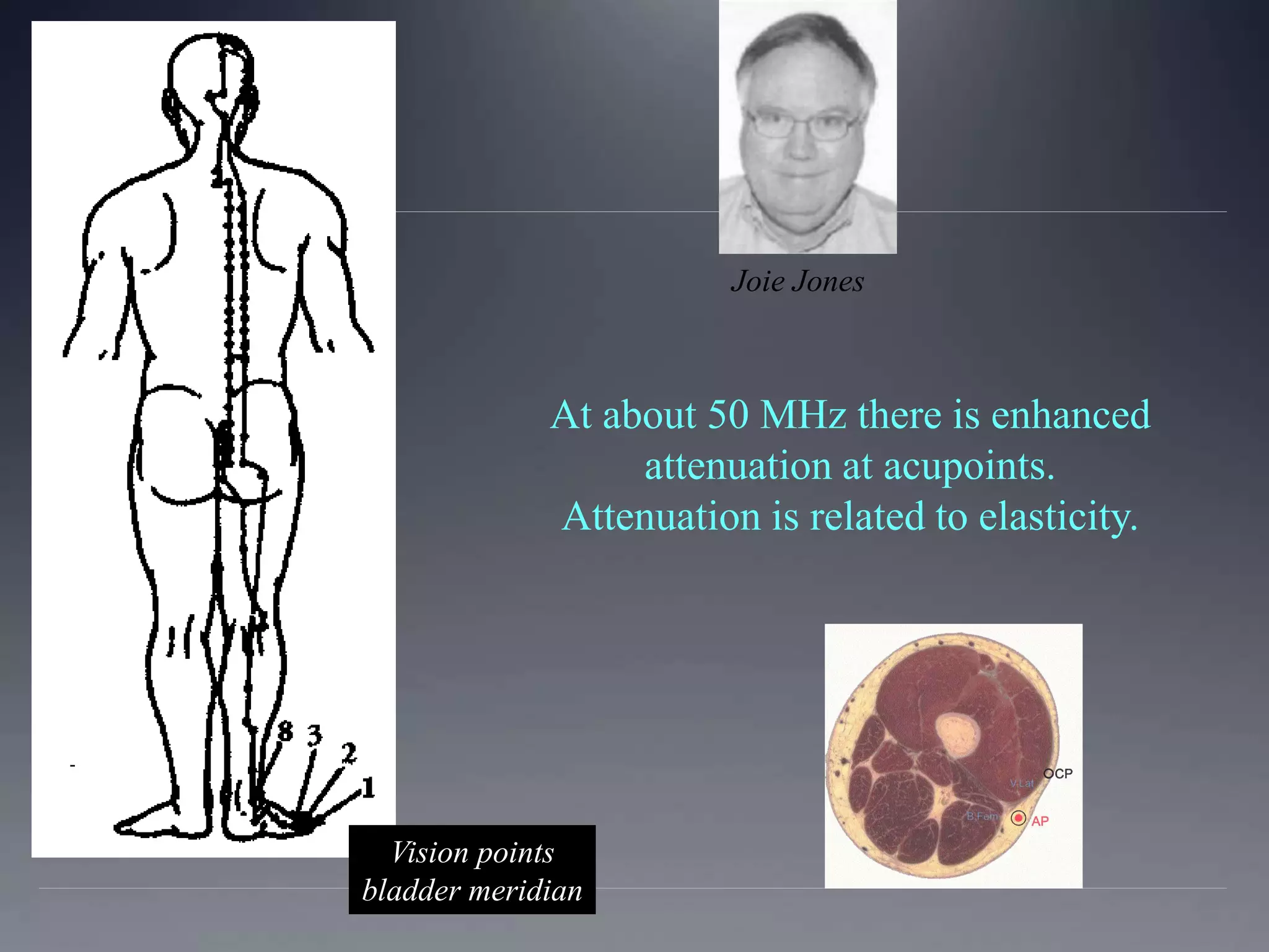
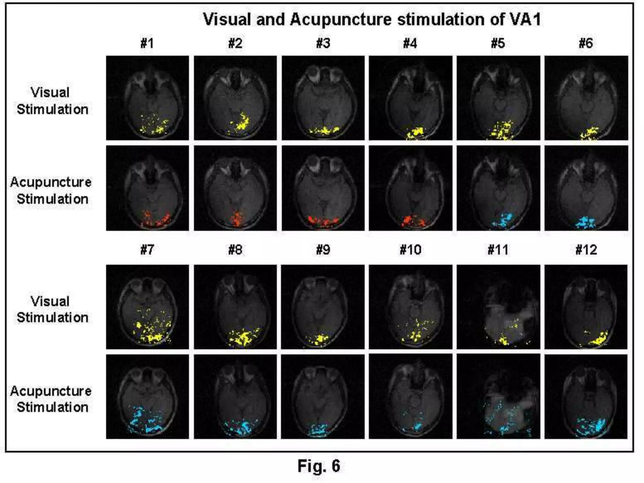
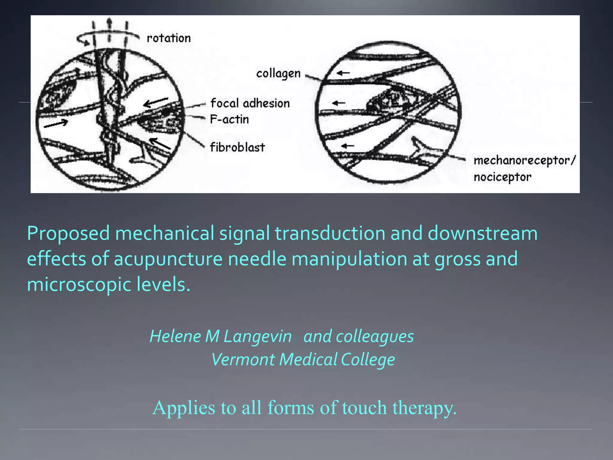
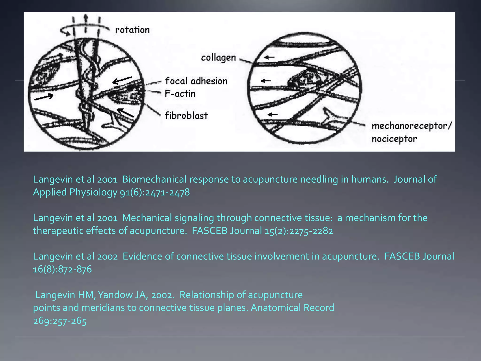
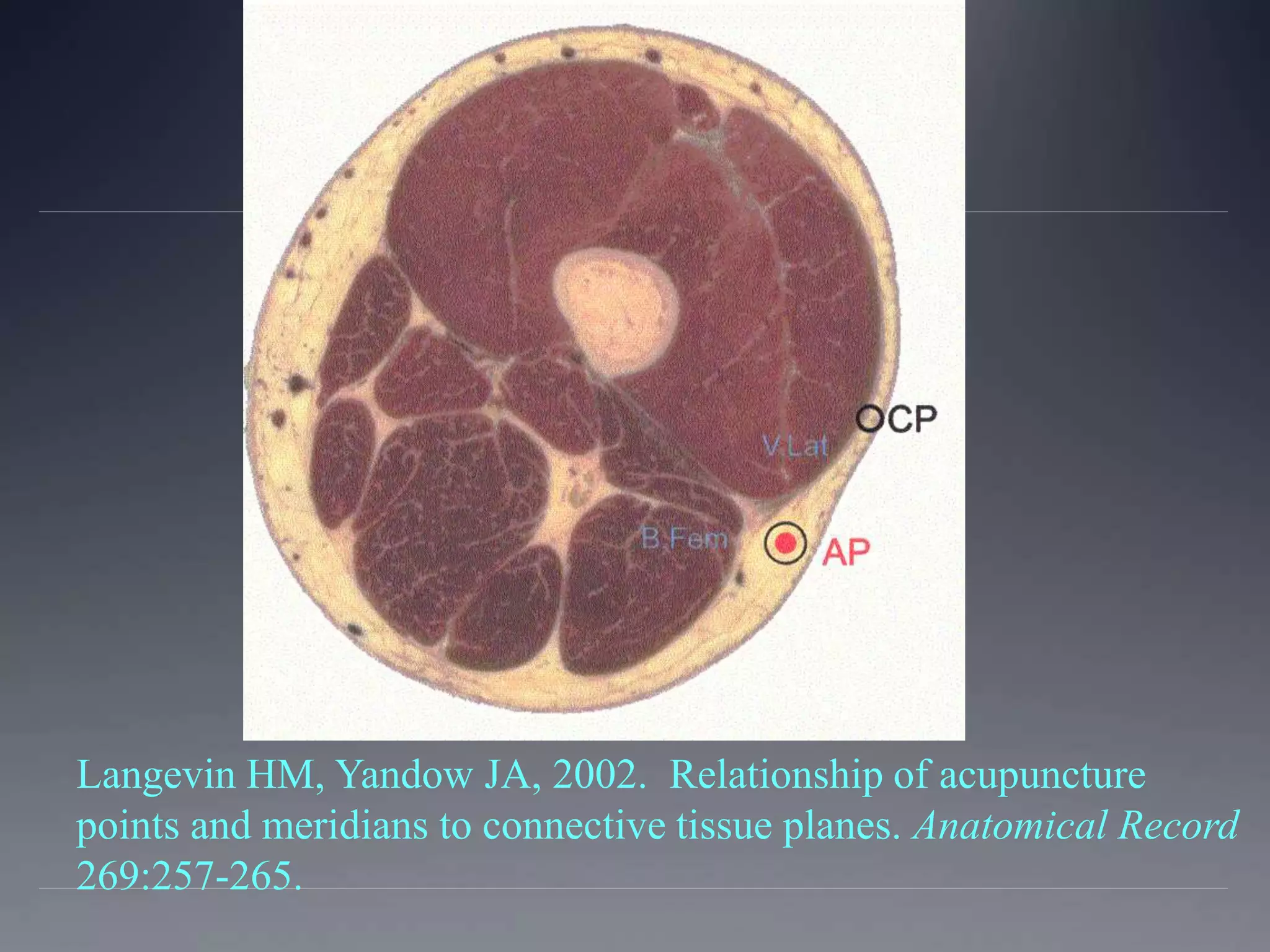
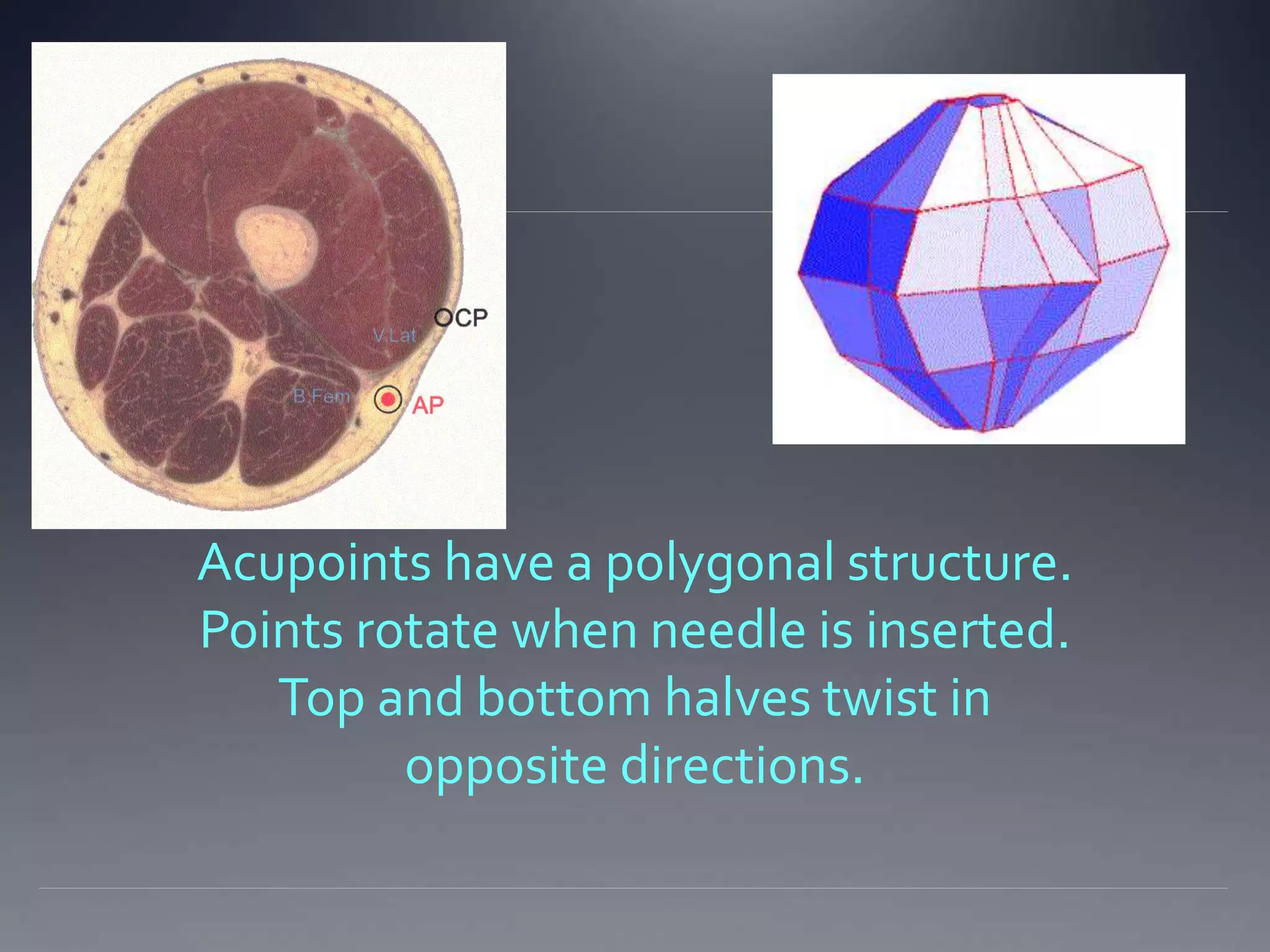
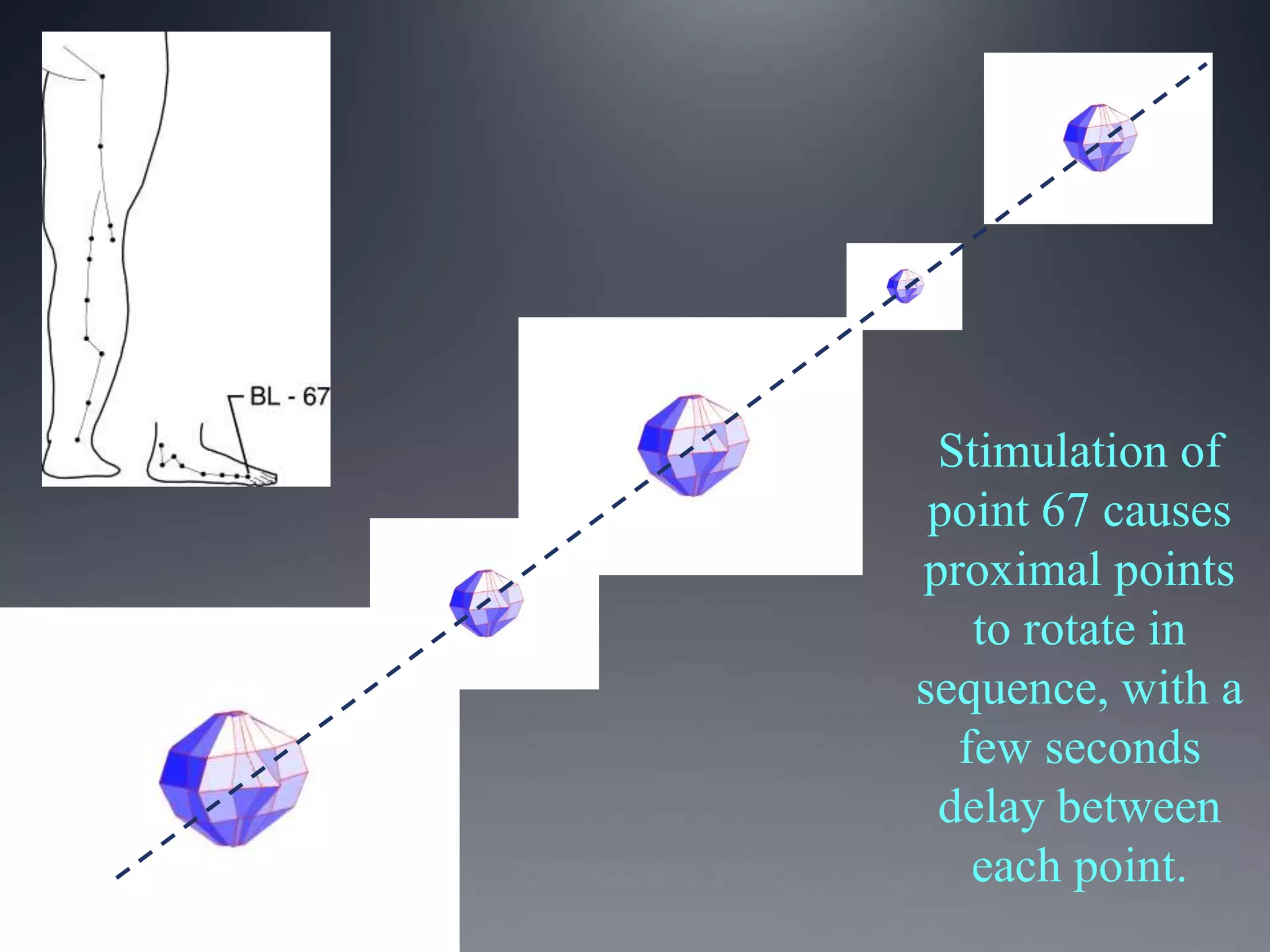
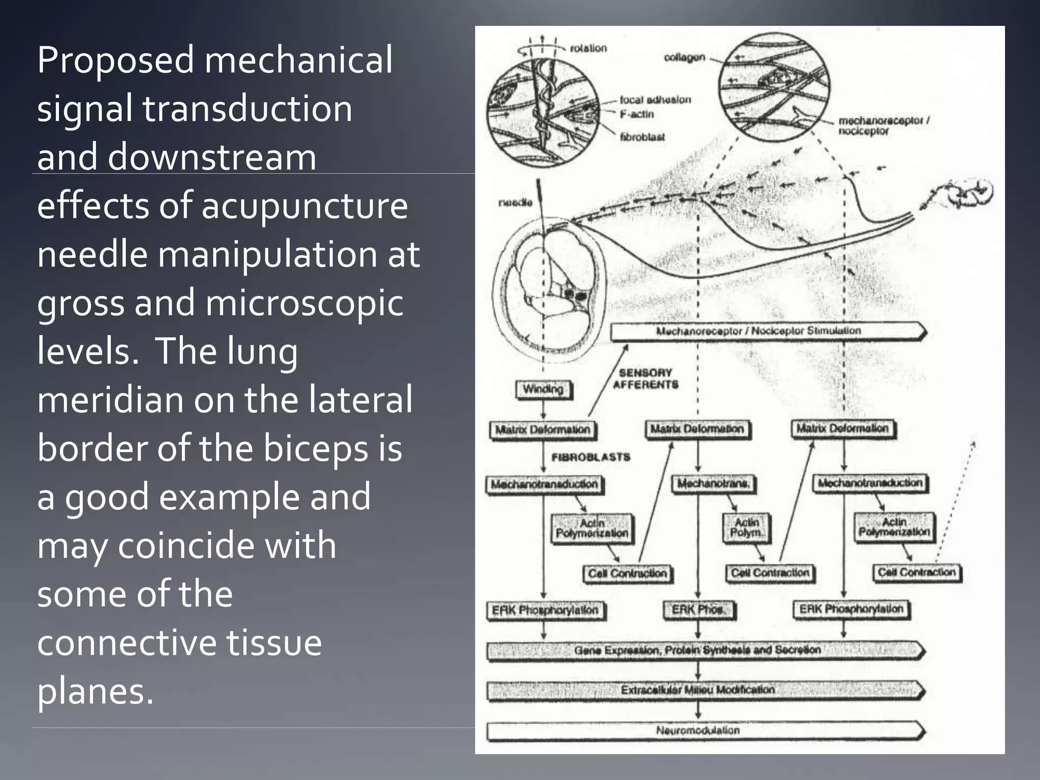
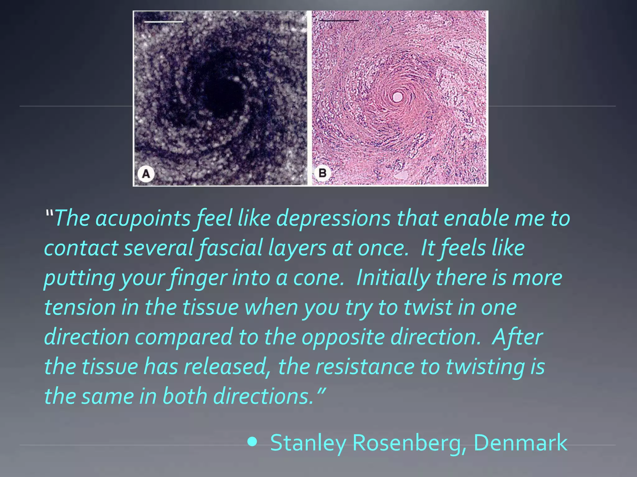
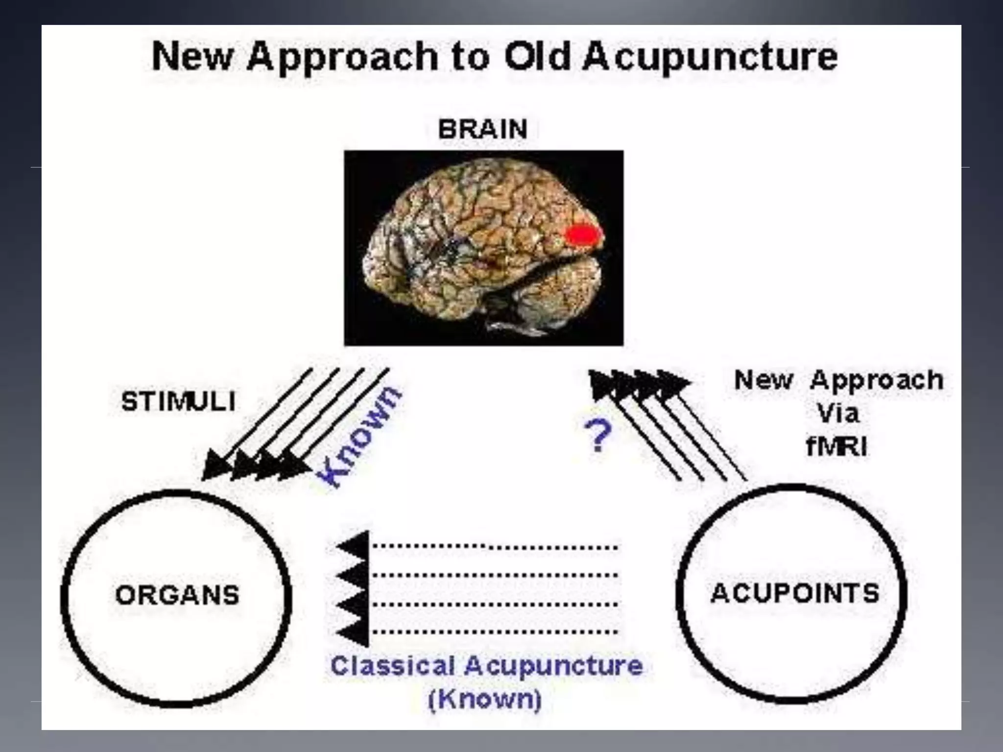
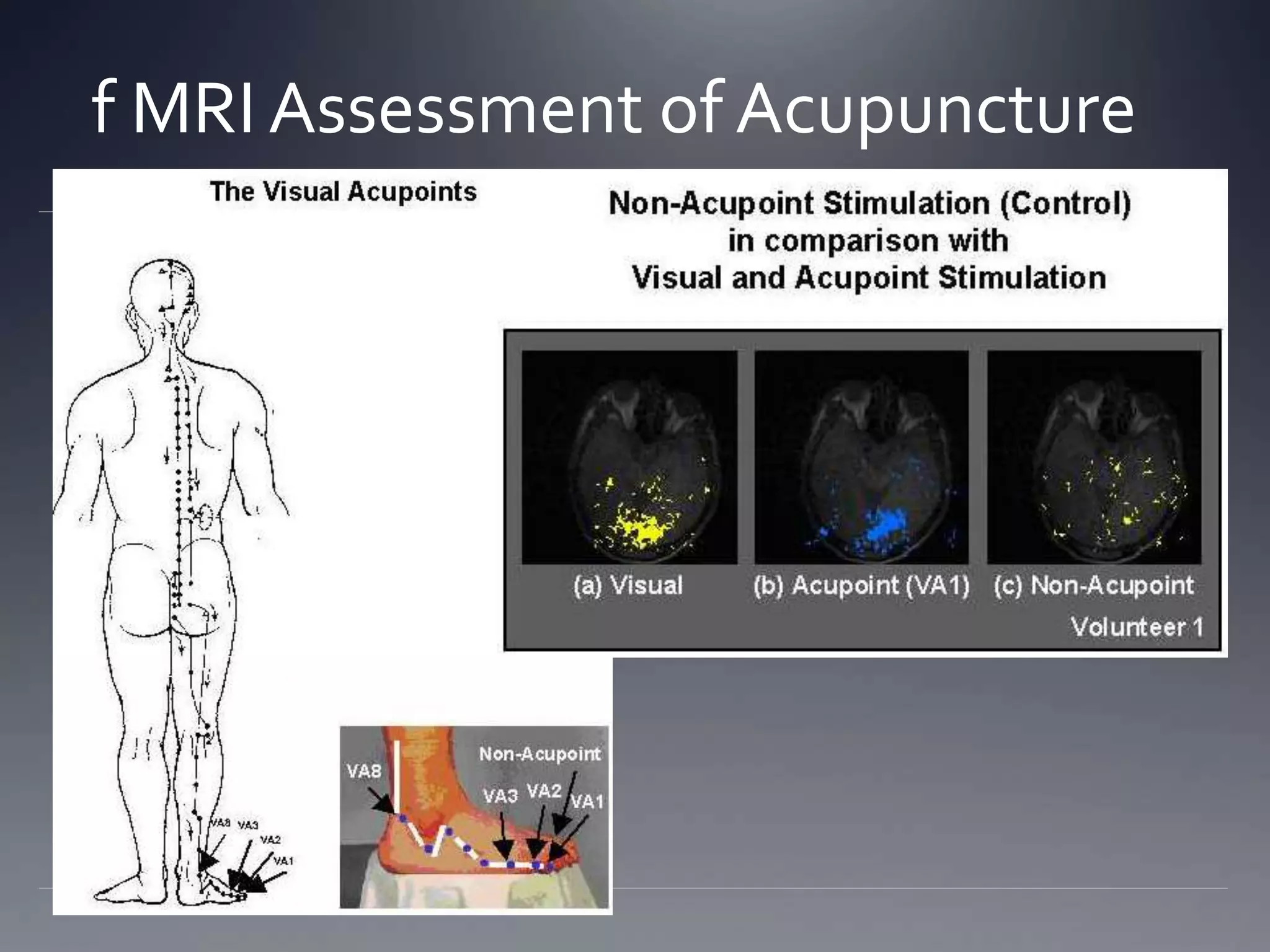
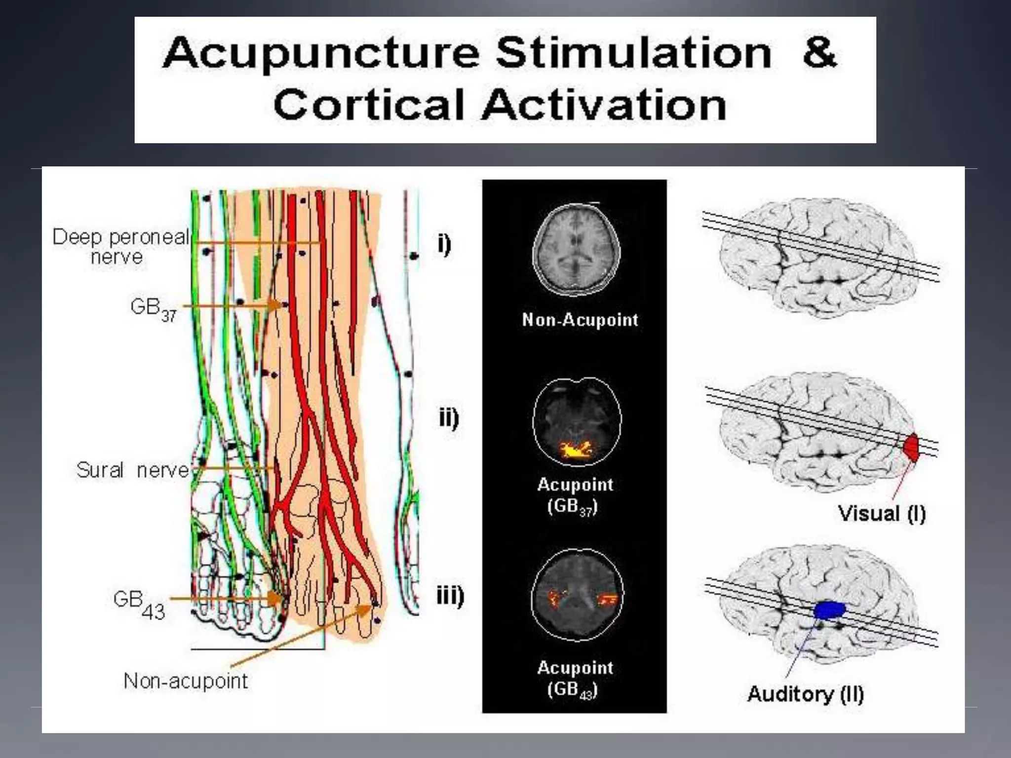
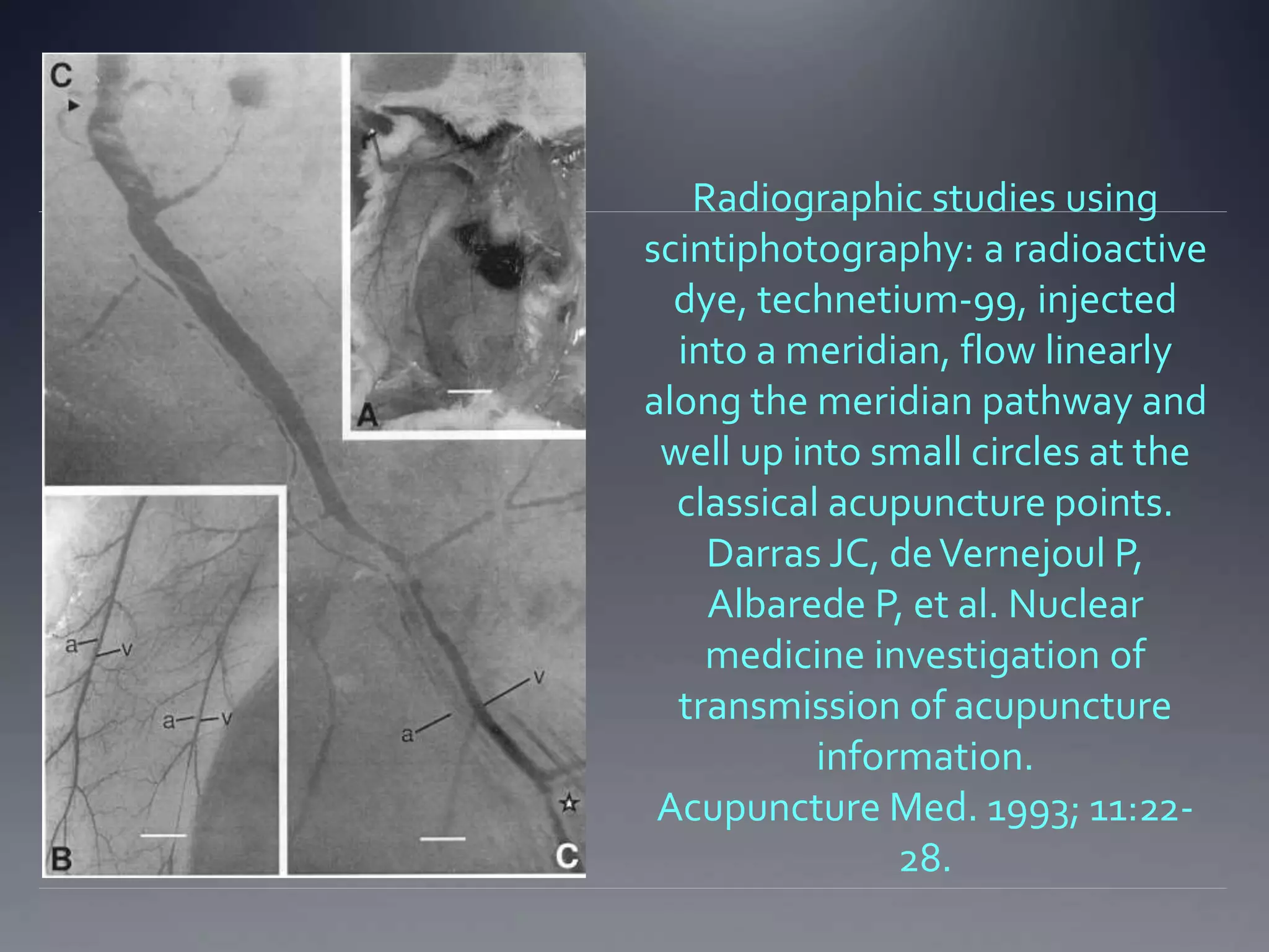
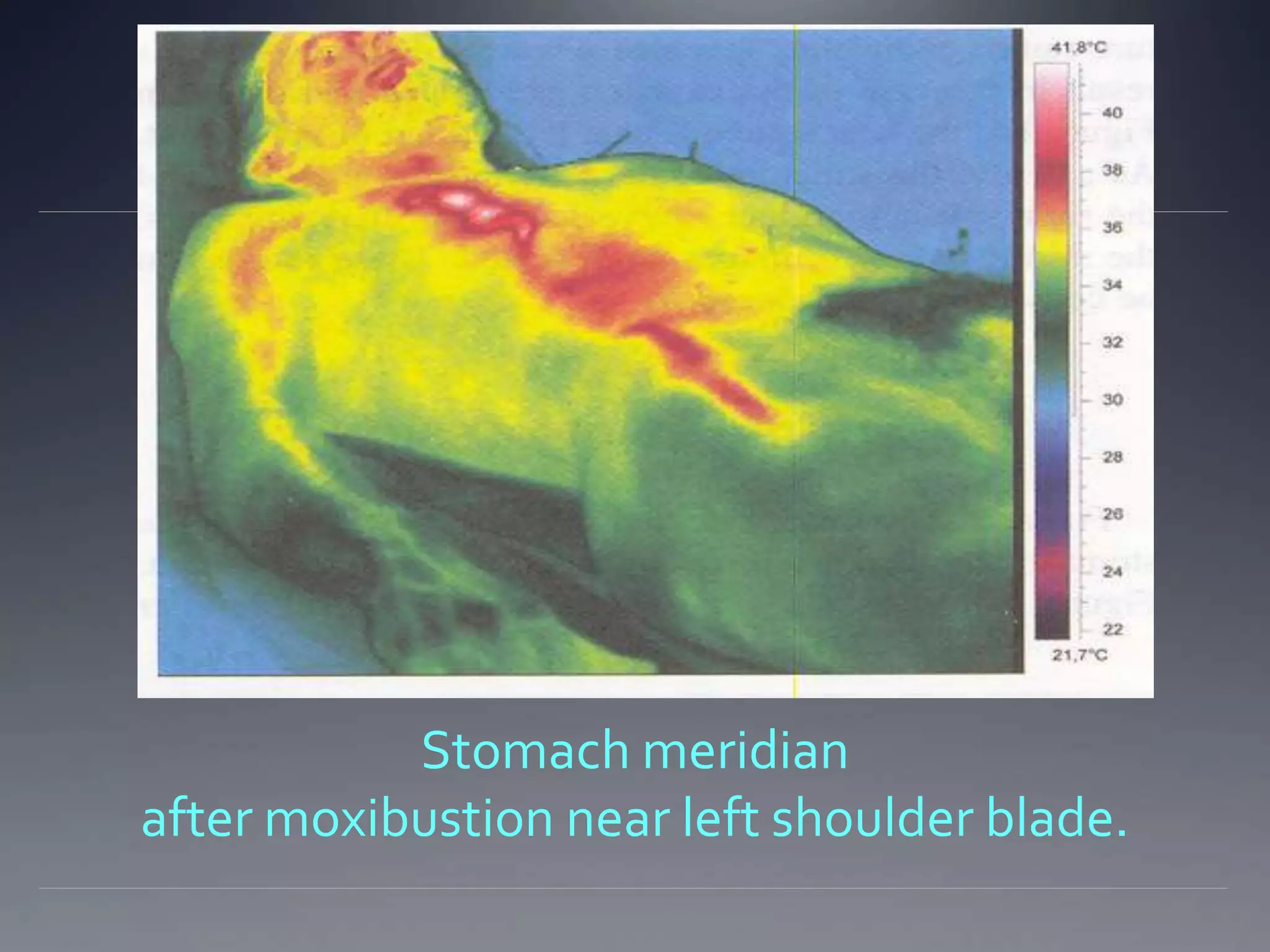
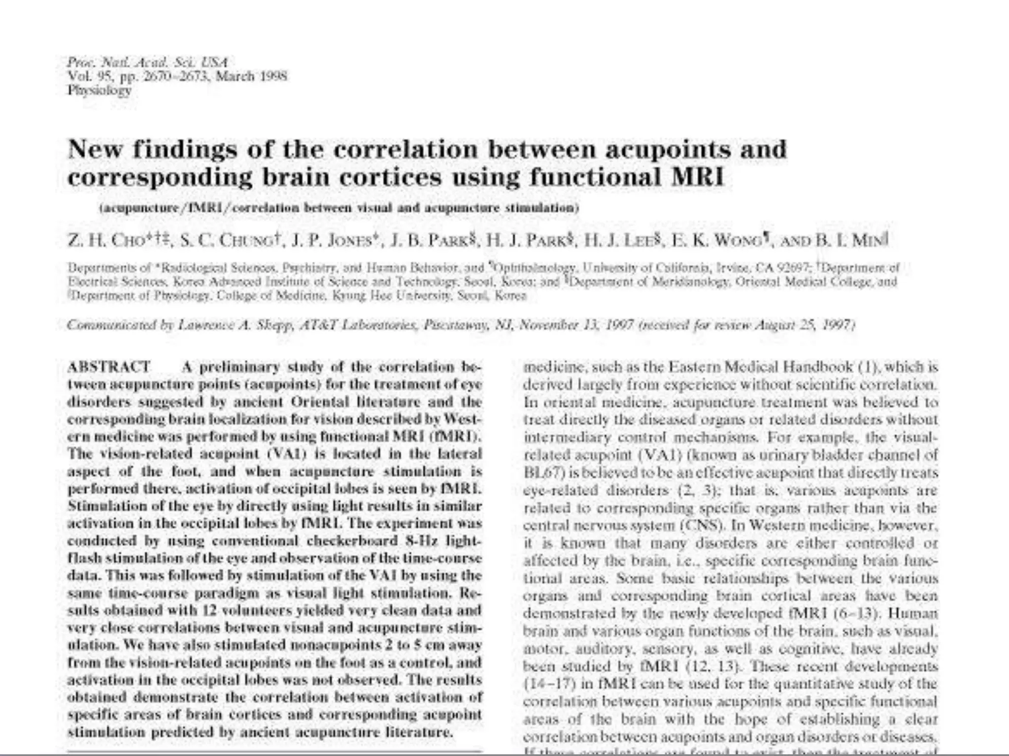
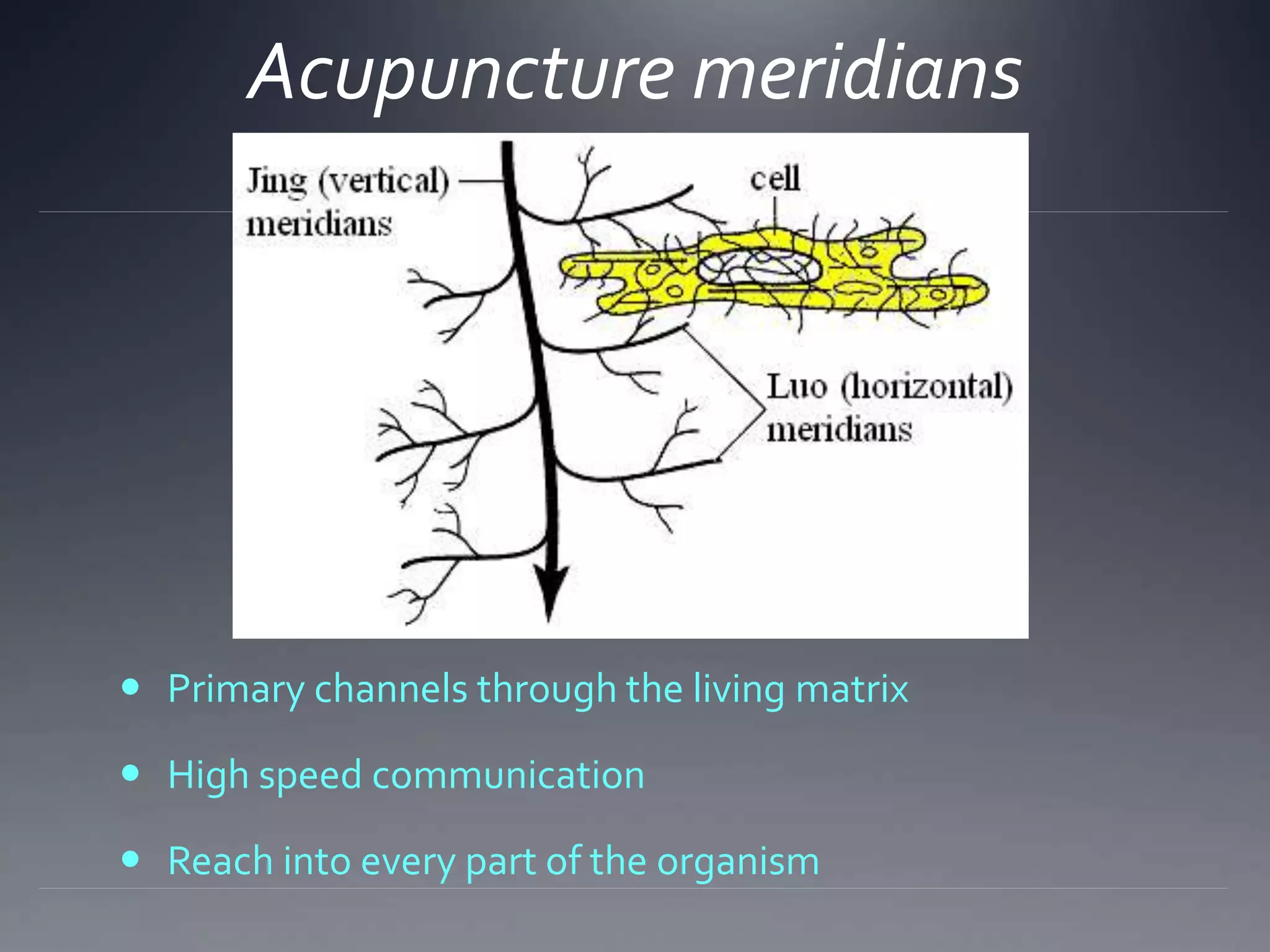
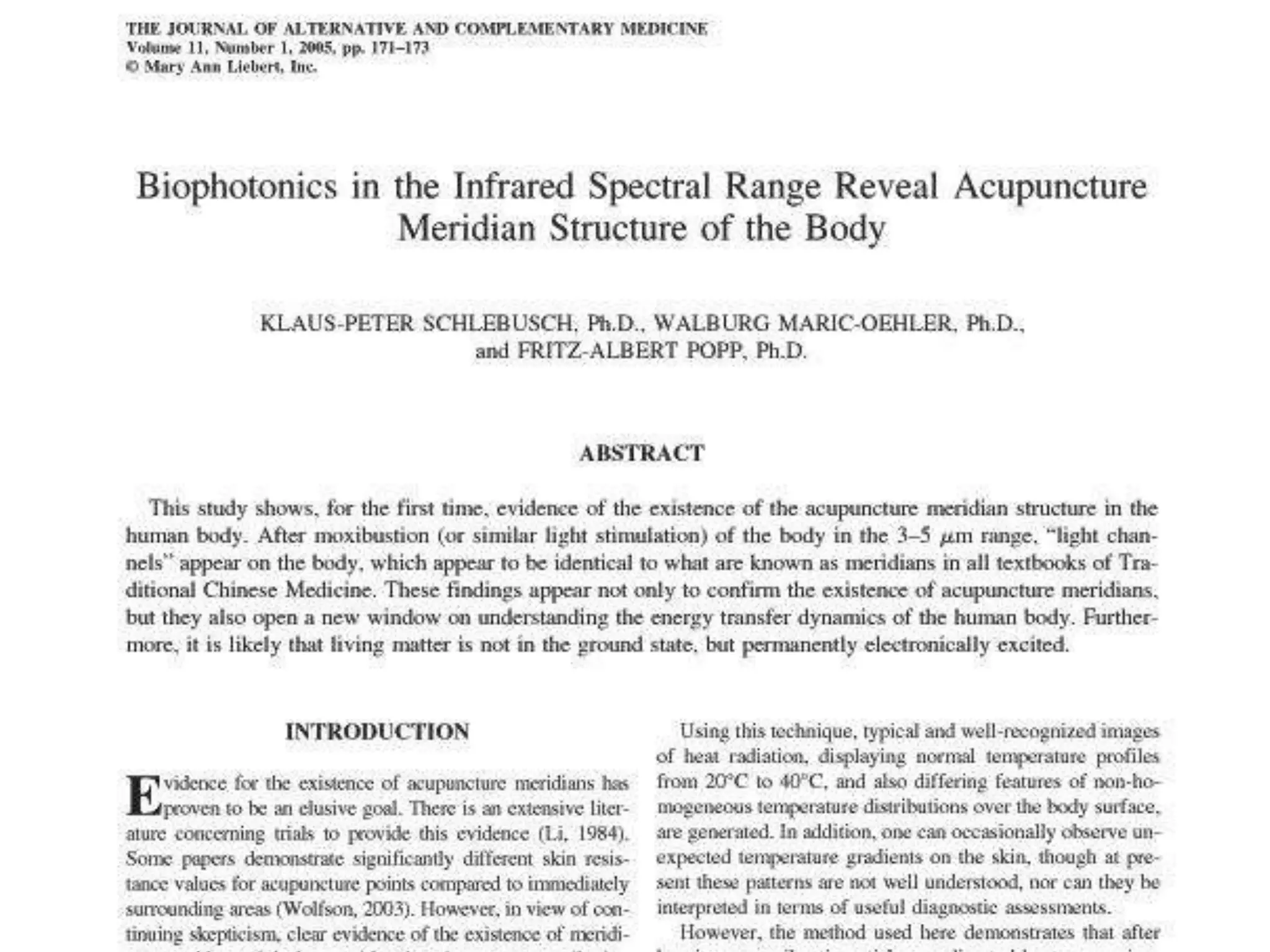


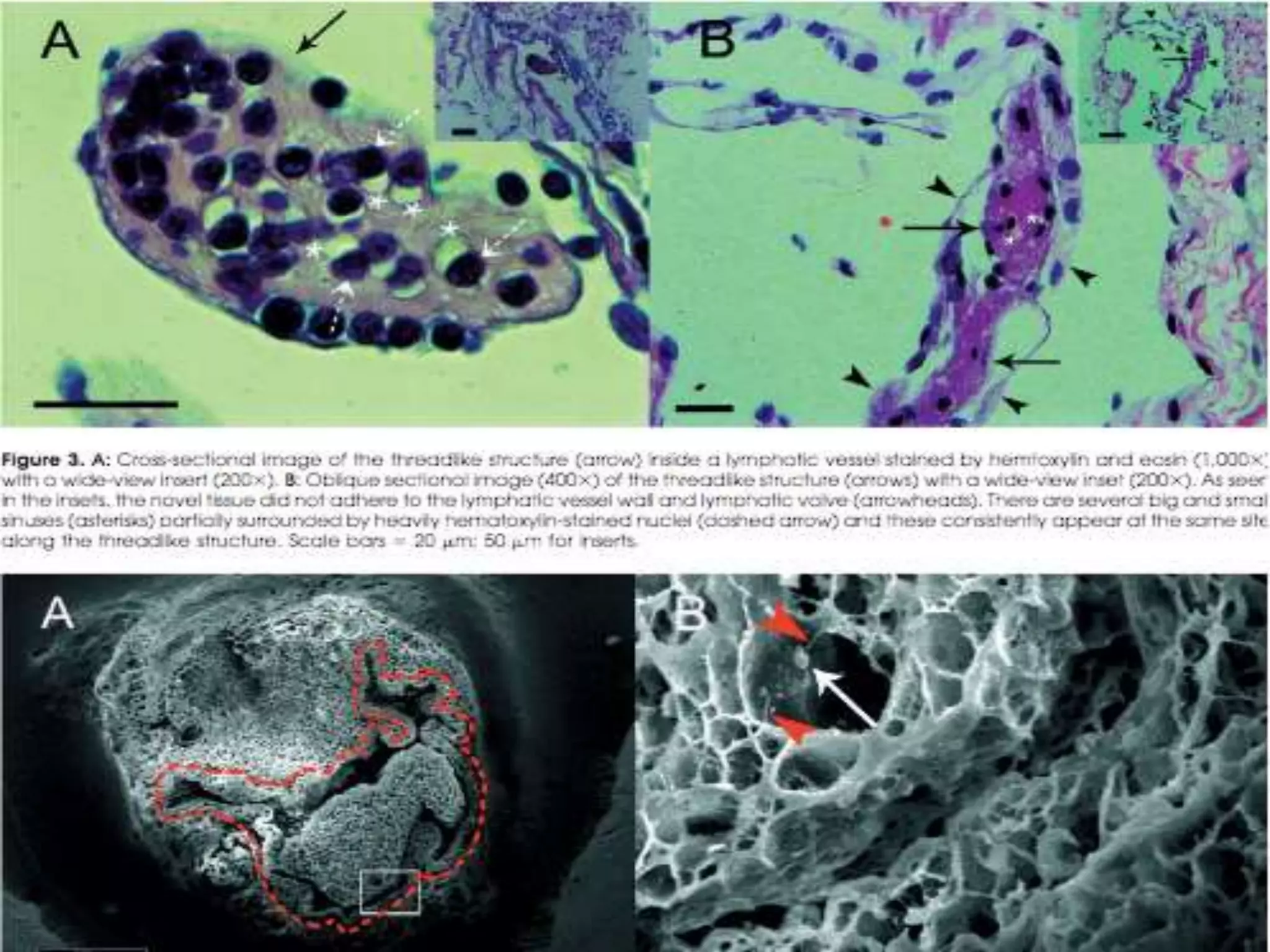


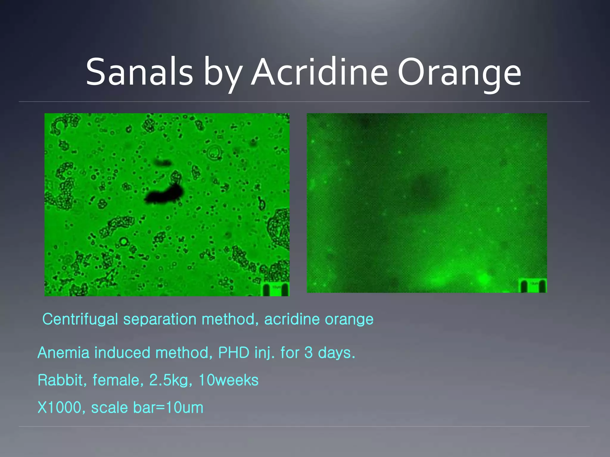

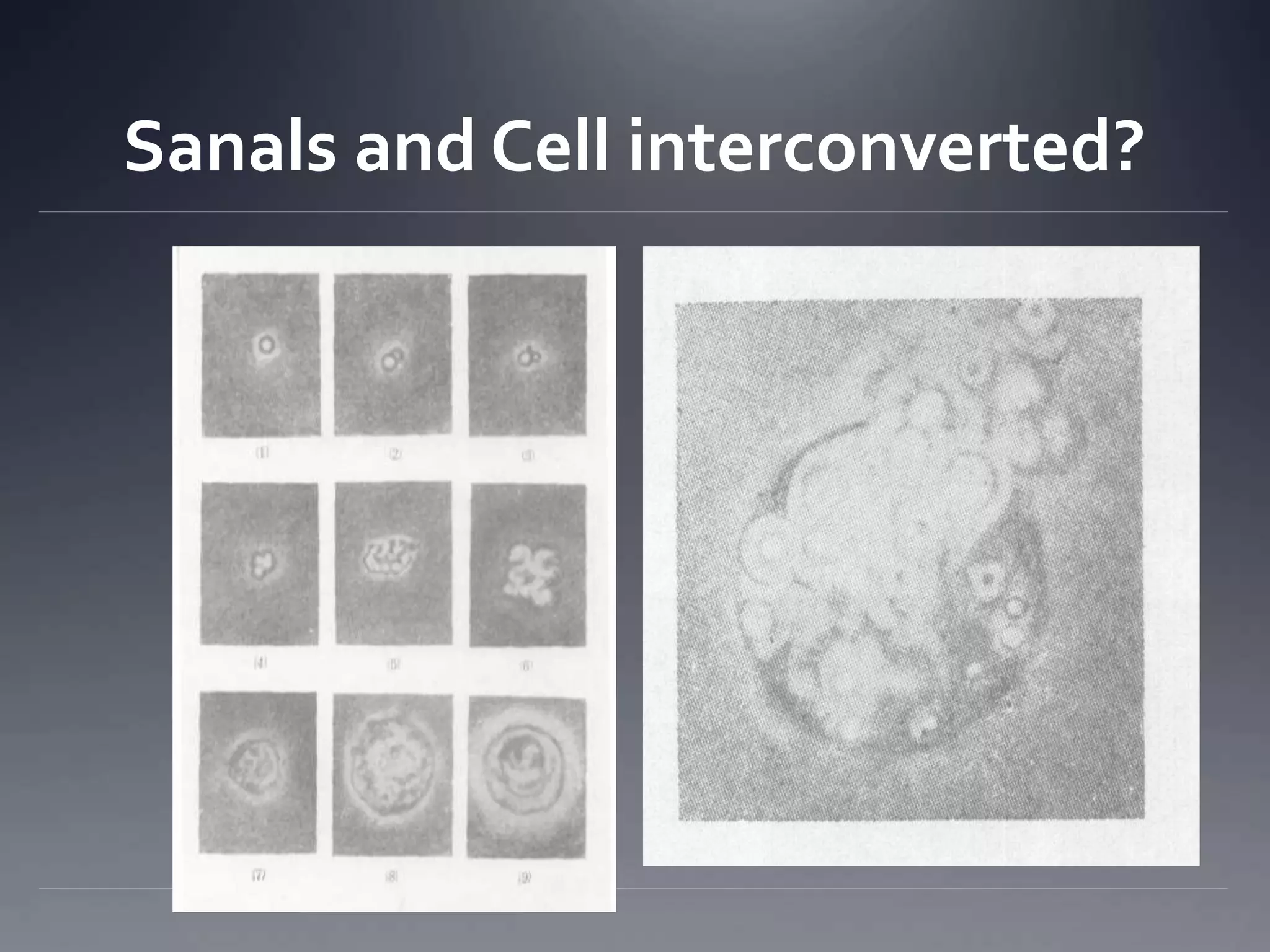
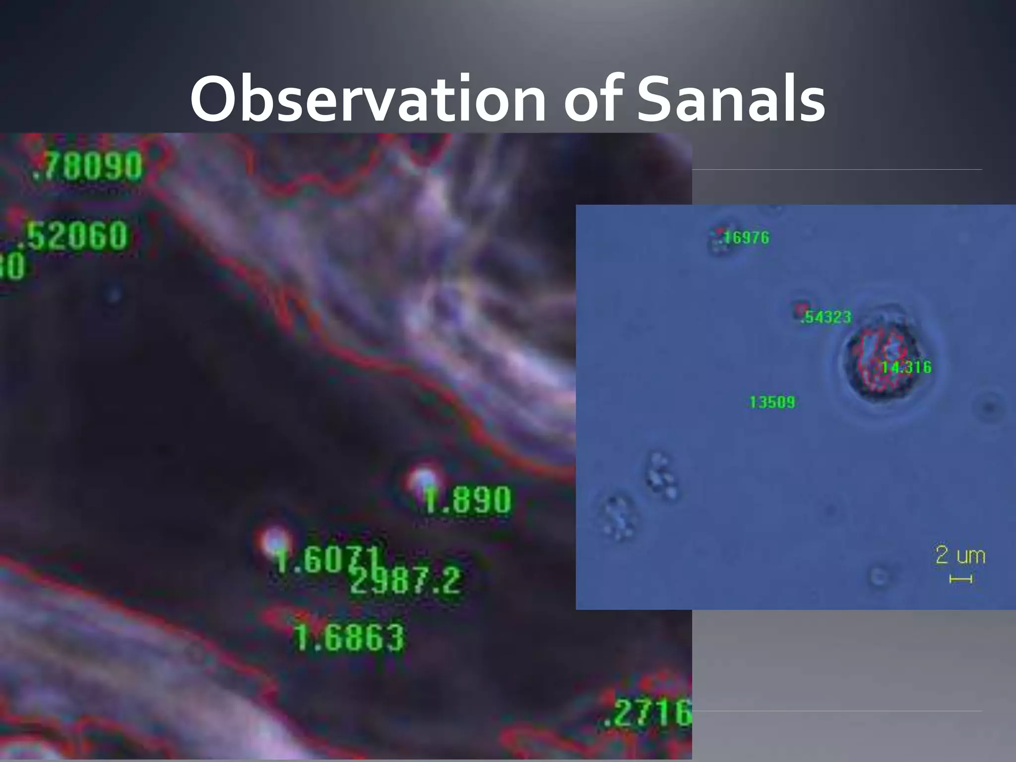
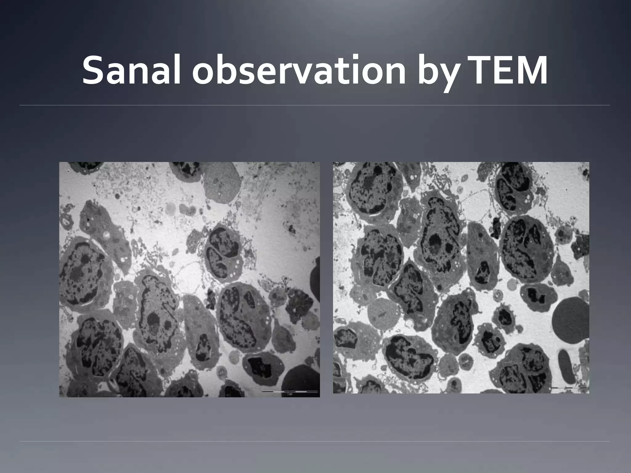
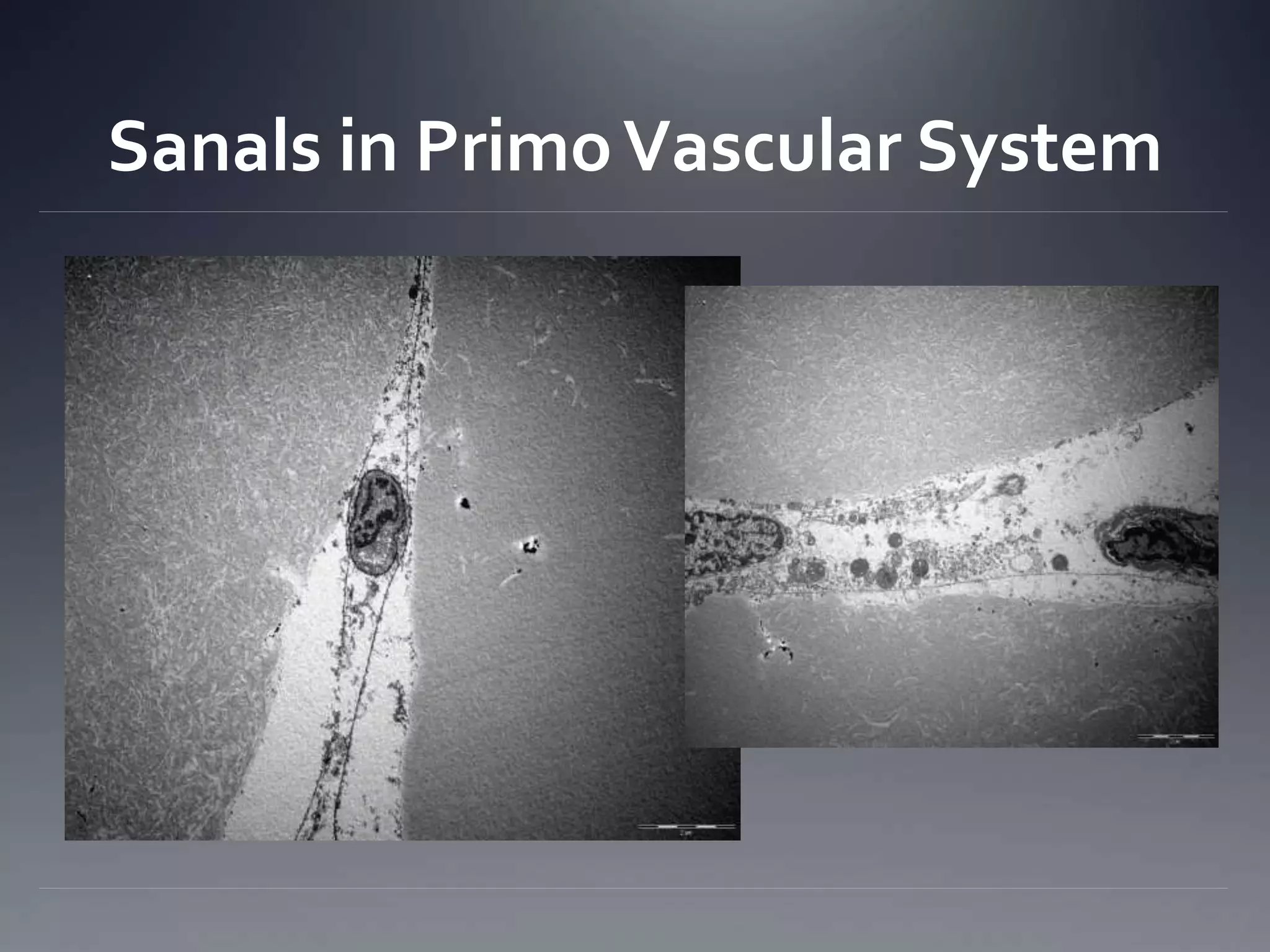
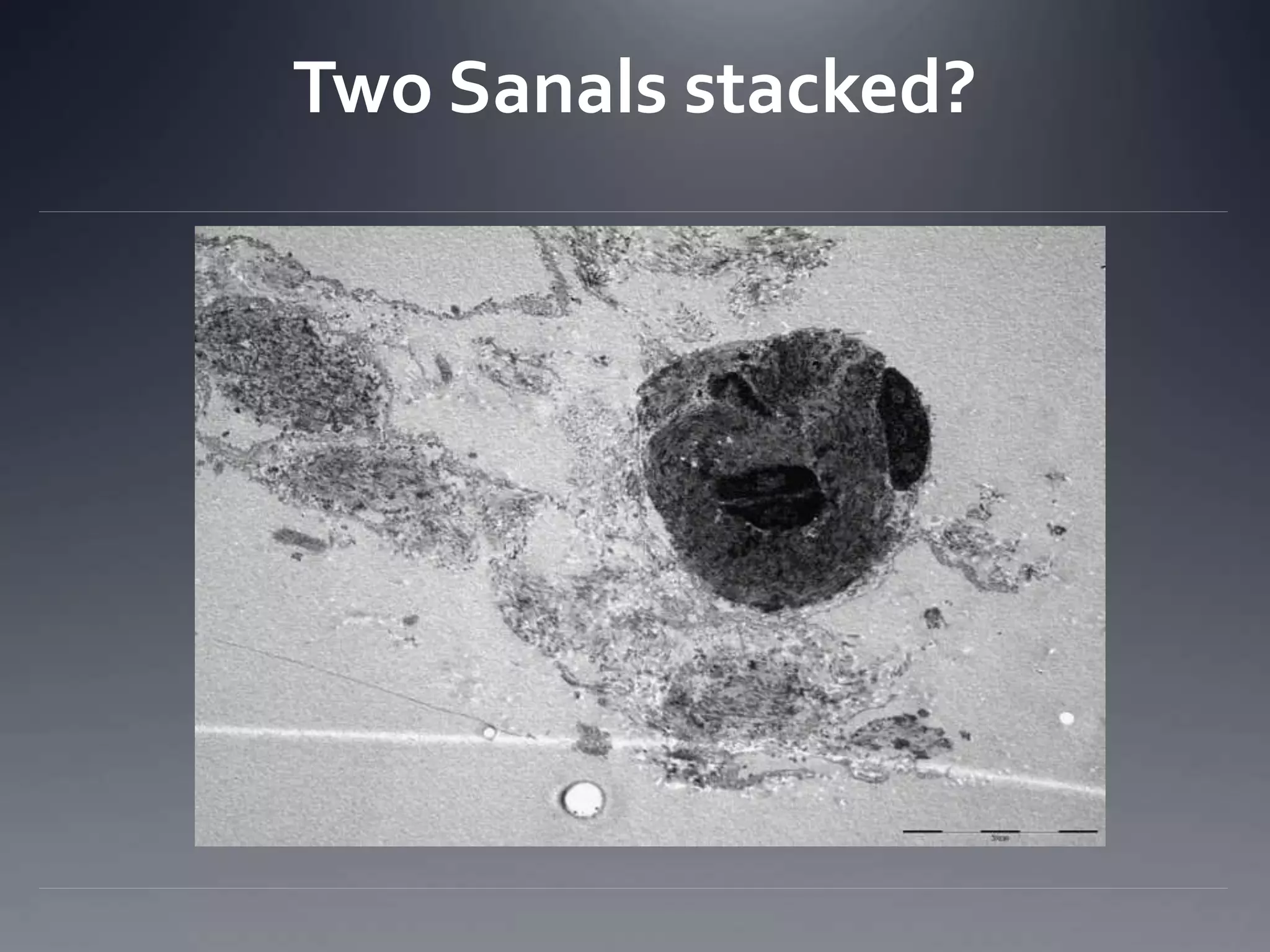
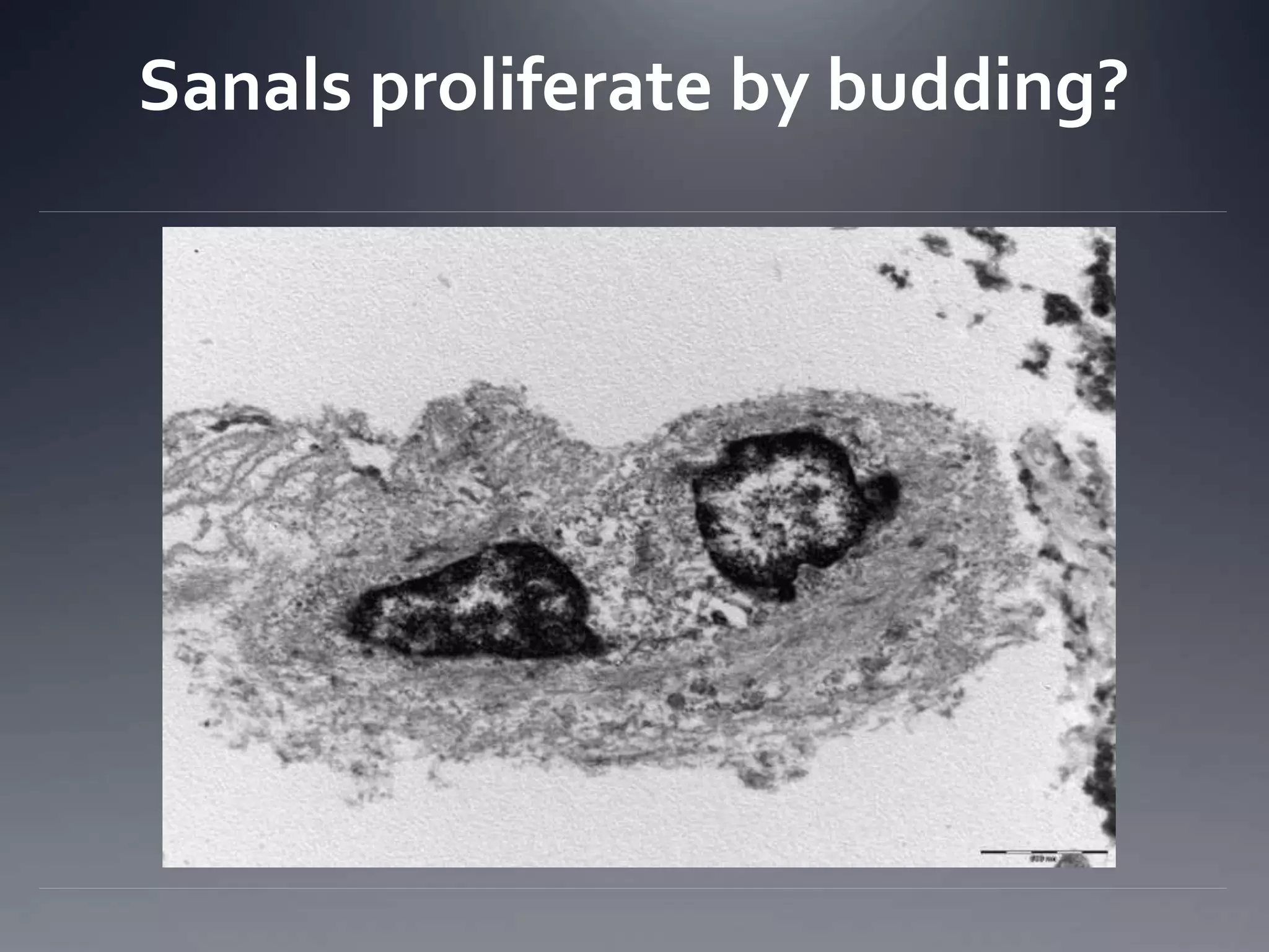

The document discusses the primovascular system, which is a network of channels associated with acupuncture points that facilitates communication and energy flow between organs and the environment. It highlights pioneering research by Bong Han Kim and others, demonstrating the physiological significance of the primovascular system and its components, including sanals, that serve as totipotent stem cells. The text further delves into the mechanical and biochemical mechanisms underlying acupuncture and its therapeutic effects, supported by various experimental findings.


![Understanding Parkinson’s Disease: Causes, Symptoms, and Treatment [2025]](https://cdn.slidesharecdn.com/ss_thumbnails/understandingparkinson-251208102525-80ba3223-thumbnail.jpg?width=640&height=640&fit=bounds)


