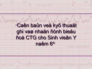
3. 1 July 2017 CTG Au Nhut Luan
3
OÁng nghe Pinard
Doppler
Fetal monitor
Caùc phöông tieän theo doõi tim thai
4. 1 July 2017 CTG Au Nhut Luan
4
„ Khaûo saùt bieán ñoäng tim
thai theo côn co töû cung
nhaèm phaùt hieän sôùm caùc
bieåu hieän khoâng bình
thöôøng cuûa tim thai
Muïc tieâu cuûa fetal monitoring
5. 1 July 2017 CTG Au Nhut Luan
5
Söû duïng hieäu öùng Doppler theo
doõi hoaït ñoäng cuûa tim thai
Maùy Monitor khoâng phaûi laø moät
micro khueách ñaïi tieáng tim thai
Tim thai ñöôïc ghi nhö theá naøo ?
6. 1 July 2017 CTG Au Nhut Luan
6
Nguoàn sieâu aâm
ñöùng yeân
Vaät phaûn hoài
di chuyeån
Vaät phaûn hoài
di chuyeån
F1
F2
Hieäu öùng Doppler laø gì ?
7. 1 July 2017 CTG Au Nhut Luan
7
Phöông cuûa soùng sieâu aâm
8. 1 July 2017 CTG Au Nhut Luan
8
Khi vaät di chuyeån, taàn soá hoài aâm thay ñoåi
Moãi laàn tim cöû ñoäng seõ gaây ra moät laàn thay
ñoåi taàn soá cuûa hoài aâm
Soá laàn hoài aâm thay ñoåi taàn soá trong moät phu
töông öùng vôùi nhòp tim moãi phuùt
Bieát khoaûng thôøi gian giöõa hai laàn tim ñaäp
seõ tính ñöôïc trò soá nhòp tim thai / phuùt
Maùy seõ cho giaù trò töùc thôøi cuûa tim thai
Maùy tính tim thai nhö theá naøo ?
9. 1 July 2017 CTG Au Nhut Luan
9
Bieåu ñoà tim thai
„ Laø taäp hôïp caùc dots bieåu thò trò soá töùc thôøi cuûa
tim thai
10. 1 July 2017 CTG Au Nhut Luan
10
Thaân maùy laø moät maùy tính coù chöùc naêng
tính toaùn söï khaùc bieät taàn soá cuûa sieâu aâm
gôûi vaø hoài aâm töø ñoù cho bieát giaù trò töùc
thôøi cuûa tim thai
Boä phaän doø tim thai laø moät ñaàu phaùt - thu
soùng sieâu aâm
Boä phaän doø côn co laø moät caûm bieán cô hoïc
ghi aùp löïc taùc ñoäng treân maøng ghi
Maùy monitor caáu taïo nhö theá naøo ?
11. 1 July 2017 CTG Au Nhut Luan
11
„ 2 hình thöùc ghi
CTG
CTG ngoaøi
CTG trong
Hình thöùc ghi CTG
12. 1 July 2017 CTG Au Nhut Luan
12
Ñoä nhaïy cao 95%
Ñoä ñaëc hieäu thaáp 50%
Giaù trò cuûa Fetal monitoring
35. 1 July 2017 CTG Au Nhut Luan
35
Nhòp chaäm traàm troïng
36. 1 July 2017 CTG Au Nhut Luan
36
Ñònh baseline coù ñôn giaûn khoâng ?
„ Severe tachycardia keøm nhòp giaûm
37. 1 July 2017 CTG Au Nhut Luan
37
Theå hieän söï ñieàu phoái cuûa haønh naõo
Caân baèng giöõa hai can thieäp giao caûm vaø
ñoái giao caûm
Goàm
Long term variability
Short term variability
Dao ñoäng noäi taïi (Variability)
38. 1 July 2017 CTG Au Nhut Luan
38
Short-term variability
„ Bieán ñoäng cuûa trò soá töùc thôøi töø chu chuyeån tim
naøy sang chu chuyeån tim ngay lieàn keà
39. 1 July 2017 CTG Au Nhut Luan
39
Long-term variability
„ Dao ñoäng taïo daïng hình soùng cho baseline, coù taàn
soá khoaûng 3-5 ñænh phuùt
40. 1 July 2017 CTG Au Nhut Luan
40
Caùc kieåu dao ñoäng noäi taïi
51. 1 July 2017 CTG Au Nhut Luan
51
Caùc bieán ñoäng cuûa nhòp tim thai
Nhòp tim thai goïi laø TAÊNG hay GIAÛM khi
noù TAÊNG LEÂN hay GIAÛM ÑI so vôùi
ñöôøng tim thai caên baûn
Hieän töôïng naøy xaûy ra trong nhöõng thôøi
ñieåm nhaát ñònh, coù theå coù hay khoâng coù
lieân quan vôùi côn co töû cung
57. Nhaän daïng
Haèng ñònh veà hình daïng vaø söï xuaát hieän
Hình soùng
Giaûm khi coù côn co
Goàm
Nhòp giaûm sôùm
Nhòp giaûm muoän
Caùc nhòp giaûm haèng ñònh
58. Nhaän daïng
Hình soùng, ñoái xöùng göông vôùi côn co
Giaûm khi baét ñaàu côn co
Ñaït cöïc tieåu khi côn co ñaït cöïc ñaïi
Trôû veà baseline ngay khi heát côn co
Do phaûn xaï qua trung gian daây X
Thöôøng xuaát hieän TREÃ (Freeman, 1991)
Nhòp giaûm sôùm (Early deceleration)
59. 1 July 2017 CTG Au Nhut Luan
59
„ Lieân quan ñeán aùp
suaát treân ñaàu thai
„ Khoâng lieân quan
Hypoxia
Acidemia
Apgar thaáp
Nhòp giaûm sôùm (Early deceleration)
60. 1 July 2017 CTG Au Nhut Luan
60
Nhòp giaûm sôùm (Early deceleration)
61. 1 July 2017 CTG Au Nhut Luan
61
„ Trong giai ñoaïn
soå thai, coù hình
thaùi khoâng haèng
ñònh nhöng vaãn
lieân heä maät thieát
vôùi côn co
„ (Ball & Parer,
1992)
Nhòp giaûm sôùm (Early deceleration)
62. „ Nhaän daïng
Hình soùng, leäch vôùi
côn co
Cöïc tieåu chaäm hôn
ñænh côn co 15“
Veà baseline muoän
khi heát côn co 15”
Nhòp giaûm muoän (Late deceleration)
63. Nhòp giaûm muoän (Late deceleration)
„ Nhaän daïng
Hình soùng, leäch vôùi
côn co
Cöïc tieåu chaäm hôn
ñænh côn co 15“
Veà baseline muoän
khi heát côn co 15”
64. 1 July 2017 CTG Au Nhut Luan
64
Nhòp giaûm muoän
65. Nhòp giaûm muoän (Late deceleration)
„ Nhaän daïng
Hình soùng, leäch
vôùi côn co
Cöïc tieåu chaäm hôn
ñænh côn co 15“
Veà baseline muoän
khi heát côn co
15”
66. Nhòp giaûm muoän (Late deceleration)
„ Nhaän daïng
Hình soùng, leäch
vôùi côn co
Bieân ñoä khoâng
phaûi laø yeáu toá
chính trong ñaùnh
giaù nhòp giaûm
muoän
67. Daáu hieäu cuûa roái loaïn trao ñoåi TC - nhau
Theå hieän thieáu Oxygen thai traàm troïng
Cô cheá
Thoâng qua hoùa caûm thuï quan - tkX
AÛnh höôûng tröïc tieáp cuûa thieáu oxy cô tim
Caàn thieát nhaän ñònh theâm Variability
Nhòp giaûm muoän (Late deceleration)
68. 1 July 2017 CTG Au Nhut Luan
68
Nhòp giaûm muoän (Late deceleration)
81. 1 July 2017 CTG Au Nhut Luan
81
Nhòp giaûm keùo daøi
„ Management of isolated
prolonged decelerations is
based on bedside clinical
judgment, which will
inevitably be imperfect
given the unpredictability of
these decelerations. Harsh
“morning after” criticisms of
such clinical judgments are
frequently inappropriate.
82. 1 July 2017 CTG Au Nhut Luan
82
„ “Phaân tích bieåu ñoà CTG theo
ñuùng trình töï vaø ñaày ñuû, ñaët trong
moät boái caûnh laâm saøng cuï theå laø
chìa khoaù ñeå lyù giaûi moät caùch
ñuùng ñaén vaán ñeà löôïng giaù thai
nhi ngoaøi vaø trong chuyeån daï”
Keát luaän
83. 1 July 2017 CTG Au Nhut Luan
83
„ So saùnh theo doõi baèng CTG lieân tuïc vaø baèng oáng
nghe Pinard cho BN nguy cô thaáp trong 18,561
tröôøng hôïp
Co giaät sô sinh OR=0.51 (0.32-0.82)
Moå sanh OR=1.41 (1.23-1.61)
Sanh thuû thuaät OR=1.20 (1.10-1.30)
Apgar score, nhaäp ICU, töû vong chu sinh:
khoâng coù söï khaùc bieät
Cochrane (1999, Nov )
Keát luaän
84. 1 July 2017 CTG Au Nhut Luan
84
Quiz 4, keát cuïc
„ CTG
Côn co toát, 5 côn co 10’
Baseline 150-155 nhòp 1’
Variability (+), Nhòp taêng (+)
Nhòp giaûm baát ñònh type tröông löïc
„ Vaøo PS Nghi daây roán quaán coå
„ Sanh huùt sau 2 giôø vì cheøn eùp roán
„ sau sanh: Roán quaán coå, Apgar 8/9
Editor's Notes
An acceleration is an increase in the fetal heart rate of at least 15 beats/min, usually of 15 to 20 seconds duration. According to Freeman and co-authors (1991), accelerations occur most commonly antepartum, in early labor, and in association with variable decelerations. Proposed explanations for intrapartum acceleration include fetal movement, stimulation by uterine contractions, umbilical cord occlusion, and fetal stimulation during pelvic examination. Fetal scalp blood sampling and acoustic stimulation both incite fetal heart rate acceleration (Clark and co-workers, 1982). Finally, acceleration can also occur during labor without any apparent stimulus. Indeed, accelerations are common in labor and nearly always associated with fetal movement. These accelerations are virtually always reassuring and almost always confirm that the fetus is not acidotic at that time.
Accelerations seem to have the same physiological explanations as beat-to-beat variability in that they represent intact neurohormonal cardiovascular control mechanisms linked to fetal behavioral states. Krebs and co-workers (1982a) analyzed electronic heart rate tracings in nearly 2000 fetuses and found sporadic accelerations during labor in 99.8 percent. Accelerations during the first and/or last 30 minutes was a favorable sign for fetal well-being. The absence of fetal heart accelerations during labor, however, is not necessarily an unfavorablesign unless coincidental with other nonreassuring changes. There is about a 50 percent chance of acidosis in the fetus who fails to respond to stimulation in the presence of an otherwise nonreassuring pattern (Clark and colleagues, 1984; Smith and colleagues, 1986).
An acceleration is an increase in the fetal heart rate of at least 15 beats/min, usually of 15 to 20 seconds duration.
An acceleration is an increase in the fetal heart rate of at least 15 beats/min, usually of 15 to 20 seconds duration.
According to Freeman and co-authors (1991), accelerations occur most commonly antepartum, in early labor, and in association with variable decelerations. Accelerations seem to have the same physiological explanations as beat-to-beat variability in that they represent intact neurohormonal cardiovascular control mechanisms linked to fetal behavioral states. Krebs and co-workers (1982a) analyzed electronic heart rate tracings in nearly 2000 fetuses and found sporadic accelerations during labor in 99.8 percent. Accelerations during the first and/or last 30 minutes was a favorable sign for fetal well-being.
Early deceleration of the fetal heart rate was first described by Hon (1958). He observed that there was a drop in heart rate with uterine contractions, and that this was related to cervical dilatation. He considered these physiological. Compressing the fetal head produced variable type decelerations in 18 of 19 attempts (Ball and Parer, 1992). Similar decelerations were elicited by locking of forceps and initiation of traction.
Freeman and co-authors (1991) defined early decelerations as those generally seen in active labor between 4 and 7 cm dilatation. In their definition, the degree of deceleration is generally proportional to the contraction strength and rarely falls below 100 to 110 beats/min or 20 to 30 beats/min below baseline. An example consistent with this definition is shown in Figure 14–17. Such decelerations are uncommon during active labor and are not associated with baseline changes. Importantly, early decelerations are not associated with fetal hypoxia, acidemia, or low Apgar scores.
Head compression probably causes vagal nerve activation due to dural stimulation that mediates heart rate deceleration (Paul and co-workers, 1964). Ball and Parer (1992) concluded that fetal head compression is a likely cause not only for the decelerations shown in Figure 14–17 but also for those shown in Figure 14–18 , which typically occur during second-stage labor. Indeed, they observed that head compression is the likely cause of many variable decelerations classically attributed to cord compression.
Hon considered these physiological. Compressing the fetal head produced variable type decelerations in 18 of 19 attempts (Ball and Parer, 1992). Similar decelerations were elicited by locking of forceps and initiation of traction. Importantly, early decelerations are not associated with fetal hypoxia, acidemia, or low Apgar scores.
Head compression probably causes vagal nerve activation due to dural stimulation that mediates heart rate deceleration (Paul and co-workers, 1964). Ball and Parer (1992) concluded that fetal head compression is a likely cause not only for the decelerations shown in Figure 14–17 but also for those shown in Figure 14–18 , which typically occur during second-stage labor. Indeed, they observed that head compression is the likely cause of many variable decelerations classically attributed to cord compression.
Descent and return of the fetal heart rate are gradual and smooth. The magnitude of late decelerations reportedly is rarely more than 30 to 40 beats/min below baseline, and typically not more than 10 to 20 beats/min in intensity. Late decelerations are usually not accompanied by accelerations.
VARIABLE DECELERATIONS. The most common deceleration patterns encountered during labor are variable decelerations attributed to umbilical cord occlusion. Release of amnionic fluid and fetal descent during parturition are conducive to umbilical cord entrapment. One fourth of fetuses have one or more loops of cord wound around the neck. Similarly, short (less than 35 cm) and long (more than 80 cm) cords are found in 6 percent of births and are associated with variable decelerations (Rayburn and associates, 1981). Melchior and Bernard (1985) identified variable decelerations in 40 percent of over 7000 monitor tracings when labor had progressed to 5 cm dilatation and in 83 percent by the end of the first stage.
Very early in the development of electronic monitoring, Hon (1959) tested the effects of umbilical cord compression on fetal heart rate (Fig. 14–22 ). Similar complete occlusion of the umbilical cord in experimental animals produces abrupt, jagged-appearing deceleration of the fetal heart rate (Fig. 14–23 ). Concomitantly, fetal aortic pressure increases. Itskovitz and co-workers (1983) observed that variable decelerations in fetal lambs occurred only after umbilical blood flow was reduced by at least 50 percent.
Two types of variable decelerations are shown in Figure 14–24. The deceleration denoted by A is very much like that seen with complete umbilical cord occlusion in experimental animals (Fig. 14–23 ). Deceleration B, however, has a different configuration because of the “shoulders” of acceleration before and after the deceleration component. Lee and co-workers (1975) proposed that the variation of variable decelerations was caused by differing degrees of partial cord occlusion. In this physiological scheme (Fig. 14–25 ), occlusion of only the vein reduces fetal blood return, thereby triggering a baroreceptor-mediated acceleration. Subsequent complete occlusion results in fetal systemic hypertension due to obstruction of umbilical artery flow. This stimulates a baroreceptor-mediated deceleration. Presumably, the aftercoming shoulder of acceleration represents the same events occurring in reverse.
Ball and Parer (1992) concluded that variable decelerations are vagally mediated and that the vagal response may be due to chemoreceptor or baroreceptor activity or both. Partial or complete cord occlusion (baroreceptor) produces afterload increase, hypertension, and decreases in fetal arterial oxygen content (chemoreceptor), both of which result in vagal activity leading to deceleration. In fetal monkeys the baroreceptor reflexes appear to be operative during the first 15 to 20 seconds of umbilical cord occlusion followed by decline in PO2 at approximately 30 seconds, which then serves as a chemoreceptor stimulus (Mueller-Heubach and Battelli, 1982). Salafia and colleagues (1996) have suggested that cord vessel vasculitis may be caused by vasospasm induced by compression.
Thus, variable decelerations represent fetal heart rate reflexes that reflect either blood pressure changes due to interruption of umbilical flow or changes in oxygenation. It is likely that most fetuses have experienced brief but recurrent periods of hypoxia due to umbilical cord compression during gestation. The frequency and inevitability of cord occlusion has undoubtedly provided the fetus with these physiological mechanisms as a means of coping. Hence, we have elected to term these reflexes “physiological” rather than pathophysiological. The great dilemma for the obstetrician in managing variable fetal heart rate decelerations is determining when variable decelerations are pathological. The American College of Obstetricians and Gynecologists (1995b) has defined significant variable decelerations as those decreasing to less than 70 beats/min and lasting more than 60 seconds.
Other fetal heart rate patterns have been associated with umbilical cord compression. Saltatory baseline heart rate (Fig. 14–26 ) was first described by Hammacher and co-workers (1968) and linked to umbilical cord complications during labor. The pattern is considered due to rapidly recurring couplets of acceleration and deceleration causing relatively large oscillations of the baseline fetal heart rate. We also observed a relationship between cord occlusion and the saltatory pattern (Leveno and associates, 1984). In the absence of other fetal heart rate findings, these do not signal fetal compromise.
Goldkrand and Speichinger (1975) described a mixed cord compression pattern consisting of an acceleration immediately followed by a deceleration associated with abnormal cord positions at delivery. Aladjem and associates (1977) subsequently termed this acceleration–deceleration combination the lambda pattern and attributed it to fetal movement (Fig. 14–27 ). Brubaker and Garite (1988) identified the lambda pattern in4 percent of labors and concluded that it was not associated with adverse outcomes.
Prolonged Deceleration
PROLONGED DECELERATION. Prolonged decelerations (Fig. 14–28 ) are defined as isolated decelerations lasting more than 60 to 90 seconds (Freeman and co-authors, 1991). However, this description does not define the maximum duration. Put another way, when does a prolonged deceleration cease being a periodic heart rate change and become a rate bradycardia? Because baseline rate refers to a baseline lasting 15 minutes or longer, then prolonged decelerations would be those lasting more than 60 and 90 seconds and less than 15 minutes. Their incidence during first-stage labor is unclear; however, Melchior and Bernard (1985) described them in approximately one third of second-stage labors. The significance of the amplitude of prolonged decelerations is also unclear; presumably, guidelines for interpretation of baseline bradycardias should prevail.
Prolonged decelerations are difficult to interpret because they are seen in many different clinical situations. Some of the more common causes include cervical examination, uterine hyperactivity, cord entanglement, and maternal supine hypotension. In a study by Tejani and associates (1975), the longest prolonged deceleration was 12 minutes. Only one of the fetuses was mildly acidemic (pH 7.18) measured by scalp sampling 20 minutes following recovery from the prolonged deceleration. They concluded that prolonged decelerations are temporary and are typically followed by fetal recovery.
Other causes of prolonged deceleration include epidural, spinal, or paracervical analgesia; maternal hypoperfusion or hypoxia due to any cause; placental abruption; umbilical cord knots or prolapse; maternal seizures including eclampsia and epilepsy; application of a fetal scalp electrode; impending birth; or even maternal valsalva maneuver.
The placenta is very effective in resuscitating the fetus if the original insult does not recur immediately. Occasionally, such self-limited prolonged decelerations are followed by loss of beat-to-beat variability, baseline tachycardia, and even a period of late decelerations; all of which resolve as the fetus recovers. Freeman and co-authors (1991) emphasize rightfully that the fetus may die during prolonged decelerations. Thus, management of prolonged decelerations can be extremely tenuous. Management of isolated prolonged decelerations is based on bedside clinical judgment, which will inevitably be imperfect given the unpredictability of these decelerations. Harsh “morning after” criticisms of such clinical judgments are frequently inappropriate.
In this physiological scheme (Fig. 14–25 ), occlusion of only the vein reduces fetal blood return, thereby triggering a baroreceptor-mediated acceleration. Subsequent complete occlusion results in fetal systemic hypertension due to obstruction of umbilical artery flow. This stimulates a baroreceptor-mediated deceleration. Presumably, the aftercoming shoulder of acceleration represents the same events occurring in reverse.
Ball and Parer (1992) concluded that variable decelerations are vagally mediated and that the vagal response may be due to chemoreceptor or baroreceptor activity or both. Partial or complete cord occlusion (baroreceptor) produces afterload increase, hypertension, and decreases in fetal arterial oxygen content (chemoreceptor), both of which result in vagal activity leading to deceleration. In fetal monkeys the baroreceptor reflexes appear to be operative during the first 15 to 20 seconds of umbilical cord occlusion followed by decline in PO2 at approximately 30 seconds, which then serves as a chemoreceptor stimulus (Mueller-Heubach and Battelli, 1982). Salafia and colleagues (1996) have suggested that cord vessel vasculitis may be caused by vasospasm induced by compression.
Thus, variable decelerations represent fetal heart rate reflexes that reflect either blood pressure changes due to interruption of umbilical flow or changes in oxygenation. It is likely that most fetuses have experienced brief but recurrent periods of hypoxia due to umbilical cord compression during gestation. The frequency and inevitability of cord occlusion has undoubtedly provided the fetus with these physiological mechanisms as a means of coping. Hence, we have elected to term these reflexes “physiological” rather than pathophysiological. The great dilemma for the obstetrician in managing variable fetal heart rate decelerations is determining when variable decelerations are pathological. The American College of Obstetricians and Gynecologists (1995b) has defined significant variable decelerations as those decreasing to less than 70 beats/min and lasting more than 60 seconds.
Goldkrand and Speichinger (1975) described a mixed cord compression pattern consisting of an acceleration immediately followed by a deceleration associated with abnormal cord positions at delivery. Aladjem and associates (1977) subsequently termed this acceleration–deceleration combination the lambda pattern and attributed it to fetal movement (Fig. 14–27 ). Brubaker and Garite (1988) identified the lambda pattern in4 percent of labors and concluded that it was not associated with adverse outcomes.
Other fetal heart rate patterns have been associated with umbilical cord compression. Saltatory baseline heart rate (Fig. 14–26 ) was first described by Hammacher and co-workers (1968) and linked to umbilical cord complications during labor. The pattern is considered due to rapidly recurring couplets of acceleration and deceleration causing relatively large oscillations of the baseline fetal heart rate. We also observed a relationship between cord occlusion and the saltatory pattern (Leveno and associates, 1984). In the absence of other fetal heart rate findings, these do not signal fetal compromise.
PROLONGED DECELERATION are defined as isolated decelerations lasting more than 60 to 90 seconds (Freeman and co-authors, 1991). However, this description does not define the maximum duration. Put another way, when does a prolonged deceleration cease being a periodic heart rate change and become a rate bradycardia? Because baseline rate refers to a baseline lasting 15 minutes or longer, then prolonged decelerations would be those lasting more than 60 and 90 seconds and less than 15 minutes. Their incidence during first-stage labor is unclear; however, Melchior and Bernard (1985) described them in approximately one third of second-stage labors. The significance of the amplitude of prolonged decelerations is also unclear; presumably, guidelines for interpretation of baseline bradycardias should prevail.
Prolonged decelerations are difficult to interpret because they are seen in many different clinical situations. Some of the more common causes include cervical examination, uterine hyperactivity, cord entanglement, and maternal supine hypotension. In a study by Tejani and associates (1975), the longest prolonged deceleration was 12 minutes. Only one of the fetuses was mildly acidemic (pH 7.18) measured by scalp sampling 20 minutes following recovery from the prolonged deceleration. They concluded that prolonged decelerations are temporary and are typically followed by fetal recovery.
Other causes of prolonged deceleration include epidural, spinal, or paracervical analgesia; maternal hypoperfusion or hypoxia due to any cause; placental abruption; umbilical cord knots or prolapse; maternal seizures including eclampsia and epilepsy; application of a fetal scalp electrode; impending birth; or even maternal valsalva maneuver.
The placenta is very effective in resuscitating the fetus if the original insult does not recur immediately. Occasionally, such self-limited prolonged decelerations are followed by loss of beat-to-beat variability, baseline tachycardia, and even a period of late decelerations; all of which resolve as the fetus recovers. Freeman and co-authors (1991) emphasize rightfully that the fetus may die during prolonged decelerations. Thus, management of prolonged decelerations can be extremely tenuous. Management of isolated prolonged decelerations is based on bedside clinical judgment, which will inevitably be imperfect given the unpredictability of these decelerations. Harsh “morning after” criticisms of such clinical judgments are frequently inappropriate.