
Recommended
Recommended
More Related Content
What's hot
What's hot (20)
Similar to Suresh yadav
Similar to Suresh yadav (20)
More from surehuasb
Recently uploaded
Recently uploaded (20)
Suresh yadav
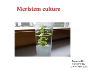
- 1. Presented by: Suresh Yadav I.D No.- Palm 6005
- 3. Production of disease free seedlings through tissue culture Presented by :- Suresh Yadav I.D No:- PALM 6005
- 4. Tissue culture :-Morel and Martin (1952) developed the technique of meristem culture for in vivo virus eradication of Dahlia. Shoots of all angiosperms and gymnosperms grow by virtue of their apical meristems. The apical meristem is usually a dome of tissue located at the extreme tip of a shoot and measures approx. 0.1 mm in diameter and 0.25 to 0.3 mm in length. Meristem or shoot tip is isolated from stem by applying a V- shaped cut. MS medium salts have been very satisfactory for such cultures though White’s and Gautheret’s were the most widely used media during the early days of meristem culture. INTRODUCTION
- 7. PROTOCOL Remove the young twigs from a healthy plant. Cut the tip (1 cm) portion of the twig Surface sterilize the shoot apices by incubation in a sodium hypochlorite solution (1% available chlorine) for 10 minutes. The explants are thoroughly rinsed 4 times in distilled water Transfer each explants to a sterilized petri dish.
- 8. Remove the outer leaves from each shoot After the removal of all outer leaves, the apex is exposed. Cut off the ultimate apex with the help of scalpel and transfer only those less than 1 mm in length Incubate the culture under 16hrs light at 25°C As soon as the growing single leafy shoot or multiple shoots obtained from single shoot tip or meristem, transfer them to hormone free medium to develop roots. The plants are later transferred to pots containing compost and kept under green house condition for hardening.
- 9. Application of Shoot-tip or Meristem Culture 1. Virus Elimination • Plants are often infected with more than one type of virus, including some even not known. • A general term virus- free is used by commercial horticulturist by this method.
- 10. 2. Micro Propagation • Asexual or vegetative propagation (vegetative part) of whole plants using tissue culture techniques referred to as micro propagation. 3. Storage of Genetic Resources • Many plants produce seeds that are highly heterozygous in nature or that is recalcitrant. Such seeds are not accepted for storing genetic resources. So , the meristem from such plants can be stored in vitro.
- 11. 4. Use in Plant Breeding: •In many plant breeding experiments the hybrid plants produce abortive seeds or non viable seeds. As a result, it makes a barrier to crossibility in plants where non-viable seeds are unable to develop into mature plants. Shoot-tip or meristem from such hybrid plant can be cultured to speed up breeding programme. 5. Quarantine • Plantlets derived from shoot-tip or meristem cultures are easily accepted by the quarantine authority for international exchange without any checking. • Therefore, using this technique , crop plants can be easily exchanged in crop improvement programmes that are based on materials from different parts of the world.
- 12. List of the plants from which viruses have been eliminated by meristem cultures
- 13. ADVANTAGES: Lack of vascular tissue. High auxin concentration. Production of virus free plants Facilitation of exchange between locations Cryopreservation or in-vitro conservation of germplasm DISADVANTAGES: Isolation is difficult Low survival rate & regeneration time for explants may be long(about 8 months for potato explant) Removal of explant causes a setback in the growth of mother plant.
- 15. Content • Introduction • Objectives • Seed Health Testing for Fungi • Detecting Seed Borne Bacterial Plant Pathogens • Detecting Seed Borne Plant Viruses • Detection of Insects and Nematodes • Conclusion • References
- 16. Introduction • Seed Health Testing is a Science of determining the presence or absence of disease causing agents, such as fungi, bacteria and viruses. • Animal pests like eelworms, insects in the seed samples.
- 17. Objectives • To determine the health status of a seed lot which in turn establishes the sanitary condition of the seed in commerce. • To obtain objective proof of whether the seed lot meets the requisite certification standards or not. • To obtain objective proof of whether the lot meets requisite quarantine requirements.
- 18. Methods of seed Heath Testing for Fungi • Visual examination – with or without stereomicroscope. • Inspected in dry state for the presence of impurities such as ergots or other sclerotia.
- 19. Determination of Karnalbunt (Neovassia indica) of wheat • Infected seeds have a characteristic black powdery mass generally along with the suture. • Severe infection – most of the endosperm together with pericarp and aleurone layer gets destroyed giving grains a boat like appearance.
- 20. Determination of bunt (Neovossia horrida) of Rice •Seeds are opened with the help of the blade •Whole grains transformed into black powdery mass
- 21. • Alternate procedure : Paddy seeds are soaked in 0.2% solution of sodium hydroxide for 24 hours at 18 -25°C . • Swollen seeds are visually examined for shiny jet black colour.
- 22. Determination of Loose smut infection in Wheat • Soak seeds for 24hours at 20°C in Sodium Hydroxide and Trypan Blue. • Separate the embryos by passing the soaked material along with warm water (50-60°C) • Embryos are washed in wire basket and dehydrated in 95% alcohol for 2min and later transferred to a mixture of lactophenol and water.
- 23. Continue:- • Further separation of embryos can be made. • Embryos are placed in lactophenol and boiled. • Embryos are examined in microscope. • Infected fungus with loose smut fungus is present as hyphal strands. • Golden brown thread like mycelium which is septate with non uniform thickness and swellings are classified as infected embryos.
- 24. UV Examination • Toxins in Fungi gives Fluoroscence appearance. • This represents presence of Fungi
- 25. Blotter Method •Seeds are placed on moistened blotters, filter papers at least 20mm apart. •Blotters are placed in closed containers and incubated for certain number of days and later examined for pathogens
- 26. Agar Plate Method •After pretreatment seeds are spaced on the surface of 2% malt extract sterilised agar. •After plating dishes are incubated at 20-25°C for 5-8 days.
- 27. Wash Method • Seeds are immersed in water with a wetting agent and shaked vigorously to remove fungal spores, hyphae etc. • Excess liquid is removed and material is examined.
- 28. Examination of Imbibed Seeds • Seeds are immersed in water, or other liquid to make fruiting bodies, symptoms more easily viable or to encourage the liberation of spores. • After imbibition the seeds are examined with a stereoscopic microscope
- 29. PCR Method • It is a kind of molecular method. • Specific primers are used in this method. • PCR is a promising tool for distinguishing specific sequences from a complex mixture of DNA and therefore is useful for determining the presence and quantity of pathogen- specific or other unique sequences within a sample
- 30. Methods to detect seed borne bacterial pathogens • Various methods are been developed. • Some of the methods are simple and some are specialized namely serological methods.
- 31. Growing out test The ‘growing out’ bioassay of a working seed sample involves the sowing of test seeds into seedlings under conditions optimal for the disease development in glass house or closed environmental chambers. ‘Growing out’ test has been successfully used for a large number of Xanthomonads and pseudomonas.
- 32. Indicator test Working seed sample is sterilized with (2.6%) sodium- hypochlorite for 15 min, and rinsed with sterile water. The seed sample is incubated for 18-24 h in sterile water. The water suspension is inoculated by infiltration into the primary leaf node of 10 day old bean seedlings. The appearance of lesions followed systemic necrosis is positive reaction.
- 33. Serological Technique Serological tests are based on ‘In vitro’ reactions between antigens and antibodies. This specific recognition of antigens by antibody has offered the basic principle for the development of various serological methods for detection and identification of phytobacteria. The washing of the working seed samples are cultured for 36 hr using sterile distilled water. The supernatant is tested with antiserum of the suspected pathogen.
- 34. Culture Method • By culturing the samples on the particular media. • Presence of bacteria can be observed on the media.
- 35. Methods to detect seed borne Plant Viruses Biological Method a) By growing seeds: The seeds are examined and abnormal seeds are separated. b) Both normal looking and abnormal looking seeds are grown separately and the seedings are observed for the characteristic symptom of the virus disease. c) Plants are kept free of insect vectors such as mites and white fly.
- 36. • Direct seed Test a) Seed are examined and abnormal seeds are separated and handled separately. b) Seeds are soaked in aqueous medium and then triturated. c) The slurries produced are then applied to indicator or test plants. d) The slurries containing virus produce symptoms typical for a given virus on the indicator plant.
- 37. Serological Tests • Double diffusion test a) Seeds to be tested are soaked in tapwater. b) Seed or part (embryo) of it is triturated and triturate is transferred to a well cut in a diffusion media(agar gel). c) Antiserum specific for a suspect virus in a seed is placed in separate well.
- 38. • In time the virus particles(antigen) and antibody molecules diffuse towards one another. • Since diffusion is in two direction it is called Double diffusion.
- 39. ELISA(Enzyme linked immunosobent assay) • In this procedure , antiserum specific for a given virus is used to coat the polystyrene plate. • Antibody molecules become absorbed. • Then seed sample is added to the plate. • It is followed by adding enzyme labelled specific antibody to the plate.
- 40. • The enzyme alkaline phosphates is conjugated to the antibody molecules specific for the virus under examination. • Finally enzyme substrate is added to the plate. • Hydrolyzed substrate is determined by measuring the extinction spectrophotometrically or by visual observation.
- 41. Detection of Insects and Nematodes Detection of presence of Insects • Examined under magnification or stereoscopic microscope • Result is mentioned as number of insects per weight of the sample
- 42. Detection of external insect injury • Working sample is Examined under magnification(10x) or stereoscopic microscope. • The absence of insects does not however guarantee that the seed lot is free from insect infestation, i.e internal injury to the seeds or internal infestation.
- 43. Detection of Nematode Infestation • Working sample is visually examined for the presence of ear cocke galls. (hard, small, dark purplish- black colour structures) • Galls are separated and soaked in water for 30 minutes. and are cut in water in petridish for observing the release of nematode larvae, galls releasing nematode are counted and result is reported in percentage. Ear cockle of wheat (Anguina tritici)
- 44. Galls on wheat Anguina tritici
- 45. Root knot Nematode of Sweet Potato (Meloidogyne incognita) • Entire submitted sample is examined for visible symptoms of nematode infestation. Galls on Sweet Potato
- 46. • Nematode infested tubers look ugly and disfigured. • When tubers are cut across the knot , small glistening round pin head shaped bodies are seen and such tubers are separated and the results are reported.
- 47. POTATO CERTIFIED SEED STANDARDS Disease Incidence India Holland* Canada UK Mild mosaics 3 2 1 5 SM and PLRV 1 0.25 1 2 Brown rot 0.014 0 0 0 LB, Dry rot 1 NP NP NP Wet rot 0 NP NP NP Black scurf 5** NP NP NP Common scab 5** NP NP NP Total tuber Disease 5 - - - Root knot nematode Nil NP NP Nil Cyst nematode Nil NP NP Nil NP : No limit prescribed * : Brown rot prevalent since 1995 ** : Tuber treatment compulsory
- 48. SEED CERTIFICATION STANDARDS FOR FOUNDATION AND CERTIFIED SEEDS Permissible DI limits for foliar diseases Seed grade % DI limits Off typeMild mosaic Severe mosaic/PLRV/PVT/ LST/ yellows PSTV FS-1 1.0 0.5 - 0.05 FS-II 2.0 0.75 - 0.05 Certified 3.0 1.0 - 0.1
- 49. PERMISSIBLE LIMITS FOR TUBER DISEASES Grade % incidence Common scab Black scurf Cut/ bruised Late blight Dry rot FS-1 5.0 5.0 1.0 1.0 1.0 FS-II 5.0 5.0 1.0 1.0 1.0 Certified 5.0 5.0 5.0 1.0 1.0
- 50. Eligibility criteria for production of minitubers through tissue culture Basic Material The basic tissue culture material must be of a notified variety and the plantlet should be tested by the accredited laboratory for freedom from PVA, PVS, PVX, PVY, PVM, PLRV, PALCV, PSTVd, endophytic bacteria and fungi 10 plantlet to be tested for each variety Method ELISA, RTPC Light microscopy Culturing on media
- 51. Laboratory & Green House Lab and net house facilities free from insect pests Growth and Potting medium – sterilized Net house facility Insect proof Double door with changing space
- 52. All micro-propagation and green house facilities must meet the prescribed standards Net house soil should be free from Wart Brown rot Non-cyst forming nematodes Cyst nematodes Common scab CONTROLLED ENVIRONMENT REQUIREMENT
- 53. The seed producer should inform the competent authority well in advance Source of seed has to be tissue culture grown plantlets/ micro tubers A minimum of three inspections shall be made First inspection (35 days : plains) (45 days : hills) - For diseases, off types & isolation (1 m) Second inspection Besides general inspection pathogen testing (viruses) 5 (minimum) – 25 (maximum) plants/varieties Third inspection (immediately after haulm cutting) - To check date of haulm cutting? - Re-growth INSPECTION OF NET HOUSE FACILITY
- 54. Use of effective sanitation Sterilization of nursery beds The net house grown crop should be 100% free from all viruses If any of the virus is detected, the crop will be rejected INSPECTION OF NET HOUSE FACILITY
- 55. Certification standards for net house grown crop Factor Maximum Permissible limits (%) Off types 0.05 Mild mosaic 0.05 Severe mosaics, Apical Leaf Curl, PLRV 0.05 Bacterial wilt Nil
- 56. Standards for mini tubers Factor Standards Weight (minimum) >1.0 g Germination (minimum) 90% Varietal purity (minimum) 99% Virus (all) 0.1