SlideShare a Scribd company logo
1 of 10
Download to read offline
Figure:
Immunohistochemical
analysis of paraffin
embedded Human colon
tissue. 1: CK8
Monoclonal
Antibody(8G8) was
diluted at 1:200 (4
degree
Celsius,overnight). 2:
Sodium citrate pH 6.0
was used for antibody
retrieval (>98 degree
Celsius,20min). 3:
Secondary antibody was
diluted at 1:200 (room
temperature, 30min).
Negative control was
used by secondary
antibody only.
Report Number 96957-a Host Mouse
Application IHC-P Clonality Monoclonal
Model Number STJ96957 Clone ID 8G8
Antibody Name Anti-CK8 antibody
Testing Species HUMAN Testing Tissue COLON
ANTIBODY VALIDATION REPORT
a. (A small amount of distilled water was added into the incubation
box to prevent evaporation of antibody).
40. Secondary antibody incubation
a. Slides were washed 3 times, with PBS on a shaker for 5min.
Shortly after the slides were dried the corresponding secondary
antibody solution was added (HRP labelled), covering the
tissues, and incubated at room temperature for 30min.
b.
41. DAB staining
a. Slides were washed 3 times, with PBS on a shaker for 5min.
b. Shortly after, the slides were dried and fresh DAB staining buffer
was added inside the circles. The staining time was adjusted
under a microscope. Yellow-brown colour represented a positive
result. Slides were washed with water to stop the staining.
c.
42. Haematoxylin staining
a. Haematoxylin was used to counter-staining for 1min, and then
the slides were washed with water. 1% Hydrochloric acid and
alcohol was added for several seconds and then washed with
water. Ammonia was used to reveal blue colour, and then
flushed with water.
b.
43. Desolation and Clearing
i. Slides were incubated sequentially into: 75% alcohol 5min, 85%
alcohol 5min, Anhydrous ethanol - 5min, Anhydrous ethanol -
5min & Xylene - 5min. Shortly after slides were dried and neutral
gum was used to seal the slides.
ii.
44. Visualization
a. Results were validated with microscope, and the slides were
scanned.
Paraffin-Embedded
Immunohistochemistry Protocol
34.
35. Tissue processing
a. Slides were incubated sequentially into Xylene; 15min –
Xylene, 15min - Anhydrous ethanol, 5min - Anhydrous
ethanol, 5min - 85% alcohol, 5min - 75% alcohol & 5min –
wash in distilled water.
b.
36. Antigen retrieval
a. Tissue slides were incubated with citric acid (PH6.0) antigen
retrieval buffer and microwaved for antigen retrieval (heated
until boiled and then stopped heating) for 8min. Slides were
then heated with medium power for 7min. During this
process slides were kept from drying out. After cooling down
at room temperature, slides were washed with PBS on
shaker for 5min, repeated for 3 times.
b.
37. Inhibition of endogenous peroxidase
a. Slides were placed in 3% Hydrogen peroxide solution, and
incubated for 10 min at room temperature without light
exposure. Slides were then washed 3 times with PBS on a
shaker for 5mins.
b.
38. BSA Blocking
a. Shortly after slides were dried, a PAP pen was used to draw
circles around the tissue sections (and to prevent draining of
the antibody solution). Inside the circles, BSA was used to
cover the tissue evenly, blocking for 30min.
b.
39. Primary antibody incubation
After blocking solution was removed a 1:200 solution of
primary antibody/PBS was added on the slide, and incubated
overnight at 4°C.
St John's Laboratory Ltd.
www.stjohnslabs.com
Figure:
Immunohistochemical
analysis of paraffin
embedded Human liver
cancer tissue. 1: CK8
Monoclonal
Antibody(8G8) was
diluted at 1:200 (4
degree
Celsius,overnight). 2:
Sodium citrate pH 6.0
was used for antibody
retrieval (>98 degree
Celsius,20min). 3:
Secondary antibody was
diluted at 1:200 (room
temperature, 30min).
Negative control was
used by secondary
antibody only.
Report Number 96957-b Host Mouse
Application IHC-P Clonality Monoclonal
Model Number STJ96957 Clone ID 8G8
Antibody Name Anti-CK8 antibody
Testing Species HUMAN Testing Tissue LIVER CANCER
ANTIBODY VALIDATION REPORT
b. (A small amount of distilled water was added into the incubation
box to prevent evaporation of antibody).
29. Secondary antibody incubation
a. Slides were washed 3 times, with PBS on a shaker for 5min.
Shortly after the slides were dried the corresponding secondary
antibody solution was added (HRP labelled), covering the
tissues, and incubated at room temperature for 30min.
b.
30. DAB staining
a. Slides were washed 3 times, with PBS on a shaker for 5min.
b. Shortly after, the slides were dried and fresh DAB staining buffer
was added inside the circles. The staining time was adjusted
under a microscope. Yellow-brown colour represented a positive
result. Slides were washed with water to stop the staining.
c.
31. Haematoxylin staining
a. Haematoxylin was used to counter-staining for 1min, and then
the slides were washed with water. 1% Hydrochloric acid and
alcohol was added for several seconds and then washed with
water. Ammonia was used to reveal blue colour, and then
flushed with water.
b.
32. Desolation and Clearing
i. Slides were incubated sequentially into: 75% alcohol 5min, 85%
alcohol 5min, Anhydrous ethanol - 5min, Anhydrous ethanol -
5min & Xylene - 5min. Shortly after slides were dried and neutral
gum was used to seal the slides.
ii.
33. Visualization
a. Results were validated with microscope, and the slides were
scanned.
Paraffin-Embedded
Immunohistochemistry Protocol
23.
24. Tissue processing
a. Slides were incubated sequentially into Xylene; 15min –
Xylene, 15min - Anhydrous ethanol, 5min - Anhydrous
ethanol, 5min - 85% alcohol, 5min - 75% alcohol & 5min –
wash in distilled water.
b.
25. Antigen retrieval
a. Tissue slides were incubated with citric acid (PH6.0) antigen
retrieval buffer and microwaved for antigen retrieval (heated
until boiled and then stopped heating) for 8min. Slides were
then heated with medium power for 7min. During this
process slides were kept from drying out. After cooling down
at room temperature, slides were washed with PBS on
shaker for 5min, repeated for 3 times.
b.
26. Inhibition of endogenous peroxidase
a. Slides were placed in 3% Hydrogen peroxide solution, and
incubated for 10 min at room temperature without light
exposure. Slides were then washed 3 times with PBS on a
shaker for 5mins.
b.
27. BSA Blocking
a. Shortly after slides were dried, a PAP pen was used to draw
circles around the tissue sections (and to prevent draining of
the antibody solution). Inside the circles, BSA was used to
cover the tissue evenly, blocking for 30min.
b.
28. Primary antibody incubation
After blocking solution was removed a 1:200 solution of
primary antibody/PBS was added on the slide, and incubated
overnight at 4°C.
St John's Laboratory Ltd.
www.stjohnslabs.com
Figure:
Immunohistochemical
analysis of paraffin
embedded Human
stomach tissue. 1: CK8
Monoclonal
Antibody(8G8) was
diluted at 1:200 (4
degree
Celsius,overnight). 2:
Sodium citrate pH 6.0
was used for antibody
retrieval (>98 degree
Celsius,20min). 3:
Secondary antibody was
diluted at 1:200 (room
temperature, 30min).
Negative control was
used by secondary
antibody only.
Report Number 96957-c Host Mouse
Application IHC-P Clonality Monoclonal
Model Number STJ96957 Clone ID 8G8
Antibody Name Anti-CK8 antibody
Testing Species HUMAN Testing Tissue STOMACH
ANTIBODY VALIDATION REPORT
b. (A small amount of distilled water was added into the incubation
box to prevent evaporation of antibody).
18. Secondary antibody incubation
a. Slides were washed 3 times, with PBS on a shaker for 5min.
Shortly after the slides were dried the corresponding secondary
antibody solution was added (HRP labelled), covering the
tissues, and incubated at room temperature for 30min.
b.
19. DAB staining
a. Slides were washed 3 times, with PBS on a shaker for 5min.
b. Shortly after, the slides were dried and fresh DAB staining buffer
was added inside the circles. The staining time was adjusted
under a microscope. Yellow-brown colour represented a positive
result. Slides were washed with water to stop the staining.
c.
20. Haematoxylin staining
a. Haematoxylin was used to counter-staining for 1min, and then
the slides were washed with water. 1% Hydrochloric acid and
alcohol was added for several seconds and then washed with
water. Ammonia was used to reveal blue colour, and then
flushed with water.
b.
21. Desolation and Clearing
i. Slides were incubated sequentially into: 75% alcohol 5min, 85%
alcohol 5min, Anhydrous ethanol - 5min, Anhydrous ethanol -
5min & Xylene - 5min. Shortly after slides were dried and neutral
gum was used to seal the slides.
ii.
22. Visualization
a. Results were validated with microscope, and the slides were
scanned.
Paraffin-Embedded
Immunohistochemistry Protocol
12.
13. Tissue processing
a. Slides were incubated sequentially into Xylene; 15min –
Xylene, 15min - Anhydrous ethanol, 5min - Anhydrous
ethanol, 5min - 85% alcohol, 5min - 75% alcohol & 5min –
wash in distilled water.
b.
14. Antigen retrieval
a. Tissue slides were incubated with citric acid (PH6.0) antigen
retrieval buffer and microwaved for antigen retrieval (heated
until boiled and then stopped heating) for 8min. Slides were
then heated with medium power for 7min. During this
process slides were kept from drying out. After cooling down
at room temperature, slides were washed with PBS on
shaker for 5min, repeated for 3 times.
b.
15. Inhibition of endogenous peroxidase
a. Slides were placed in 3% Hydrogen peroxide solution, and
incubated for 10 min at room temperature without light
exposure. Slides were then washed 3 times with PBS on a
shaker for 5mins.
b.
16. BSA Blocking
a. Shortly after slides were dried, a PAP pen was used to draw
circles around the tissue sections (and to prevent draining of
the antibody solution). Inside the circles, BSA was used to
cover the tissue evenly, blocking for 30min.
b.
17. Primary antibody incubation
After blocking solution was removed a 1:200 solution of
primary antibody/PBS was added on the slide, and incubated
overnight at 4°C.
St John's Laboratory Ltd.
www.stjohnslabs.com
Figure:
Immunohistochemical
analysis of paraffin
embedded Human
stomach cancer tissue.
1: CK8 Monoclonal
Antibody(8G8) was
diluted at 1:200 (4
degree
Celsius,overnight). 2:
Sodium citrate pH 6.0
was used for antibody
retrieval (>98 degree
Celsius,20min). 3:
Secondary antibody was
diluted at 1:200 (room
temperature, 30min).
Negative control was
used by secondary
antibody only.
Report Number 96957-d Host Mouse
Application IHC-P Clonality Monoclonal
Model Number STJ96957 Clone ID 8G8
Antibody Name Anti-CK8 antibody
Testing Species HUMAN Testing Tissue STOMACH CANCER
ANTIBODY VALIDATION REPORT
b. (A small amount of distilled water was added into the incubation
box to prevent evaporation of antibody).
7. Secondary antibody incubation
a. Slides were washed 3 times, with PBS on a shaker for 5min.
Shortly after the slides were dried the corresponding secondary
antibody solution was added (HRP labelled), covering the
tissues, and incubated at room temperature for 30min.
b.
8. DAB staining
a. Slides were washed 3 times, with PBS on a shaker for 5min.
b. Shortly after, the slides were dried and fresh DAB staining buffer
was added inside the circles. The staining time was adjusted
under a microscope. Yellow-brown colour represented a positive
result. Slides were washed with water to stop the staining.
c.
9. Haematoxylin staining
a. Haematoxylin was used to counter-staining for 1min, and then
the slides were washed with water. 1% Hydrochloric acid and
alcohol was added for several seconds and then washed with
water. Ammonia was used to reveal blue colour, and then
flushed with water.
b.
10. Desolation and Clearing
i. Slides were incubated sequentially into: 75% alcohol 5min, 85%
alcohol 5min, Anhydrous ethanol - 5min, Anhydrous ethanol -
5min & Xylene - 5min. Shortly after slides were dried and neutral
gum was used to seal the slides.
ii.
11. Visualization
a. Results were validated with microscope, and the slides were
scanned.
Paraffin-Embedded
Immunohistochemistry Protocol
1.
2. Tissue processing
a. Slides were incubated sequentially into Xylene; 15min –
Xylene, 15min - Anhydrous ethanol, 5min - Anhydrous
ethanol, 5min - 85% alcohol, 5min - 75% alcohol & 5min –
wash in distilled water.
b.
3. Antigen retrieval
a. Tissue slides were incubated with citric acid (PH6.0) antigen
retrieval buffer and microwaved for antigen retrieval (heated
until boiled and then stopped heating) for 8min. Slides were
then heated with medium power for 7min. During this
process slides were kept from drying out. After cooling down
at room temperature, slides were washed with PBS on
shaker for 5min, repeated for 3 times.
b.
4. Inhibition of endogenous peroxidase
a. Slides were placed in 3% Hydrogen peroxide solution, and
incubated for 10 min at room temperature without light
exposure. Slides were then washed 3 times with PBS on a
shaker for 5mins.
b.
5. BSA Blocking
a. Shortly after slides were dried, a PAP pen was used to draw
circles around the tissue sections (and to prevent draining of
the antibody solution). Inside the circles, BSA was used to
cover the tissue evenly, blocking for 30min.
b.
6. Primary antibody incubation
After blocking solution was removed a 1:200 solution of
primary antibody/PBS was added on the slide, and incubated
overnight at 4°C.
St John's Laboratory Ltd.
www.stjohnslabs.com
Figure:
Immunohistochemical
analysis of paraffin
embedded Rat liver
tissue. 1: CK8
Monoclonal
Antibody(8G8) was
diluted at 1:200 (4
degree
Celsius,overnight). 2:
Sodium citrate pH 6.0
was used for antibody
retrieval (>98 degree
Celsius,20min). 3:
Secondary antibody was
diluted at 1:200 (room
temperature, 30min).
Negative control was
used by secondary
antibody only.
Report Number 96957-e Host Mouse
Application IHC-P Clonality Monoclonal
Model Number STJ96957 Clone ID 8G8
Antibody Name Anti-CK8 antibody
Testing Species RAT Testing Tissue LIVER
ANTIBODY VALIDATION REPORT
c. (A small amount of distilled water was added into the incubation
box to prevent evaporation of antibody).
45. Secondary antibody incubation
a. Slides were washed 3 times, with PBS on a shaker for 5min.
Shortly after the slides were dried the corresponding secondary
antibody solution was added (HRP labelled), covering the
tissues, and incubated at room temperature for 30min.
b.
46. DAB staining
a. Slides were washed 3 times, with PBS on a shaker for 5min.
b. Shortly after, the slides were dried and fresh DAB staining buffer
was added inside the circles. The staining time was adjusted
under a microscope. Yellow-brown colour represented a positive
result. Slides were washed with water to stop the staining.
c.
47. Haematoxylin staining
a. Haematoxylin was used to counter-staining for 1min, and then
the slides were washed with water. 1% Hydrochloric acid and
alcohol was added for several seconds and then washed with
water. Ammonia was used to reveal blue colour, and then
flushed with water.
b.
48. Desolation and Clearing
i. Slides were incubated sequentially into: 75% alcohol 5min, 85%
alcohol 5min, Anhydrous ethanol - 5min, Anhydrous ethanol -
5min & Xylene - 5min. Shortly after slides were dried and neutral
gum was used to seal the slides.
ii.
49. Visualization
a. Results were validated with microscope, and the slides were
scanned.
Paraffin-Embedded
Immunohistochemistry Protocol
50.
51. Tissue processing
a. Slides were incubated sequentially into Xylene; 15min –
Xylene, 15min - Anhydrous ethanol, 5min - Anhydrous
ethanol, 5min - 85% alcohol, 5min - 75% alcohol & 5min –
wash in distilled water.
b.
52. Antigen retrieval
a. Tissue slides were incubated with citric acid (PH6.0) antigen
retrieval buffer and microwaved for antigen retrieval (heated
until boiled and then stopped heating) for 8min. Slides were
then heated with medium power for 7min. During this
process slides were kept from drying out. After cooling down
at room temperature, slides were washed with PBS on
shaker for 5min, repeated for 3 times.
b.
53. Inhibition of endogenous peroxidase
a. Slides were placed in 3% Hydrogen peroxide solution, and
incubated for 10 min at room temperature without light
exposure. Slides were then washed 3 times with PBS on a
shaker for 5mins.
b.
54. BSA Blocking
a. Shortly after slides were dried, a PAP pen was used to draw
circles around the tissue sections (and to prevent draining of
the antibody solution). Inside the circles, BSA was used to
cover the tissue evenly, blocking for 30min.
b.
55. Primary antibody incubation
After blocking solution was removed a 1:200 solution of
primary antibody/PBS was added on the slide, and incubated
overnight at 4°C.
St John's Laboratory Ltd.
www.stjohnslabs.com
Figure:
Immunohistochemical
analysis of paraffin
embedded Rat lung
tissue. 1: CK8
Monoclonal
Antibody(8G8) was
diluted at 1:200 (4
degree
Celsius,overnight). 2:
Sodium citrate pH 6.0
was used for antibody
retrieval (>98 degree
Celsius,20min). 3:
Secondary antibody was
diluted at 1:200 (room
temperature, 30min).
Negative control was
used by secondary
antibody only.
Report Number 96957-f Host Mouse
Application IHC-P Clonality Monoclonal
Model Number STJ96957 Clone ID 8G8
Antibody Name Anti-CK8 antibody
Testing Species RAT Testing Tissue LUNG
ANTIBODY VALIDATION REPORT
b. (A small amount of distilled water was added into the incubation
box to prevent evaporation of antibody).
56. Secondary antibody incubation
a. Slides were washed 3 times, with PBS on a shaker for 5min.
Shortly after the slides were dried the corresponding secondary
antibody solution was added (HRP labelled), covering the
tissues, and incubated at room temperature for 30min.
b.
57. DAB staining
a. Slides were washed 3 times, with PBS on a shaker for 5min.
b. Shortly after, the slides were dried and fresh DAB staining buffer
was added inside the circles. The staining time was adjusted
under a microscope. Yellow-brown colour represented a positive
result. Slides were washed with water to stop the staining.
c.
58. Haematoxylin staining
a. Haematoxylin was used to counter-staining for 1min, and then
the slides were washed with water. 1% Hydrochloric acid and
alcohol was added for several seconds and then washed with
water. Ammonia was used to reveal blue colour, and then
flushed with water.
b.
59. Desolation and Clearing
i. Slides were incubated sequentially into: 75% alcohol 5min, 85%
alcohol 5min, Anhydrous ethanol - 5min, Anhydrous ethanol -
5min & Xylene - 5min. Shortly after slides were dried and neutral
gum was used to seal the slides.
ii.
60. Visualization
a. Results were validated with microscope, and the slides were
scanned.
Paraffin-Embedded
Immunohistochemistry Protocol
61.
62. Tissue processing
a. Slides were incubated sequentially into Xylene; 15min –
Xylene, 15min - Anhydrous ethanol, 5min - Anhydrous
ethanol, 5min - 85% alcohol, 5min - 75% alcohol & 5min –
wash in distilled water.
b.
63. Antigen retrieval
a. Tissue slides were incubated with citric acid (PH6.0) antigen
retrieval buffer and microwaved for antigen retrieval (heated
until boiled and then stopped heating) for 8min. Slides were
then heated with medium power for 7min. During this
process slides were kept from drying out. After cooling down
at room temperature, slides were washed with PBS on
shaker for 5min, repeated for 3 times.
b.
64. Inhibition of endogenous peroxidase
a. Slides were placed in 3% Hydrogen peroxide solution, and
incubated for 10 min at room temperature without light
exposure. Slides were then washed 3 times with PBS on a
shaker for 5mins.
b.
65. BSA Blocking
a. Shortly after slides were dried, a PAP pen was used to draw
circles around the tissue sections (and to prevent draining of
the antibody solution). Inside the circles, BSA was used to
cover the tissue evenly, blocking for 30min.
b.
66. Primary antibody incubation
After blocking solution was removed a 1:200 solution of
primary antibody/PBS was added on the slide, and incubated
overnight at 4°C.
St John's Laboratory Ltd.
www.stjohnslabs.com
Figure:
Immunohistochemical
analysis of paraffin
embedded Rat kidney
tissue. 1: CK8
Monoclonal
Antibody(8G8) was
diluted at 1:200 (4
degree
Celsius,overnight). 2:
Sodium citrate pH 6.0
was used for antibody
retrieval (>98 degree
Celsius,20min). 3:
Secondary antibody was
diluted at 1:200 (room
temperature, 30min).
Negative control was
used by secondary
antibody only.
Report Number 96957-g Host Mouse
Application IHC-P Clonality Monoclonal
Model Number STJ96957 Clone ID 8G8
Antibody Name Anti-CK8 antibody
Testing Species RAT Testing Tissue KIDNEY
ANTIBODY VALIDATION REPORT
b. (A small amount of distilled water was added into the incubation
box to prevent evaporation of antibody).
67. Secondary antibody incubation
a. Slides were washed 3 times, with PBS on a shaker for 5min.
Shortly after the slides were dried the corresponding secondary
antibody solution was added (HRP labelled), covering the
tissues, and incubated at room temperature for 30min.
b.
68. DAB staining
a. Slides were washed 3 times, with PBS on a shaker for 5min.
b. Shortly after, the slides were dried and fresh DAB staining buffer
was added inside the circles. The staining time was adjusted
under a microscope. Yellow-brown colour represented a positive
result. Slides were washed with water to stop the staining.
c.
69. Haematoxylin staining
a. Haematoxylin was used to counter-staining for 1min, and then
the slides were washed with water. 1% Hydrochloric acid and
alcohol was added for several seconds and then washed with
water. Ammonia was used to reveal blue colour, and then
flushed with water.
b.
70. Desolation and Clearing
i. Slides were incubated sequentially into: 75% alcohol 5min, 85%
alcohol 5min, Anhydrous ethanol - 5min, Anhydrous ethanol -
5min & Xylene - 5min. Shortly after slides were dried and neutral
gum was used to seal the slides.
ii.
71. Visualization
a. Results were validated with microscope, and the slides were
scanned.
Paraffin-Embedded
Immunohistochemistry Protocol
72.
73. Tissue processing
a. Slides were incubated sequentially into Xylene; 15min –
Xylene, 15min - Anhydrous ethanol, 5min - Anhydrous
ethanol, 5min - 85% alcohol, 5min - 75% alcohol & 5min –
wash in distilled water.
b.
74. Antigen retrieval
a. Tissue slides were incubated with citric acid (PH6.0) antigen
retrieval buffer and microwaved for antigen retrieval (heated
until boiled and then stopped heating) for 8min. Slides were
then heated with medium power for 7min. During this
process slides were kept from drying out. After cooling down
at room temperature, slides were washed with PBS on
shaker for 5min, repeated for 3 times.
b.
75. Inhibition of endogenous peroxidase
a. Slides were placed in 3% Hydrogen peroxide solution, and
incubated for 10 min at room temperature without light
exposure. Slides were then washed 3 times with PBS on a
shaker for 5mins.
b.
76. BSA Blocking
a. Shortly after slides were dried, a PAP pen was used to draw
circles around the tissue sections (and to prevent draining of
the antibody solution). Inside the circles, BSA was used to
cover the tissue evenly, blocking for 30min.
b.
77. Primary antibody incubation
After blocking solution was removed a 1:200 solution of
primary antibody/PBS was added on the slide, and incubated
overnight at 4°C.
St John's Laboratory Ltd.
www.stjohnslabs.com
Figure:
Immunohistochemical
analysis of paraffin
embedded Mouse liver
tissue. 1: CK8
Monoclonal
Antibody(8G8) was
diluted at 1:200 (4
degree
Celsius,overnight). 2:
Sodium citrate pH 6.0
was used for antibody
retrieval (>98 degree
Celsius,20min). 3:
Secondary antibody was
diluted at 1:200 (room
temperature, 30min).
Negative control was
used by secondary
antibody only.
Report Number 96957-h Host Mouse
Application IHC-P Clonality Monoclonal
Model Number STJ96957 Clone ID 8G8
Antibody Name Anti-CK8 antibody
Testing Species MOUSE Testing Tissue LIVER
ANTIBODY VALIDATION REPORT
b. (A small amount of distilled water was added into the incubation
box to prevent evaporation of antibody).
78. Secondary antibody incubation
a. Slides were washed 3 times, with PBS on a shaker for 5min.
Shortly after the slides were dried the corresponding secondary
antibody solution was added (HRP labelled), covering the
tissues, and incubated at room temperature for 30min.
b.
79. DAB staining
a. Slides were washed 3 times, with PBS on a shaker for 5min.
b. Shortly after, the slides were dried and fresh DAB staining buffer
was added inside the circles. The staining time was adjusted
under a microscope. Yellow-brown colour represented a positive
result. Slides were washed with water to stop the staining.
c.
80. Haematoxylin staining
a. Haematoxylin was used to counter-staining for 1min, and then
the slides were washed with water. 1% Hydrochloric acid and
alcohol was added for several seconds and then washed with
water. Ammonia was used to reveal blue colour, and then
flushed with water.
b.
81. Desolation and Clearing
i. Slides were incubated sequentially into: 75% alcohol 5min, 85%
alcohol 5min, Anhydrous ethanol - 5min, Anhydrous ethanol -
5min & Xylene - 5min. Shortly after slides were dried and neutral
gum was used to seal the slides.
ii.
82. Visualization
a. Results were validated with microscope, and the slides were
scanned.
Paraffin-Embedded
Immunohistochemistry Protocol
83.
84. Tissue processing
a. Slides were incubated sequentially into Xylene; 15min –
Xylene, 15min - Anhydrous ethanol, 5min - Anhydrous
ethanol, 5min - 85% alcohol, 5min - 75% alcohol & 5min –
wash in distilled water.
b.
85. Antigen retrieval
a. Tissue slides were incubated with citric acid (PH6.0) antigen
retrieval buffer and microwaved for antigen retrieval (heated
until boiled and then stopped heating) for 8min. Slides were
then heated with medium power for 7min. During this
process slides were kept from drying out. After cooling down
at room temperature, slides were washed with PBS on
shaker for 5min, repeated for 3 times.
b.
86. Inhibition of endogenous peroxidase
a. Slides were placed in 3% Hydrogen peroxide solution, and
incubated for 10 min at room temperature without light
exposure. Slides were then washed 3 times with PBS on a
shaker for 5mins.
b.
87. BSA Blocking
a. Shortly after slides were dried, a PAP pen was used to draw
circles around the tissue sections (and to prevent draining of
the antibody solution). Inside the circles, BSA was used to
cover the tissue evenly, blocking for 30min.
b.
88. Primary antibody incubation
After blocking solution was removed a 1:200 solution of
primary antibody/PBS was added on the slide, and incubated
overnight at 4°C.
St John's Laboratory Ltd.
www.stjohnslabs.com
Figure:
Immunohistochemical
analysis of paraffin
embedded Mouse lung
tissue. 1: CK8
Monoclonal
Antibody(8G8) was
diluted at 1:200 (4
degree
Celsius,overnight). 2:
Sodium citrate pH 6.0
was used for antibody
retrieval (>98 degree
Celsius,20min). 3:
Secondary antibody was
diluted at 1:200 (room
temperature, 30min).
Negative control was
used by secondary
antibody only.
Report Number 96957-i Host Mouse
Application IHC-P Clonality Monoclonal
Model Number STJ96957 Clone ID 8G8
Antibody Name Anti-CK8 antibody
Testing Species MOUSE Testing Tissue LUNG
ANTIBODY VALIDATION REPORT
b. (A small amount of distilled water was added into the incubation
box to prevent evaporation of antibody).
89. Secondary antibody incubation
a. Slides were washed 3 times, with PBS on a shaker for 5min.
Shortly after the slides were dried the corresponding secondary
antibody solution was added (HRP labelled), covering the
tissues, and incubated at room temperature for 30min.
b.
90. DAB staining
a. Slides were washed 3 times, with PBS on a shaker for 5min.
b. Shortly after, the slides were dried and fresh DAB staining buffer
was added inside the circles. The staining time was adjusted
under a microscope. Yellow-brown colour represented a positive
result. Slides were washed with water to stop the staining.
c.
91. Haematoxylin staining
a. Haematoxylin was used to counter-staining for 1min, and then
the slides were washed with water. 1% Hydrochloric acid and
alcohol was added for several seconds and then washed with
water. Ammonia was used to reveal blue colour, and then
flushed with water.
b.
92. Desolation and Clearing
i. Slides were incubated sequentially into: 75% alcohol 5min, 85%
alcohol 5min, Anhydrous ethanol - 5min, Anhydrous ethanol -
5min & Xylene - 5min. Shortly after slides were dried and neutral
gum was used to seal the slides.
ii.
93. Visualization
a. Results were validated with microscope, and the slides were
scanned.
Paraffin-Embedded
Immunohistochemistry Protocol
94.
95. Tissue processing
a. Slides were incubated sequentially into Xylene; 15min –
Xylene, 15min - Anhydrous ethanol, 5min - Anhydrous
ethanol, 5min - 85% alcohol, 5min - 75% alcohol & 5min –
wash in distilled water.
b.
96. Antigen retrieval
a. Tissue slides were incubated with citric acid (PH6.0) antigen
retrieval buffer and microwaved for antigen retrieval (heated
until boiled and then stopped heating) for 8min. Slides were
then heated with medium power for 7min. During this
process slides were kept from drying out. After cooling down
at room temperature, slides were washed with PBS on
shaker for 5min, repeated for 3 times.
b.
97. Inhibition of endogenous peroxidase
a. Slides were placed in 3% Hydrogen peroxide solution, and
incubated for 10 min at room temperature without light
exposure. Slides were then washed 3 times with PBS on a
shaker for 5mins.
b.
98. BSA Blocking
a. Shortly after slides were dried, a PAP pen was used to draw
circles around the tissue sections (and to prevent draining of
the antibody solution). Inside the circles, BSA was used to
cover the tissue evenly, blocking for 30min.
b.
99. Primary antibody incubation
After blocking solution was removed a 1:200 solution of
primary antibody/PBS was added on the slide, and incubated
overnight at 4°C.
St John's Laboratory Ltd.
www.stjohnslabs.com
Figure:
Immunohistochemical
analysis of paraffin
embedded Mouse
kidney tissue. 1: CK8
Monoclonal
Antibody(8G8) was
diluted at 1:200 (4
degree
Celsius,overnight). 2:
Sodium citrate pH 6.0
was used for antibody
retrieval (>98 degree
Celsius,20min). 3:
Secondary antibody was
diluted at 1:200 (room
temperature, 30min).
Negative control was
used by secondary
antibody only.
Report Number 96957-j Host Mouse
Application IHC-P Clonality Monoclonal
Model Number STJ96957 Clone ID 8G8
Antibody Name Anti-CK8 antibody
Testing Species MOUSE Testing Tissue KIDNEY
ANTIBODY VALIDATION REPORT
b. (A small amount of distilled water was added into the incubation
box to prevent evaporation of antibody).
100. Secondary antibody incubation
a. Slides were washed 3 times, with PBS on a shaker for 5min.
Shortly after the slides were dried the corresponding secondary
antibody solution was added (HRP labelled), covering the
tissues, and incubated at room temperature for 30min.
b.
101. DAB staining
a. Slides were washed 3 times, with PBS on a shaker for 5min.
b. Shortly after, the slides were dried and fresh DAB staining buffer
was added inside the circles. The staining time was adjusted
under a microscope. Yellow-brown colour represented a positive
result. Slides were washed with water to stop the staining.
c.
102. Haematoxylin staining
a. Haematoxylin was used to counter-staining for 1min, and then
the slides were washed with water. 1% Hydrochloric acid and
alcohol was added for several seconds and then washed with
water. Ammonia was used to reveal blue colour, and then
flushed with water.
b.
103. Desolation and Clearing
i. Slides were incubated sequentially into: 75% alcohol 5min, 85%
alcohol 5min, Anhydrous ethanol - 5min, Anhydrous ethanol -
5min & Xylene - 5min. Shortly after slides were dried and neutral
gum was used to seal the slides.
ii.
104. Visualization
a. Results were validated with microscope, and the slides were
scanned.
Paraffin-Embedded
Immunohistochemistry Protocol
105.
106. Tissue processing
a. Slides were incubated sequentially into Xylene; 15min –
Xylene, 15min - Anhydrous ethanol, 5min - Anhydrous
ethanol, 5min - 85% alcohol, 5min - 75% alcohol & 5min –
wash in distilled water.
b.
107. Antigen retrieval
a. Tissue slides were incubated with citric acid (PH6.0) antigen
retrieval buffer and microwaved for antigen retrieval (heated
until boiled and then stopped heating) for 8min. Slides were
then heated with medium power for 7min. During this
process slides were kept from drying out. After cooling down
at room temperature, slides were washed with PBS on
shaker for 5min, repeated for 3 times.
b.
108. Inhibition of endogenous peroxidase
a. Slides were placed in 3% Hydrogen peroxide solution, and
incubated for 10 min at room temperature without light
exposure. Slides were then washed 3 times with PBS on a
shaker for 5mins.
b.
109. BSA Blocking
a. Shortly after slides were dried, a PAP pen was used to draw
circles around the tissue sections (and to prevent draining of
the antibody solution). Inside the circles, BSA was used to
cover the tissue evenly, blocking for 30min.
b.
110. Primary antibody incubation
After blocking solution was removed a 1:200 solution of
primary antibody/PBS was added on the slide, and incubated
overnight at 4°C.
St John's Laboratory Ltd.
www.stjohnslabs.com

More Related Content

What's hot

Immunohistochemistry Antibody Validation Report for Anti-Catalase Antibody (S...
Immunohistochemistry Antibody Validation Report for Anti-Catalase Antibody (S...Immunohistochemistry Antibody Validation Report for Anti-Catalase Antibody (S...
Immunohistochemistry Antibody Validation Report for Anti-Catalase Antibody (S...St John's Laboratory Ltd
 
Immunohistochemistry Antibody Validation Report for Anti-Cytokeratin 19 Antib...
Immunohistochemistry Antibody Validation Report for Anti-Cytokeratin 19 Antib...Immunohistochemistry Antibody Validation Report for Anti-Cytokeratin 19 Antib...
Immunohistochemistry Antibody Validation Report for Anti-Cytokeratin 19 Antib...St John's Laboratory Ltd
 
Immunohistochemistry Antibody Validation Report for Anti-Collagen I Antibody ...
Immunohistochemistry Antibody Validation Report for Anti-Collagen I Antibody ...Immunohistochemistry Antibody Validation Report for Anti-Collagen I Antibody ...
Immunohistochemistry Antibody Validation Report for Anti-Collagen I Antibody ...St John's Laboratory Ltd
 
Immunohistochemistry Antibody Validation Report for Anti-Claudin-5 Antibody (...
Immunohistochemistry Antibody Validation Report for Anti-Claudin-5 Antibody (...Immunohistochemistry Antibody Validation Report for Anti-Claudin-5 Antibody (...
Immunohistochemistry Antibody Validation Report for Anti-Claudin-5 Antibody (...St John's Laboratory Ltd
 
Immunohistochemistry Antibody Validation Report for Anti-Cytokeratin 18 Antib...
Immunohistochemistry Antibody Validation Report for Anti-Cytokeratin 18 Antib...Immunohistochemistry Antibody Validation Report for Anti-Cytokeratin 18 Antib...
Immunohistochemistry Antibody Validation Report for Anti-Cytokeratin 18 Antib...St John's Laboratory Ltd
 
Immunohistochemistry Antibody Validation Report for Anti-CD1 Antibody (STJ96970)
Immunohistochemistry Antibody Validation Report for Anti-CD1 Antibody (STJ96970)Immunohistochemistry Antibody Validation Report for Anti-CD1 Antibody (STJ96970)
Immunohistochemistry Antibody Validation Report for Anti-CD1 Antibody (STJ96970)St John's Laboratory Ltd
 
Immunohistochemistry Antibody Validation Report for Anti-FH Antibody (STJ96975)
Immunohistochemistry Antibody Validation Report for Anti-FH Antibody (STJ96975)Immunohistochemistry Antibody Validation Report for Anti-FH Antibody (STJ96975)
Immunohistochemistry Antibody Validation Report for Anti-FH Antibody (STJ96975)St John's Laboratory Ltd
 
Immunohistochemistry Antibody Validation Report for Anti-Tau Antibody (STJ98827)
Immunohistochemistry Antibody Validation Report for Anti-Tau Antibody (STJ98827)Immunohistochemistry Antibody Validation Report for Anti-Tau Antibody (STJ98827)
Immunohistochemistry Antibody Validation Report for Anti-Tau Antibody (STJ98827)St John's Laboratory Ltd
 
Immunohistochemistry Antibody Validation Report for Anti-COL2A1 Antibody (STJ...
Immunohistochemistry Antibody Validation Report for Anti-COL2A1 Antibody (STJ...Immunohistochemistry Antibody Validation Report for Anti-COL2A1 Antibody (STJ...
Immunohistochemistry Antibody Validation Report for Anti-COL2A1 Antibody (STJ...St John's Laboratory Ltd
 
Immunohistochemistry Antibody Validation Report for Anti-PR Antibody (STJ97365)
Immunohistochemistry Antibody Validation Report for Anti-PR Antibody (STJ97365)Immunohistochemistry Antibody Validation Report for Anti-PR Antibody (STJ97365)
Immunohistochemistry Antibody Validation Report for Anti-PR Antibody (STJ97365)St John's Laboratory Ltd
 
Immunohistochemistry Antibody Validation Report for Anti-β II tubulin Antibod...
Immunohistochemistry Antibody Validation Report for Anti-β II tubulin Antibod...Immunohistochemistry Antibody Validation Report for Anti-β II tubulin Antibod...
Immunohistochemistry Antibody Validation Report for Anti-β II tubulin Antibod...St John's Laboratory Ltd
 
Immunohistochemistry Antibody Validation Report for Anti-NGF Antibody (STJ97260)
Immunohistochemistry Antibody Validation Report for Anti-NGF Antibody (STJ97260)Immunohistochemistry Antibody Validation Report for Anti-NGF Antibody (STJ97260)
Immunohistochemistry Antibody Validation Report for Anti-NGF Antibody (STJ97260)St John's Laboratory Ltd
 
Immunohistochemistry Antibody Validation Report for Anti-Phospho-CREB-1 (S133...
Immunohistochemistry Antibody Validation Report for Anti-Phospho-CREB-1 (S133...Immunohistochemistry Antibody Validation Report for Anti-Phospho-CREB-1 (S133...
Immunohistochemistry Antibody Validation Report for Anti-Phospho-CREB-1 (S133...St John's Laboratory Ltd
 
Immunohistochemistry Antibody Validation Report for Anti-ATG5 Antibody (STJ98...
Immunohistochemistry Antibody Validation Report for Anti-ATG5 Antibody (STJ98...Immunohistochemistry Antibody Validation Report for Anti-ATG5 Antibody (STJ98...
Immunohistochemistry Antibody Validation Report for Anti-ATG5 Antibody (STJ98...St John's Laboratory Ltd
 
Immunohistochemistry Antibody Validation Report for Anti-Caveolin-1 Antibody ...
Immunohistochemistry Antibody Validation Report for Anti-Caveolin-1 Antibody ...Immunohistochemistry Antibody Validation Report for Anti-Caveolin-1 Antibody ...
Immunohistochemistry Antibody Validation Report for Anti-Caveolin-1 Antibody ...St John's Laboratory Ltd
 
Immunohistochemistry Antibody Validation Report for Anti-COX IV Antibody (STJ...
Immunohistochemistry Antibody Validation Report for Anti-COX IV Antibody (STJ...Immunohistochemistry Antibody Validation Report for Anti-COX IV Antibody (STJ...
Immunohistochemistry Antibody Validation Report for Anti-COX IV Antibody (STJ...St John's Laboratory Ltd
 
Immunohistochemistry Antibody Validation Report for Anti-Cystatin C Antibody ...
Immunohistochemistry Antibody Validation Report for Anti-Cystatin C Antibody ...Immunohistochemistry Antibody Validation Report for Anti-Cystatin C Antibody ...
Immunohistochemistry Antibody Validation Report for Anti-Cystatin C Antibody ...St John's Laboratory Ltd
 
Immunohistochemistry Antibody Validation Report for Anti-Luciferase Antibody ...
Immunohistochemistry Antibody Validation Report for Anti-Luciferase Antibody ...Immunohistochemistry Antibody Validation Report for Anti-Luciferase Antibody ...
Immunohistochemistry Antibody Validation Report for Anti-Luciferase Antibody ...St John's Laboratory Ltd
 
Immunohistochemistry Antibody Validation Report for Anti-CD23 Antibody (STJ96...
Immunohistochemistry Antibody Validation Report for Anti-CD23 Antibody (STJ96...Immunohistochemistry Antibody Validation Report for Anti-CD23 Antibody (STJ96...
Immunohistochemistry Antibody Validation Report for Anti-CD23 Antibody (STJ96...St John's Laboratory Ltd
 
Immunohistochemistry Antibody Validation Report for Anti-Survivin Antibody (S...
Immunohistochemistry Antibody Validation Report for Anti-Survivin Antibody (S...Immunohistochemistry Antibody Validation Report for Anti-Survivin Antibody (S...
Immunohistochemistry Antibody Validation Report for Anti-Survivin Antibody (S...St John's Laboratory Ltd
 

What's hot (20)

Immunohistochemistry Antibody Validation Report for Anti-Catalase Antibody (S...
Immunohistochemistry Antibody Validation Report for Anti-Catalase Antibody (S...Immunohistochemistry Antibody Validation Report for Anti-Catalase Antibody (S...
Immunohistochemistry Antibody Validation Report for Anti-Catalase Antibody (S...
 
Immunohistochemistry Antibody Validation Report for Anti-Cytokeratin 19 Antib...
Immunohistochemistry Antibody Validation Report for Anti-Cytokeratin 19 Antib...Immunohistochemistry Antibody Validation Report for Anti-Cytokeratin 19 Antib...
Immunohistochemistry Antibody Validation Report for Anti-Cytokeratin 19 Antib...
 
Immunohistochemistry Antibody Validation Report for Anti-Collagen I Antibody ...
Immunohistochemistry Antibody Validation Report for Anti-Collagen I Antibody ...Immunohistochemistry Antibody Validation Report for Anti-Collagen I Antibody ...
Immunohistochemistry Antibody Validation Report for Anti-Collagen I Antibody ...
 
Immunohistochemistry Antibody Validation Report for Anti-Claudin-5 Antibody (...
Immunohistochemistry Antibody Validation Report for Anti-Claudin-5 Antibody (...Immunohistochemistry Antibody Validation Report for Anti-Claudin-5 Antibody (...
Immunohistochemistry Antibody Validation Report for Anti-Claudin-5 Antibody (...
 
Immunohistochemistry Antibody Validation Report for Anti-Cytokeratin 18 Antib...
Immunohistochemistry Antibody Validation Report for Anti-Cytokeratin 18 Antib...Immunohistochemistry Antibody Validation Report for Anti-Cytokeratin 18 Antib...
Immunohistochemistry Antibody Validation Report for Anti-Cytokeratin 18 Antib...
 
Immunohistochemistry Antibody Validation Report for Anti-CD1 Antibody (STJ96970)
Immunohistochemistry Antibody Validation Report for Anti-CD1 Antibody (STJ96970)Immunohistochemistry Antibody Validation Report for Anti-CD1 Antibody (STJ96970)
Immunohistochemistry Antibody Validation Report for Anti-CD1 Antibody (STJ96970)
 
Immunohistochemistry Antibody Validation Report for Anti-FH Antibody (STJ96975)
Immunohistochemistry Antibody Validation Report for Anti-FH Antibody (STJ96975)Immunohistochemistry Antibody Validation Report for Anti-FH Antibody (STJ96975)
Immunohistochemistry Antibody Validation Report for Anti-FH Antibody (STJ96975)
 
Immunohistochemistry Antibody Validation Report for Anti-Tau Antibody (STJ98827)
Immunohistochemistry Antibody Validation Report for Anti-Tau Antibody (STJ98827)Immunohistochemistry Antibody Validation Report for Anti-Tau Antibody (STJ98827)
Immunohistochemistry Antibody Validation Report for Anti-Tau Antibody (STJ98827)
 
Immunohistochemistry Antibody Validation Report for Anti-COL2A1 Antibody (STJ...
Immunohistochemistry Antibody Validation Report for Anti-COL2A1 Antibody (STJ...Immunohistochemistry Antibody Validation Report for Anti-COL2A1 Antibody (STJ...
Immunohistochemistry Antibody Validation Report for Anti-COL2A1 Antibody (STJ...
 
Immunohistochemistry Antibody Validation Report for Anti-PR Antibody (STJ97365)
Immunohistochemistry Antibody Validation Report for Anti-PR Antibody (STJ97365)Immunohistochemistry Antibody Validation Report for Anti-PR Antibody (STJ97365)
Immunohistochemistry Antibody Validation Report for Anti-PR Antibody (STJ97365)
 
Immunohistochemistry Antibody Validation Report for Anti-β II tubulin Antibod...
Immunohistochemistry Antibody Validation Report for Anti-β II tubulin Antibod...Immunohistochemistry Antibody Validation Report for Anti-β II tubulin Antibod...
Immunohistochemistry Antibody Validation Report for Anti-β II tubulin Antibod...
 
Immunohistochemistry Antibody Validation Report for Anti-NGF Antibody (STJ97260)
Immunohistochemistry Antibody Validation Report for Anti-NGF Antibody (STJ97260)Immunohistochemistry Antibody Validation Report for Anti-NGF Antibody (STJ97260)
Immunohistochemistry Antibody Validation Report for Anti-NGF Antibody (STJ97260)
 
Immunohistochemistry Antibody Validation Report for Anti-Phospho-CREB-1 (S133...
Immunohistochemistry Antibody Validation Report for Anti-Phospho-CREB-1 (S133...Immunohistochemistry Antibody Validation Report for Anti-Phospho-CREB-1 (S133...
Immunohistochemistry Antibody Validation Report for Anti-Phospho-CREB-1 (S133...
 
Immunohistochemistry Antibody Validation Report for Anti-ATG5 Antibody (STJ98...
Immunohistochemistry Antibody Validation Report for Anti-ATG5 Antibody (STJ98...Immunohistochemistry Antibody Validation Report for Anti-ATG5 Antibody (STJ98...
Immunohistochemistry Antibody Validation Report for Anti-ATG5 Antibody (STJ98...
 
Immunohistochemistry Antibody Validation Report for Anti-Caveolin-1 Antibody ...
Immunohistochemistry Antibody Validation Report for Anti-Caveolin-1 Antibody ...Immunohistochemistry Antibody Validation Report for Anti-Caveolin-1 Antibody ...
Immunohistochemistry Antibody Validation Report for Anti-Caveolin-1 Antibody ...
 
Immunohistochemistry Antibody Validation Report for Anti-COX IV Antibody (STJ...
Immunohistochemistry Antibody Validation Report for Anti-COX IV Antibody (STJ...Immunohistochemistry Antibody Validation Report for Anti-COX IV Antibody (STJ...
Immunohistochemistry Antibody Validation Report for Anti-COX IV Antibody (STJ...
 
Immunohistochemistry Antibody Validation Report for Anti-Cystatin C Antibody ...
Immunohistochemistry Antibody Validation Report for Anti-Cystatin C Antibody ...Immunohistochemistry Antibody Validation Report for Anti-Cystatin C Antibody ...
Immunohistochemistry Antibody Validation Report for Anti-Cystatin C Antibody ...
 
Immunohistochemistry Antibody Validation Report for Anti-Luciferase Antibody ...
Immunohistochemistry Antibody Validation Report for Anti-Luciferase Antibody ...Immunohistochemistry Antibody Validation Report for Anti-Luciferase Antibody ...
Immunohistochemistry Antibody Validation Report for Anti-Luciferase Antibody ...
 
Immunohistochemistry Antibody Validation Report for Anti-CD23 Antibody (STJ96...
Immunohistochemistry Antibody Validation Report for Anti-CD23 Antibody (STJ96...Immunohistochemistry Antibody Validation Report for Anti-CD23 Antibody (STJ96...
Immunohistochemistry Antibody Validation Report for Anti-CD23 Antibody (STJ96...
 
Immunohistochemistry Antibody Validation Report for Anti-Survivin Antibody (S...
Immunohistochemistry Antibody Validation Report for Anti-Survivin Antibody (S...Immunohistochemistry Antibody Validation Report for Anti-Survivin Antibody (S...
Immunohistochemistry Antibody Validation Report for Anti-Survivin Antibody (S...
 

Viewers also liked

Immunofluorescence Antibody Validation Report for Anti-CD63 Antibody (STJ96890)
Immunofluorescence Antibody Validation Report for Anti-CD63 Antibody (STJ96890)Immunofluorescence Antibody Validation Report for Anti-CD63 Antibody (STJ96890)
Immunofluorescence Antibody Validation Report for Anti-CD63 Antibody (STJ96890)St John's Laboratory Ltd
 
Immunofluorescence Antibody Validation Report for Anti-Caspase 9 Antibody (ST...
Immunofluorescence Antibody Validation Report for Anti-Caspase 9 Antibody (ST...Immunofluorescence Antibody Validation Report for Anti-Caspase 9 Antibody (ST...
Immunofluorescence Antibody Validation Report for Anti-Caspase 9 Antibody (ST...St John's Laboratory Ltd
 
Immunofluorescence Antibody Validation Report for Anti-CK16 Antibody (STJ96958)
Immunofluorescence Antibody Validation Report for Anti-CK16 Antibody (STJ96958)Immunofluorescence Antibody Validation Report for Anti-CK16 Antibody (STJ96958)
Immunofluorescence Antibody Validation Report for Anti-CK16 Antibody (STJ96958)St John's Laboratory Ltd
 
Immunofluorescence Antibody Validation Report for Anti-CK17 Antibody (STJ96983)
Immunofluorescence Antibody Validation Report for Anti-CK17 Antibody (STJ96983)Immunofluorescence Antibody Validation Report for Anti-CK17 Antibody (STJ96983)
Immunofluorescence Antibody Validation Report for Anti-CK17 Antibody (STJ96983)St John's Laboratory Ltd
 
Immunofluorescence Antibody Validation Report for Anti-CK8 Antibody (STJ96957)
Immunofluorescence Antibody Validation Report for Anti-CK8 Antibody (STJ96957)Immunofluorescence Antibody Validation Report for Anti-CK8 Antibody (STJ96957)
Immunofluorescence Antibody Validation Report for Anti-CK8 Antibody (STJ96957)St John's Laboratory Ltd
 
Immunofluorescence Antibody Validation Report for Anti-Cytokeratin 6 Antibody...
Immunofluorescence Antibody Validation Report for Anti-Cytokeratin 6 Antibody...Immunofluorescence Antibody Validation Report for Anti-Cytokeratin 6 Antibody...
Immunofluorescence Antibody Validation Report for Anti-Cytokeratin 6 Antibody...St John's Laboratory Ltd
 
Immunofluorescence Antibody Validation Report for Anti-alpha Lactalbumin Anti...
Immunofluorescence Antibody Validation Report for Anti-alpha Lactalbumin Anti...Immunofluorescence Antibody Validation Report for Anti-alpha Lactalbumin Anti...
Immunofluorescence Antibody Validation Report for Anti-alpha Lactalbumin Anti...St John's Laboratory Ltd
 
Immunofluorescence Antibody Validation Report for Anti-Ki 67 Antibody (STJ96966)
Immunofluorescence Antibody Validation Report for Anti-Ki 67 Antibody (STJ96966)Immunofluorescence Antibody Validation Report for Anti-Ki 67 Antibody (STJ96966)
Immunofluorescence Antibody Validation Report for Anti-Ki 67 Antibody (STJ96966)St John's Laboratory Ltd
 
Immunofluorescence Antibody Validation Report for Anti-ABCB5 Antibody (STJ96976)
Immunofluorescence Antibody Validation Report for Anti-ABCB5 Antibody (STJ96976)Immunofluorescence Antibody Validation Report for Anti-ABCB5 Antibody (STJ96976)
Immunofluorescence Antibody Validation Report for Anti-ABCB5 Antibody (STJ96976)St John's Laboratory Ltd
 
Immunofluorescence Antibody Validation Report for Anti-CD23 Antibody (STJ96951)
Immunofluorescence Antibody Validation Report for Anti-CD23 Antibody (STJ96951)Immunofluorescence Antibody Validation Report for Anti-CD23 Antibody (STJ96951)
Immunofluorescence Antibody Validation Report for Anti-CD23 Antibody (STJ96951)St John's Laboratory Ltd
 
Immunofluorescence Antibody Validation Report for Anti-Nrf2 Antibody (STJ96702)
Immunofluorescence Antibody Validation Report for Anti-Nrf2 Antibody (STJ96702)Immunofluorescence Antibody Validation Report for Anti-Nrf2 Antibody (STJ96702)
Immunofluorescence Antibody Validation Report for Anti-Nrf2 Antibody (STJ96702)St John's Laboratory Ltd
 
Immunofluorescence Antibody Validation Report for Anti-CD1 Antibody (STJ96970)
Immunofluorescence Antibody Validation Report for Anti-CD1 Antibody (STJ96970)Immunofluorescence Antibody Validation Report for Anti-CD1 Antibody (STJ96970)
Immunofluorescence Antibody Validation Report for Anti-CD1 Antibody (STJ96970)St John's Laboratory Ltd
 
Immunofluorescence Antibody Validation Report for Anti-CA IX Antibody (STJ96978)
Immunofluorescence Antibody Validation Report for Anti-CA IX Antibody (STJ96978)Immunofluorescence Antibody Validation Report for Anti-CA IX Antibody (STJ96978)
Immunofluorescence Antibody Validation Report for Anti-CA IX Antibody (STJ96978)St John's Laboratory Ltd
 
Immunofluorescence Antibody Validation Report for Anti-EPAS-1 Antibody (STJ96...
Immunofluorescence Antibody Validation Report for Anti-EPAS-1 Antibody (STJ96...Immunofluorescence Antibody Validation Report for Anti-EPAS-1 Antibody (STJ96...
Immunofluorescence Antibody Validation Report for Anti-EPAS-1 Antibody (STJ96...St John's Laboratory Ltd
 
Immunofluorescence Antibody Validation Report for Anti-CD5 Antibody (STJ96973)
Immunofluorescence Antibody Validation Report for Anti-CD5 Antibody (STJ96973)Immunofluorescence Antibody Validation Report for Anti-CD5 Antibody (STJ96973)
Immunofluorescence Antibody Validation Report for Anti-CD5 Antibody (STJ96973)St John's Laboratory Ltd
 
Immunofluorescence Antibody Validation Report for Anti-AFP alpha 1 Fetoprotei...
Immunofluorescence Antibody Validation Report for Anti-AFP alpha 1 Fetoprotei...Immunofluorescence Antibody Validation Report for Anti-AFP alpha 1 Fetoprotei...
Immunofluorescence Antibody Validation Report for Anti-AFP alpha 1 Fetoprotei...St John's Laboratory Ltd
 
Immunofluorescence Antibody Validation Report for Anti-CD21 Antibody (STJ96962)
Immunofluorescence Antibody Validation Report for Anti-CD21 Antibody (STJ96962)Immunofluorescence Antibody Validation Report for Anti-CD21 Antibody (STJ96962)
Immunofluorescence Antibody Validation Report for Anti-CD21 Antibody (STJ96962)St John's Laboratory Ltd
 
Immunofluorescence Antibody Validation Report for Anti-IDE Antibody (STJ96985)
Immunofluorescence Antibody Validation Report for Anti-IDE Antibody (STJ96985)Immunofluorescence Antibody Validation Report for Anti-IDE Antibody (STJ96985)
Immunofluorescence Antibody Validation Report for Anti-IDE Antibody (STJ96985)St John's Laboratory Ltd
 

Viewers also liked (20)

Applying QbD to Biotech Process Validation
Applying QbD to Biotech Process ValidationApplying QbD to Biotech Process Validation
Applying QbD to Biotech Process Validation
 
Biotech Product
Biotech ProductBiotech Product
Biotech Product
 
Immunofluorescence Antibody Validation Report for Anti-CD63 Antibody (STJ96890)
Immunofluorescence Antibody Validation Report for Anti-CD63 Antibody (STJ96890)Immunofluorescence Antibody Validation Report for Anti-CD63 Antibody (STJ96890)
Immunofluorescence Antibody Validation Report for Anti-CD63 Antibody (STJ96890)
 
Immunofluorescence Antibody Validation Report for Anti-Caspase 9 Antibody (ST...
Immunofluorescence Antibody Validation Report for Anti-Caspase 9 Antibody (ST...Immunofluorescence Antibody Validation Report for Anti-Caspase 9 Antibody (ST...
Immunofluorescence Antibody Validation Report for Anti-Caspase 9 Antibody (ST...
 
Immunofluorescence Antibody Validation Report for Anti-CK16 Antibody (STJ96958)
Immunofluorescence Antibody Validation Report for Anti-CK16 Antibody (STJ96958)Immunofluorescence Antibody Validation Report for Anti-CK16 Antibody (STJ96958)
Immunofluorescence Antibody Validation Report for Anti-CK16 Antibody (STJ96958)
 
Immunofluorescence Antibody Validation Report for Anti-CK17 Antibody (STJ96983)
Immunofluorescence Antibody Validation Report for Anti-CK17 Antibody (STJ96983)Immunofluorescence Antibody Validation Report for Anti-CK17 Antibody (STJ96983)
Immunofluorescence Antibody Validation Report for Anti-CK17 Antibody (STJ96983)
 
Immunofluorescence Antibody Validation Report for Anti-CK8 Antibody (STJ96957)
Immunofluorescence Antibody Validation Report for Anti-CK8 Antibody (STJ96957)Immunofluorescence Antibody Validation Report for Anti-CK8 Antibody (STJ96957)
Immunofluorescence Antibody Validation Report for Anti-CK8 Antibody (STJ96957)
 
Immunofluorescence Antibody Validation Report for Anti-Cytokeratin 6 Antibody...
Immunofluorescence Antibody Validation Report for Anti-Cytokeratin 6 Antibody...Immunofluorescence Antibody Validation Report for Anti-Cytokeratin 6 Antibody...
Immunofluorescence Antibody Validation Report for Anti-Cytokeratin 6 Antibody...
 
Immunofluorescence Antibody Validation Report for Anti-alpha Lactalbumin Anti...
Immunofluorescence Antibody Validation Report for Anti-alpha Lactalbumin Anti...Immunofluorescence Antibody Validation Report for Anti-alpha Lactalbumin Anti...
Immunofluorescence Antibody Validation Report for Anti-alpha Lactalbumin Anti...
 
Immunofluorescence Antibody Validation Report for Anti-Ki 67 Antibody (STJ96966)
Immunofluorescence Antibody Validation Report for Anti-Ki 67 Antibody (STJ96966)Immunofluorescence Antibody Validation Report for Anti-Ki 67 Antibody (STJ96966)
Immunofluorescence Antibody Validation Report for Anti-Ki 67 Antibody (STJ96966)
 
Immunofluorescence Antibody Validation Report for Anti-ABCB5 Antibody (STJ96976)
Immunofluorescence Antibody Validation Report for Anti-ABCB5 Antibody (STJ96976)Immunofluorescence Antibody Validation Report for Anti-ABCB5 Antibody (STJ96976)
Immunofluorescence Antibody Validation Report for Anti-ABCB5 Antibody (STJ96976)
 
Immunofluorescence Antibody Validation Report for Anti-CD23 Antibody (STJ96951)
Immunofluorescence Antibody Validation Report for Anti-CD23 Antibody (STJ96951)Immunofluorescence Antibody Validation Report for Anti-CD23 Antibody (STJ96951)
Immunofluorescence Antibody Validation Report for Anti-CD23 Antibody (STJ96951)
 
Immunofluorescence Antibody Validation Report for Anti-Nrf2 Antibody (STJ96702)
Immunofluorescence Antibody Validation Report for Anti-Nrf2 Antibody (STJ96702)Immunofluorescence Antibody Validation Report for Anti-Nrf2 Antibody (STJ96702)
Immunofluorescence Antibody Validation Report for Anti-Nrf2 Antibody (STJ96702)
 
Immunofluorescence Antibody Validation Report for Anti-CD1 Antibody (STJ96970)
Immunofluorescence Antibody Validation Report for Anti-CD1 Antibody (STJ96970)Immunofluorescence Antibody Validation Report for Anti-CD1 Antibody (STJ96970)
Immunofluorescence Antibody Validation Report for Anti-CD1 Antibody (STJ96970)
 
Immunofluorescence Antibody Validation Report for Anti-CA IX Antibody (STJ96978)
Immunofluorescence Antibody Validation Report for Anti-CA IX Antibody (STJ96978)Immunofluorescence Antibody Validation Report for Anti-CA IX Antibody (STJ96978)
Immunofluorescence Antibody Validation Report for Anti-CA IX Antibody (STJ96978)
 
Immunofluorescence Antibody Validation Report for Anti-EPAS-1 Antibody (STJ96...
Immunofluorescence Antibody Validation Report for Anti-EPAS-1 Antibody (STJ96...Immunofluorescence Antibody Validation Report for Anti-EPAS-1 Antibody (STJ96...
Immunofluorescence Antibody Validation Report for Anti-EPAS-1 Antibody (STJ96...
 
Immunofluorescence Antibody Validation Report for Anti-CD5 Antibody (STJ96973)
Immunofluorescence Antibody Validation Report for Anti-CD5 Antibody (STJ96973)Immunofluorescence Antibody Validation Report for Anti-CD5 Antibody (STJ96973)
Immunofluorescence Antibody Validation Report for Anti-CD5 Antibody (STJ96973)
 
Immunofluorescence Antibody Validation Report for Anti-AFP alpha 1 Fetoprotei...
Immunofluorescence Antibody Validation Report for Anti-AFP alpha 1 Fetoprotei...Immunofluorescence Antibody Validation Report for Anti-AFP alpha 1 Fetoprotei...
Immunofluorescence Antibody Validation Report for Anti-AFP alpha 1 Fetoprotei...
 
Immunofluorescence Antibody Validation Report for Anti-CD21 Antibody (STJ96962)
Immunofluorescence Antibody Validation Report for Anti-CD21 Antibody (STJ96962)Immunofluorescence Antibody Validation Report for Anti-CD21 Antibody (STJ96962)
Immunofluorescence Antibody Validation Report for Anti-CD21 Antibody (STJ96962)
 
Immunofluorescence Antibody Validation Report for Anti-IDE Antibody (STJ96985)
Immunofluorescence Antibody Validation Report for Anti-IDE Antibody (STJ96985)Immunofluorescence Antibody Validation Report for Anti-IDE Antibody (STJ96985)
Immunofluorescence Antibody Validation Report for Anti-IDE Antibody (STJ96985)
 

Similar to Immunohistochemistry Antibody Validation Report for Anti-CK8 Antibody (STJ96957)

Immunohistochemistry Antibody Validation Report for Anti-Ki 67 Antibody (STJ9...
Immunohistochemistry Antibody Validation Report for Anti-Ki 67 Antibody (STJ9...Immunohistochemistry Antibody Validation Report for Anti-Ki 67 Antibody (STJ9...
Immunohistochemistry Antibody Validation Report for Anti-Ki 67 Antibody (STJ9...St John's Laboratory Ltd
 
Immunohistochemistry Antibody Validation Report for Anti-Bad Antibody (STJ91800)
Immunohistochemistry Antibody Validation Report for Anti-Bad Antibody (STJ91800)Immunohistochemistry Antibody Validation Report for Anti-Bad Antibody (STJ91800)
Immunohistochemistry Antibody Validation Report for Anti-Bad Antibody (STJ91800)St John's Laboratory Ltd
 
Immunohistochemistry Antibody Validation Report for Anti-SIRT1 Antibody (STJ9...
Immunohistochemistry Antibody Validation Report for Anti-SIRT1 Antibody (STJ9...Immunohistochemistry Antibody Validation Report for Anti-SIRT1 Antibody (STJ9...
Immunohistochemistry Antibody Validation Report for Anti-SIRT1 Antibody (STJ9...St John's Laboratory Ltd
 
Immunohistochemistry Antibody Validation Report for Anti-Akt Antibody (STJ91550)
Immunohistochemistry Antibody Validation Report for Anti-Akt Antibody (STJ91550)Immunohistochemistry Antibody Validation Report for Anti-Akt Antibody (STJ91550)
Immunohistochemistry Antibody Validation Report for Anti-Akt Antibody (STJ91550)St John's Laboratory Ltd
 
Immunohistochemistry Antibody Validation Report for Anti-CD10 Antibody (STJ96...
Immunohistochemistry Antibody Validation Report for Anti-CD10 Antibody (STJ96...Immunohistochemistry Antibody Validation Report for Anti-CD10 Antibody (STJ96...
Immunohistochemistry Antibody Validation Report for Anti-CD10 Antibody (STJ96...St John's Laboratory Ltd
 
Immunohistochemistry Antibody Validation Report for Anti-Amyloid-β Antibody (...
Immunohistochemistry Antibody Validation Report for Anti-Amyloid-β Antibody (...Immunohistochemistry Antibody Validation Report for Anti-Amyloid-β Antibody (...
Immunohistochemistry Antibody Validation Report for Anti-Amyloid-β Antibody (...St John's Laboratory Ltd
 
Immunohistochemistry Antibody Validation Report for Anti-Ki67 Antibody (STJ93...
Immunohistochemistry Antibody Validation Report for Anti-Ki67 Antibody (STJ93...Immunohistochemistry Antibody Validation Report for Anti-Ki67 Antibody (STJ93...
Immunohistochemistry Antibody Validation Report for Anti-Ki67 Antibody (STJ93...St John's Laboratory Ltd
 
Immunohistochemistry Antibody Validation Report for Anti-GAPDH Antibody (STJ9...
Immunohistochemistry Antibody Validation Report for Anti-GAPDH Antibody (STJ9...Immunohistochemistry Antibody Validation Report for Anti-GAPDH Antibody (STJ9...
Immunohistochemistry Antibody Validation Report for Anti-GAPDH Antibody (STJ9...St John's Laboratory Ltd
 
Immunohistochemistry Antibody Validation Report for Anti-Annexin I Antibody (...
Immunohistochemistry Antibody Validation Report for Anti-Annexin I Antibody (...Immunohistochemistry Antibody Validation Report for Anti-Annexin I Antibody (...
Immunohistochemistry Antibody Validation Report for Anti-Annexin I Antibody (...St John's Laboratory Ltd
 
Immunohistochemistry Antibody Validation Report for Anti-Cytokeratin 6 Antibo...
Immunohistochemistry Antibody Validation Report for Anti-Cytokeratin 6 Antibo...Immunohistochemistry Antibody Validation Report for Anti-Cytokeratin 6 Antibo...
Immunohistochemistry Antibody Validation Report for Anti-Cytokeratin 6 Antibo...St John's Laboratory Ltd
 
Immunohistochemistry Antibody Validation Report for Anti-CD68 Antibody (STJ96...
Immunohistochemistry Antibody Validation Report for Anti-CD68 Antibody (STJ96...Immunohistochemistry Antibody Validation Report for Anti-CD68 Antibody (STJ96...
Immunohistochemistry Antibody Validation Report for Anti-CD68 Antibody (STJ96...St John's Laboratory Ltd
 
Immunohistochemistry Antibody Validation Report for Anti-Akt1 Antibody (STJ91...
Immunohistochemistry Antibody Validation Report for Anti-Akt1 Antibody (STJ91...Immunohistochemistry Antibody Validation Report for Anti-Akt1 Antibody (STJ91...
Immunohistochemistry Antibody Validation Report for Anti-Akt1 Antibody (STJ91...St John's Laboratory Ltd
 
Immunohistochemistry Antibody Validation Report for Anti-CD41 Antibody (STJ97...
Immunohistochemistry Antibody Validation Report for Anti-CD41 Antibody (STJ97...Immunohistochemistry Antibody Validation Report for Anti-CD41 Antibody (STJ97...
Immunohistochemistry Antibody Validation Report for Anti-CD41 Antibody (STJ97...St John's Laboratory Ltd
 
Immunohistochemistry Antibody Validation Report for Anti-OPG Antibody (STJ94831)
Immunohistochemistry Antibody Validation Report for Anti-OPG Antibody (STJ94831)Immunohistochemistry Antibody Validation Report for Anti-OPG Antibody (STJ94831)
Immunohistochemistry Antibody Validation Report for Anti-OPG Antibody (STJ94831)St John's Laboratory Ltd
 
Immunohistochemistry Antibody Validation Report for Anti-LC3A Antibody (STJ97...
Immunohistochemistry Antibody Validation Report for Anti-LC3A Antibody (STJ97...Immunohistochemistry Antibody Validation Report for Anti-LC3A Antibody (STJ97...
Immunohistochemistry Antibody Validation Report for Anti-LC3A Antibody (STJ97...St John's Laboratory Ltd
 
Immunohistochemistry Antibody Validation Report for Anti-Synapsin I Antibody ...
Immunohistochemistry Antibody Validation Report for Anti-Synapsin I Antibody ...Immunohistochemistry Antibody Validation Report for Anti-Synapsin I Antibody ...
Immunohistochemistry Antibody Validation Report for Anti-Synapsin I Antibody ...St John's Laboratory Ltd
 
Immunohistochemistry Antibody Validation Report for Anti-Bcl-x Antibody (STJ9...
Immunohistochemistry Antibody Validation Report for Anti-Bcl-x Antibody (STJ9...Immunohistochemistry Antibody Validation Report for Anti-Bcl-x Antibody (STJ9...
Immunohistochemistry Antibody Validation Report for Anti-Bcl-x Antibody (STJ9...St John's Laboratory Ltd
 
Immunohistochemistry Antibody Validation Report for Anti-PPAR-γ Antibody (STJ...
Immunohistochemistry Antibody Validation Report for Anti-PPAR-γ Antibody (STJ...Immunohistochemistry Antibody Validation Report for Anti-PPAR-γ Antibody (STJ...
Immunohistochemistry Antibody Validation Report for Anti-PPAR-γ Antibody (STJ...St John's Laboratory Ltd
 
Immunohistochemistry Antibody Validation Report for Anti-CK17 Antibody (STJ96...
Immunohistochemistry Antibody Validation Report for Anti-CK17 Antibody (STJ96...Immunohistochemistry Antibody Validation Report for Anti-CK17 Antibody (STJ96...
Immunohistochemistry Antibody Validation Report for Anti-CK17 Antibody (STJ96...St John's Laboratory Ltd
 
Immunohistochemistry Antibody Validation Report for Anti-XRCC4 Antibody (STJ9...
Immunohistochemistry Antibody Validation Report for Anti-XRCC4 Antibody (STJ9...Immunohistochemistry Antibody Validation Report for Anti-XRCC4 Antibody (STJ9...
Immunohistochemistry Antibody Validation Report for Anti-XRCC4 Antibody (STJ9...St John's Laboratory Ltd
 

Similar to Immunohistochemistry Antibody Validation Report for Anti-CK8 Antibody (STJ96957) (20)

Immunohistochemistry Antibody Validation Report for Anti-Ki 67 Antibody (STJ9...
Immunohistochemistry Antibody Validation Report for Anti-Ki 67 Antibody (STJ9...Immunohistochemistry Antibody Validation Report for Anti-Ki 67 Antibody (STJ9...
Immunohistochemistry Antibody Validation Report for Anti-Ki 67 Antibody (STJ9...
 
Immunohistochemistry Antibody Validation Report for Anti-Bad Antibody (STJ91800)
Immunohistochemistry Antibody Validation Report for Anti-Bad Antibody (STJ91800)Immunohistochemistry Antibody Validation Report for Anti-Bad Antibody (STJ91800)
Immunohistochemistry Antibody Validation Report for Anti-Bad Antibody (STJ91800)
 
Immunohistochemistry Antibody Validation Report for Anti-SIRT1 Antibody (STJ9...
Immunohistochemistry Antibody Validation Report for Anti-SIRT1 Antibody (STJ9...Immunohistochemistry Antibody Validation Report for Anti-SIRT1 Antibody (STJ9...
Immunohistochemistry Antibody Validation Report for Anti-SIRT1 Antibody (STJ9...
 
Immunohistochemistry Antibody Validation Report for Anti-Akt Antibody (STJ91550)
Immunohistochemistry Antibody Validation Report for Anti-Akt Antibody (STJ91550)Immunohistochemistry Antibody Validation Report for Anti-Akt Antibody (STJ91550)
Immunohistochemistry Antibody Validation Report for Anti-Akt Antibody (STJ91550)
 
Immunohistochemistry Antibody Validation Report for Anti-CD10 Antibody (STJ96...
Immunohistochemistry Antibody Validation Report for Anti-CD10 Antibody (STJ96...Immunohistochemistry Antibody Validation Report for Anti-CD10 Antibody (STJ96...
Immunohistochemistry Antibody Validation Report for Anti-CD10 Antibody (STJ96...
 
Immunohistochemistry Antibody Validation Report for Anti-Amyloid-β Antibody (...
Immunohistochemistry Antibody Validation Report for Anti-Amyloid-β Antibody (...Immunohistochemistry Antibody Validation Report for Anti-Amyloid-β Antibody (...
Immunohistochemistry Antibody Validation Report for Anti-Amyloid-β Antibody (...
 
Immunohistochemistry Antibody Validation Report for Anti-Ki67 Antibody (STJ93...
Immunohistochemistry Antibody Validation Report for Anti-Ki67 Antibody (STJ93...Immunohistochemistry Antibody Validation Report for Anti-Ki67 Antibody (STJ93...
Immunohistochemistry Antibody Validation Report for Anti-Ki67 Antibody (STJ93...
 
Immunohistochemistry Antibody Validation Report for Anti-GAPDH Antibody (STJ9...
Immunohistochemistry Antibody Validation Report for Anti-GAPDH Antibody (STJ9...Immunohistochemistry Antibody Validation Report for Anti-GAPDH Antibody (STJ9...
Immunohistochemistry Antibody Validation Report for Anti-GAPDH Antibody (STJ9...
 
Immunohistochemistry Antibody Validation Report for Anti-Annexin I Antibody (...
Immunohistochemistry Antibody Validation Report for Anti-Annexin I Antibody (...Immunohistochemistry Antibody Validation Report for Anti-Annexin I Antibody (...
Immunohistochemistry Antibody Validation Report for Anti-Annexin I Antibody (...
 
Immunohistochemistry Antibody Validation Report for Anti-Cytokeratin 6 Antibo...
Immunohistochemistry Antibody Validation Report for Anti-Cytokeratin 6 Antibo...Immunohistochemistry Antibody Validation Report for Anti-Cytokeratin 6 Antibo...
Immunohistochemistry Antibody Validation Report for Anti-Cytokeratin 6 Antibo...
 
Immunohistochemistry Antibody Validation Report for Anti-CD68 Antibody (STJ96...
Immunohistochemistry Antibody Validation Report for Anti-CD68 Antibody (STJ96...Immunohistochemistry Antibody Validation Report for Anti-CD68 Antibody (STJ96...
Immunohistochemistry Antibody Validation Report for Anti-CD68 Antibody (STJ96...
 
Immunohistochemistry Antibody Validation Report for Anti-Akt1 Antibody (STJ91...
Immunohistochemistry Antibody Validation Report for Anti-Akt1 Antibody (STJ91...Immunohistochemistry Antibody Validation Report for Anti-Akt1 Antibody (STJ91...
Immunohistochemistry Antibody Validation Report for Anti-Akt1 Antibody (STJ91...
 
Immunohistochemistry Antibody Validation Report for Anti-CD41 Antibody (STJ97...
Immunohistochemistry Antibody Validation Report for Anti-CD41 Antibody (STJ97...Immunohistochemistry Antibody Validation Report for Anti-CD41 Antibody (STJ97...
Immunohistochemistry Antibody Validation Report for Anti-CD41 Antibody (STJ97...
 
Immunohistochemistry Antibody Validation Report for Anti-OPG Antibody (STJ94831)
Immunohistochemistry Antibody Validation Report for Anti-OPG Antibody (STJ94831)Immunohistochemistry Antibody Validation Report for Anti-OPG Antibody (STJ94831)
Immunohistochemistry Antibody Validation Report for Anti-OPG Antibody (STJ94831)
 
Immunohistochemistry Antibody Validation Report for Anti-LC3A Antibody (STJ97...
Immunohistochemistry Antibody Validation Report for Anti-LC3A Antibody (STJ97...Immunohistochemistry Antibody Validation Report for Anti-LC3A Antibody (STJ97...
Immunohistochemistry Antibody Validation Report for Anti-LC3A Antibody (STJ97...
 
Immunohistochemistry Antibody Validation Report for Anti-Synapsin I Antibody ...
Immunohistochemistry Antibody Validation Report for Anti-Synapsin I Antibody ...Immunohistochemistry Antibody Validation Report for Anti-Synapsin I Antibody ...
Immunohistochemistry Antibody Validation Report for Anti-Synapsin I Antibody ...
 
Immunohistochemistry Antibody Validation Report for Anti-Bcl-x Antibody (STJ9...
Immunohistochemistry Antibody Validation Report for Anti-Bcl-x Antibody (STJ9...Immunohistochemistry Antibody Validation Report for Anti-Bcl-x Antibody (STJ9...
Immunohistochemistry Antibody Validation Report for Anti-Bcl-x Antibody (STJ9...
 
Immunohistochemistry Antibody Validation Report for Anti-PPAR-γ Antibody (STJ...
Immunohistochemistry Antibody Validation Report for Anti-PPAR-γ Antibody (STJ...Immunohistochemistry Antibody Validation Report for Anti-PPAR-γ Antibody (STJ...
Immunohistochemistry Antibody Validation Report for Anti-PPAR-γ Antibody (STJ...
 
Immunohistochemistry Antibody Validation Report for Anti-CK17 Antibody (STJ96...
Immunohistochemistry Antibody Validation Report for Anti-CK17 Antibody (STJ96...Immunohistochemistry Antibody Validation Report for Anti-CK17 Antibody (STJ96...
Immunohistochemistry Antibody Validation Report for Anti-CK17 Antibody (STJ96...
 
Immunohistochemistry Antibody Validation Report for Anti-XRCC4 Antibody (STJ9...
Immunohistochemistry Antibody Validation Report for Anti-XRCC4 Antibody (STJ9...Immunohistochemistry Antibody Validation Report for Anti-XRCC4 Antibody (STJ9...
Immunohistochemistry Antibody Validation Report for Anti-XRCC4 Antibody (STJ9...
 

More from St John's Laboratory Ltd

Immunohistochemistry Customer Review Anti-Smo antibody (STJ95710)
Immunohistochemistry Customer Review Anti-Smo antibody (STJ95710)Immunohistochemistry Customer Review Anti-Smo antibody (STJ95710)
Immunohistochemistry Customer Review Anti-Smo antibody (STJ95710)St John's Laboratory Ltd
 
Western Blot Customer Review Anti Glucocorticoid Receptor antibody (STJ97101)
Western Blot Customer Review Anti Glucocorticoid Receptor antibody (STJ97101)Western Blot Customer Review Anti Glucocorticoid Receptor antibody (STJ97101)
Western Blot Customer Review Anti Glucocorticoid Receptor antibody (STJ97101)St John's Laboratory Ltd
 
Western Blot Customer Review Anti-Phospho-Cofilin (S3) Antibody (STJ90230)
Western Blot Customer Review Anti-Phospho-Cofilin (S3) Antibody (STJ90230)Western Blot Customer Review Anti-Phospho-Cofilin (S3) Antibody (STJ90230)
Western Blot Customer Review Anti-Phospho-Cofilin (S3) Antibody (STJ90230)St John's Laboratory Ltd
 
Western Blot Customer Review Anti-WAVE2 Antibody (STJ96263)
Western Blot Customer Review Anti-WAVE2 Antibody (STJ96263)Western Blot Customer Review Anti-WAVE2 Antibody (STJ96263)
Western Blot Customer Review Anti-WAVE2 Antibody (STJ96263)St John's Laboratory Ltd
 
Western Blot Customer Review Anti-Endophilin I Antibody (STJ92924)
Western Blot Customer Review Anti-Endophilin I Antibody (STJ92924)Western Blot Customer Review Anti-Endophilin I Antibody (STJ92924)
Western Blot Customer Review Anti-Endophilin I Antibody (STJ92924)St John's Laboratory Ltd
 
Western Blot Customer Review Anti-β-tubulin Antibody (STJ97037)
Western Blot Customer Review Anti-β-tubulin Antibody (STJ97037)Western Blot Customer Review Anti-β-tubulin Antibody (STJ97037)
Western Blot Customer Review Anti-β-tubulin Antibody (STJ97037)St John's Laboratory Ltd
 
Immunohistochemistry Antibody Validation Report for Anti-Gamma Tubulin Antibo...
Immunohistochemistry Antibody Validation Report for Anti-Gamma Tubulin Antibo...Immunohistochemistry Antibody Validation Report for Anti-Gamma Tubulin Antibo...
Immunohistochemistry Antibody Validation Report for Anti-Gamma Tubulin Antibo...St John's Laboratory Ltd
 
Immunohistochemistry Antibody Validation Report for Anti-Epsilon Tubulin Anti...
Immunohistochemistry Antibody Validation Report for Anti-Epsilon Tubulin Anti...Immunohistochemistry Antibody Validation Report for Anti-Epsilon Tubulin Anti...
Immunohistochemistry Antibody Validation Report for Anti-Epsilon Tubulin Anti...St John's Laboratory Ltd
 
Immunohistochemistry Antibody Validation Report for Anti-CHOP Antibody (STJ97...
Immunohistochemistry Antibody Validation Report for Anti-CHOP Antibody (STJ97...Immunohistochemistry Antibody Validation Report for Anti-CHOP Antibody (STJ97...
Immunohistochemistry Antibody Validation Report for Anti-CHOP Antibody (STJ97...St John's Laboratory Ltd
 
Immunohistochemistry Antibody Validation Report for Anti-phospho-MLKL (S358) ...
Immunohistochemistry Antibody Validation Report for Anti-phospho-MLKL (S358) ...Immunohistochemistry Antibody Validation Report for Anti-phospho-MLKL (S358) ...
Immunohistochemistry Antibody Validation Report for Anti-phospho-MLKL (S358) ...St John's Laboratory Ltd
 
Immunohistochemistry Antibody Validation Report for Anti-ERK1 Antibody (STJ97...
Immunohistochemistry Antibody Validation Report for Anti-ERK1 Antibody (STJ97...Immunohistochemistry Antibody Validation Report for Anti-ERK1 Antibody (STJ97...
Immunohistochemistry Antibody Validation Report for Anti-ERK1 Antibody (STJ97...St John's Laboratory Ltd
 
Immunohistochemistry Antibody Validation Report for Anti-PDGFRα Antibody (STJ...
Immunohistochemistry Antibody Validation Report for Anti-PDGFRα Antibody (STJ...Immunohistochemistry Antibody Validation Report for Anti-PDGFRα Antibody (STJ...
Immunohistochemistry Antibody Validation Report for Anti-PDGFRα Antibody (STJ...St John's Laboratory Ltd
 
Immunohistochemistry Antibody Validation Report for Anti-Caspase-1 Antibody (...
Immunohistochemistry Antibody Validation Report for Anti-Caspase-1 Antibody (...Immunohistochemistry Antibody Validation Report for Anti-Caspase-1 Antibody (...
Immunohistochemistry Antibody Validation Report for Anti-Caspase-1 Antibody (...St John's Laboratory Ltd
 
Immunohistochemistry Antibody Validation Report for Anti-Bcl-2 Antibody (STJ9...
Immunohistochemistry Antibody Validation Report for Anti-Bcl-2 Antibody (STJ9...Immunohistochemistry Antibody Validation Report for Anti-Bcl-2 Antibody (STJ9...
Immunohistochemistry Antibody Validation Report for Anti-Bcl-2 Antibody (STJ9...St John's Laboratory Ltd
 
Immunohistochemistry Antibody Validation Report for Anti-α-SMA Antibody (STJ9...
Immunohistochemistry Antibody Validation Report for Anti-α-SMA Antibody (STJ9...Immunohistochemistry Antibody Validation Report for Anti-α-SMA Antibody (STJ9...
Immunohistochemistry Antibody Validation Report for Anti-α-SMA Antibody (STJ9...St John's Laboratory Ltd
 
Immunohistochemistry Antibody Validation Report for Anti-E2F-1 Antibody (STJ9...
Immunohistochemistry Antibody Validation Report for Anti-E2F-1 Antibody (STJ9...Immunohistochemistry Antibody Validation Report for Anti-E2F-1 Antibody (STJ9...
Immunohistochemistry Antibody Validation Report for Anti-E2F-1 Antibody (STJ9...St John's Laboratory Ltd
 
Immunohistochemistry Antibody Validation Report for Anti-TTR Antibody (STJ98876)
Immunohistochemistry Antibody Validation Report for Anti-TTR Antibody (STJ98876)Immunohistochemistry Antibody Validation Report for Anti-TTR Antibody (STJ98876)
Immunohistochemistry Antibody Validation Report for Anti-TTR Antibody (STJ98876)St John's Laboratory Ltd
 
Immunohistochemistry Antibody Validation Report for Anti-HP-1γ Antibody (STJ9...
Immunohistochemistry Antibody Validation Report for Anti-HP-1γ Antibody (STJ9...Immunohistochemistry Antibody Validation Report for Anti-HP-1γ Antibody (STJ9...
Immunohistochemistry Antibody Validation Report for Anti-HP-1γ Antibody (STJ9...St John's Laboratory Ltd
 
Immunohistochemistry Antibody Validation Report for Anti-HP-1α Antibody (STJ9...
Immunohistochemistry Antibody Validation Report for Anti-HP-1α Antibody (STJ9...Immunohistochemistry Antibody Validation Report for Anti-HP-1α Antibody (STJ9...
Immunohistochemistry Antibody Validation Report for Anti-HP-1α Antibody (STJ9...St John's Laboratory Ltd
 

More from St John's Laboratory Ltd (20)

Immunohistochemistry Customer Review Anti-Smo antibody (STJ95710)
Immunohistochemistry Customer Review Anti-Smo antibody (STJ95710)Immunohistochemistry Customer Review Anti-Smo antibody (STJ95710)
Immunohistochemistry Customer Review Anti-Smo antibody (STJ95710)
 
Western Blot Customer Review Anti Glucocorticoid Receptor antibody (STJ97101)
Western Blot Customer Review Anti Glucocorticoid Receptor antibody (STJ97101)Western Blot Customer Review Anti Glucocorticoid Receptor antibody (STJ97101)
Western Blot Customer Review Anti Glucocorticoid Receptor antibody (STJ97101)
 
Western Blot Customer Review Anti-Phospho-Cofilin (S3) Antibody (STJ90230)
Western Blot Customer Review Anti-Phospho-Cofilin (S3) Antibody (STJ90230)Western Blot Customer Review Anti-Phospho-Cofilin (S3) Antibody (STJ90230)
Western Blot Customer Review Anti-Phospho-Cofilin (S3) Antibody (STJ90230)
 
Scientist of the Month - May Byron Baron
Scientist of the Month - May Byron BaronScientist of the Month - May Byron Baron
Scientist of the Month - May Byron Baron
 
Western Blot Customer Review Anti-WAVE2 Antibody (STJ96263)
Western Blot Customer Review Anti-WAVE2 Antibody (STJ96263)Western Blot Customer Review Anti-WAVE2 Antibody (STJ96263)
Western Blot Customer Review Anti-WAVE2 Antibody (STJ96263)
 
Western Blot Customer Review Anti-Endophilin I Antibody (STJ92924)
Western Blot Customer Review Anti-Endophilin I Antibody (STJ92924)Western Blot Customer Review Anti-Endophilin I Antibody (STJ92924)
Western Blot Customer Review Anti-Endophilin I Antibody (STJ92924)
 
Western Blot Customer Review Anti-β-tubulin Antibody (STJ97037)
Western Blot Customer Review Anti-β-tubulin Antibody (STJ97037)Western Blot Customer Review Anti-β-tubulin Antibody (STJ97037)
Western Blot Customer Review Anti-β-tubulin Antibody (STJ97037)
 
Immunohistochemistry Antibody Validation Report for Anti-Gamma Tubulin Antibo...
Immunohistochemistry Antibody Validation Report for Anti-Gamma Tubulin Antibo...Immunohistochemistry Antibody Validation Report for Anti-Gamma Tubulin Antibo...
Immunohistochemistry Antibody Validation Report for Anti-Gamma Tubulin Antibo...
 
Immunohistochemistry Antibody Validation Report for Anti-Epsilon Tubulin Anti...
Immunohistochemistry Antibody Validation Report for Anti-Epsilon Tubulin Anti...Immunohistochemistry Antibody Validation Report for Anti-Epsilon Tubulin Anti...
Immunohistochemistry Antibody Validation Report for Anti-Epsilon Tubulin Anti...
 
Immunohistochemistry Antibody Validation Report for Anti-CHOP Antibody (STJ97...
Immunohistochemistry Antibody Validation Report for Anti-CHOP Antibody (STJ97...Immunohistochemistry Antibody Validation Report for Anti-CHOP Antibody (STJ97...
Immunohistochemistry Antibody Validation Report for Anti-CHOP Antibody (STJ97...
 
Immunohistochemistry Antibody Validation Report for Anti-phospho-MLKL (S358) ...
Immunohistochemistry Antibody Validation Report for Anti-phospho-MLKL (S358) ...Immunohistochemistry Antibody Validation Report for Anti-phospho-MLKL (S358) ...
Immunohistochemistry Antibody Validation Report for Anti-phospho-MLKL (S358) ...
 
Immunohistochemistry Antibody Validation Report for Anti-ERK1 Antibody (STJ97...
Immunohistochemistry Antibody Validation Report for Anti-ERK1 Antibody (STJ97...Immunohistochemistry Antibody Validation Report for Anti-ERK1 Antibody (STJ97...
Immunohistochemistry Antibody Validation Report for Anti-ERK1 Antibody (STJ97...
 
Immunohistochemistry Antibody Validation Report for Anti-PDGFRα Antibody (STJ...
Immunohistochemistry Antibody Validation Report for Anti-PDGFRα Antibody (STJ...Immunohistochemistry Antibody Validation Report for Anti-PDGFRα Antibody (STJ...
Immunohistochemistry Antibody Validation Report for Anti-PDGFRα Antibody (STJ...
 
Immunohistochemistry Antibody Validation Report for Anti-Caspase-1 Antibody (...
Immunohistochemistry Antibody Validation Report for Anti-Caspase-1 Antibody (...Immunohistochemistry Antibody Validation Report for Anti-Caspase-1 Antibody (...
Immunohistochemistry Antibody Validation Report for Anti-Caspase-1 Antibody (...
 
Immunohistochemistry Antibody Validation Report for Anti-Bcl-2 Antibody (STJ9...
Immunohistochemistry Antibody Validation Report for Anti-Bcl-2 Antibody (STJ9...Immunohistochemistry Antibody Validation Report for Anti-Bcl-2 Antibody (STJ9...
Immunohistochemistry Antibody Validation Report for Anti-Bcl-2 Antibody (STJ9...
 
Immunohistochemistry Antibody Validation Report for Anti-α-SMA Antibody (STJ9...
Immunohistochemistry Antibody Validation Report for Anti-α-SMA Antibody (STJ9...Immunohistochemistry Antibody Validation Report for Anti-α-SMA Antibody (STJ9...
Immunohistochemistry Antibody Validation Report for Anti-α-SMA Antibody (STJ9...
 
Immunohistochemistry Antibody Validation Report for Anti-E2F-1 Antibody (STJ9...
Immunohistochemistry Antibody Validation Report for Anti-E2F-1 Antibody (STJ9...Immunohistochemistry Antibody Validation Report for Anti-E2F-1 Antibody (STJ9...
Immunohistochemistry Antibody Validation Report for Anti-E2F-1 Antibody (STJ9...
 
Immunohistochemistry Antibody Validation Report for Anti-TTR Antibody (STJ98876)
Immunohistochemistry Antibody Validation Report for Anti-TTR Antibody (STJ98876)Immunohistochemistry Antibody Validation Report for Anti-TTR Antibody (STJ98876)
Immunohistochemistry Antibody Validation Report for Anti-TTR Antibody (STJ98876)
 
Immunohistochemistry Antibody Validation Report for Anti-HP-1γ Antibody (STJ9...
Immunohistochemistry Antibody Validation Report for Anti-HP-1γ Antibody (STJ9...Immunohistochemistry Antibody Validation Report for Anti-HP-1γ Antibody (STJ9...
Immunohistochemistry Antibody Validation Report for Anti-HP-1γ Antibody (STJ9...
 
Immunohistochemistry Antibody Validation Report for Anti-HP-1α Antibody (STJ9...
Immunohistochemistry Antibody Validation Report for Anti-HP-1α Antibody (STJ9...Immunohistochemistry Antibody Validation Report for Anti-HP-1α Antibody (STJ9...
Immunohistochemistry Antibody Validation Report for Anti-HP-1α Antibody (STJ9...
 

Recently uploaded

Botany krishna series 2nd semester Only Mcq type questions
Botany krishna series 2nd semester Only Mcq type questionsBotany krishna series 2nd semester Only Mcq type questions
Botany krishna series 2nd semester Only Mcq type questionsSumit Kumar yadav
 
SOLUBLE PATTERN RECOGNITION RECEPTORS.pptx
SOLUBLE PATTERN RECOGNITION RECEPTORS.pptxSOLUBLE PATTERN RECOGNITION RECEPTORS.pptx
SOLUBLE PATTERN RECOGNITION RECEPTORS.pptxkessiyaTpeter
 
Botany 4th semester series (krishna).pdf
Botany 4th semester series (krishna).pdfBotany 4th semester series (krishna).pdf
Botany 4th semester series (krishna).pdfSumit Kumar yadav
 
Natural Polymer Based Nanomaterials
Natural Polymer Based NanomaterialsNatural Polymer Based Nanomaterials
Natural Polymer Based NanomaterialsAArockiyaNisha
 
Stunning ➥8448380779▻ Call Girls In Panchshil Enclave Delhi NCR
Stunning ➥8448380779▻ Call Girls In Panchshil Enclave Delhi NCRStunning ➥8448380779▻ Call Girls In Panchshil Enclave Delhi NCR
Stunning ➥8448380779▻ Call Girls In Panchshil Enclave Delhi NCRDelhi Call girls
 
GBSN - Microbiology (Unit 1)
GBSN - Microbiology (Unit 1)GBSN - Microbiology (Unit 1)
GBSN - Microbiology (Unit 1)Areesha Ahmad
 
Biological Classification BioHack (3).pdf
Biological Classification BioHack (3).pdfBiological Classification BioHack (3).pdf
Biological Classification BioHack (3).pdfmuntazimhurra
 
Lucknow 💋 Russian Call Girls Lucknow Finest Escorts Service 8923113531 Availa...
Lucknow 💋 Russian Call Girls Lucknow Finest Escorts Service 8923113531 Availa...Lucknow 💋 Russian Call Girls Lucknow Finest Escorts Service 8923113531 Availa...
Lucknow 💋 Russian Call Girls Lucknow Finest Escorts Service 8923113531 Availa...anilsa9823
 
Animal Communication- Auditory and Visual.pptx
Animal Communication- Auditory and Visual.pptxAnimal Communication- Auditory and Visual.pptx
Animal Communication- Auditory and Visual.pptxUmerFayaz5
 
All-domain Anomaly Resolution Office U.S. Department of Defense (U) Case: “Eg...
All-domain Anomaly Resolution Office U.S. Department of Defense (U) Case: “Eg...All-domain Anomaly Resolution Office U.S. Department of Defense (U) Case: “Eg...
All-domain Anomaly Resolution Office U.S. Department of Defense (U) Case: “Eg...Sérgio Sacani
 
Traditional Agroforestry System in India- Shifting Cultivation, Taungya, Home...
Traditional Agroforestry System in India- Shifting Cultivation, Taungya, Home...Traditional Agroforestry System in India- Shifting Cultivation, Taungya, Home...
Traditional Agroforestry System in India- Shifting Cultivation, Taungya, Home...jana861314
 
Recombinant DNA technology (Immunological screening)
Recombinant DNA technology (Immunological screening)Recombinant DNA technology (Immunological screening)
Recombinant DNA technology (Immunological screening)PraveenaKalaiselvan1
 
GFP in rDNA Technology (Biotechnology).pptx
GFP in rDNA Technology (Biotechnology).pptxGFP in rDNA Technology (Biotechnology).pptx
GFP in rDNA Technology (Biotechnology).pptxAleenaTreesaSaji
 
Isotopic evidence of long-lived volcanism on Io
Isotopic evidence of long-lived volcanism on IoIsotopic evidence of long-lived volcanism on Io
Isotopic evidence of long-lived volcanism on IoSérgio Sacani
 
VIRUSES structure and classification ppt by Dr.Prince C P
VIRUSES structure and classification ppt by Dr.Prince C PVIRUSES structure and classification ppt by Dr.Prince C P
VIRUSES structure and classification ppt by Dr.Prince C PPRINCE C P
 
Nightside clouds and disequilibrium chemistry on the hot Jupiter WASP-43b
Nightside clouds and disequilibrium chemistry on the hot Jupiter WASP-43bNightside clouds and disequilibrium chemistry on the hot Jupiter WASP-43b
Nightside clouds and disequilibrium chemistry on the hot Jupiter WASP-43bSérgio Sacani
 
Disentangling the origin of chemical differences using GHOST
Disentangling the origin of chemical differences using GHOSTDisentangling the origin of chemical differences using GHOST
Disentangling the origin of chemical differences using GHOSTSérgio Sacani
 
Nanoparticles synthesis and characterization​ ​
Nanoparticles synthesis and characterization​  ​Nanoparticles synthesis and characterization​  ​
Nanoparticles synthesis and characterization​ ​kaibalyasahoo82800
 

Recently uploaded (20)

CELL -Structural and Functional unit of life.pdf
CELL -Structural and Functional unit of life.pdfCELL -Structural and Functional unit of life.pdf
CELL -Structural and Functional unit of life.pdf
 
Botany krishna series 2nd semester Only Mcq type questions
Botany krishna series 2nd semester Only Mcq type questionsBotany krishna series 2nd semester Only Mcq type questions
Botany krishna series 2nd semester Only Mcq type questions
 
SOLUBLE PATTERN RECOGNITION RECEPTORS.pptx
SOLUBLE PATTERN RECOGNITION RECEPTORS.pptxSOLUBLE PATTERN RECOGNITION RECEPTORS.pptx
SOLUBLE PATTERN RECOGNITION RECEPTORS.pptx
 
Botany 4th semester series (krishna).pdf
Botany 4th semester series (krishna).pdfBotany 4th semester series (krishna).pdf
Botany 4th semester series (krishna).pdf
 
Natural Polymer Based Nanomaterials
Natural Polymer Based NanomaterialsNatural Polymer Based Nanomaterials
Natural Polymer Based Nanomaterials
 
Stunning ➥8448380779▻ Call Girls In Panchshil Enclave Delhi NCR
Stunning ➥8448380779▻ Call Girls In Panchshil Enclave Delhi NCRStunning ➥8448380779▻ Call Girls In Panchshil Enclave Delhi NCR
Stunning ➥8448380779▻ Call Girls In Panchshil Enclave Delhi NCR
 
GBSN - Microbiology (Unit 1)
GBSN - Microbiology (Unit 1)GBSN - Microbiology (Unit 1)
GBSN - Microbiology (Unit 1)
 
Biological Classification BioHack (3).pdf
Biological Classification BioHack (3).pdfBiological Classification BioHack (3).pdf
Biological Classification BioHack (3).pdf
 
Lucknow 💋 Russian Call Girls Lucknow Finest Escorts Service 8923113531 Availa...
Lucknow 💋 Russian Call Girls Lucknow Finest Escorts Service 8923113531 Availa...Lucknow 💋 Russian Call Girls Lucknow Finest Escorts Service 8923113531 Availa...
Lucknow 💋 Russian Call Girls Lucknow Finest Escorts Service 8923113531 Availa...
 
Animal Communication- Auditory and Visual.pptx
Animal Communication- Auditory and Visual.pptxAnimal Communication- Auditory and Visual.pptx
Animal Communication- Auditory and Visual.pptx
 
All-domain Anomaly Resolution Office U.S. Department of Defense (U) Case: “Eg...
All-domain Anomaly Resolution Office U.S. Department of Defense (U) Case: “Eg...All-domain Anomaly Resolution Office U.S. Department of Defense (U) Case: “Eg...
All-domain Anomaly Resolution Office U.S. Department of Defense (U) Case: “Eg...
 
Traditional Agroforestry System in India- Shifting Cultivation, Taungya, Home...
Traditional Agroforestry System in India- Shifting Cultivation, Taungya, Home...Traditional Agroforestry System in India- Shifting Cultivation, Taungya, Home...
Traditional Agroforestry System in India- Shifting Cultivation, Taungya, Home...
 
Recombinant DNA technology (Immunological screening)
Recombinant DNA technology (Immunological screening)Recombinant DNA technology (Immunological screening)
Recombinant DNA technology (Immunological screening)
 
GFP in rDNA Technology (Biotechnology).pptx
GFP in rDNA Technology (Biotechnology).pptxGFP in rDNA Technology (Biotechnology).pptx
GFP in rDNA Technology (Biotechnology).pptx
 
Isotopic evidence of long-lived volcanism on Io
Isotopic evidence of long-lived volcanism on IoIsotopic evidence of long-lived volcanism on Io
Isotopic evidence of long-lived volcanism on Io
 
VIRUSES structure and classification ppt by Dr.Prince C P
VIRUSES structure and classification ppt by Dr.Prince C PVIRUSES structure and classification ppt by Dr.Prince C P
VIRUSES structure and classification ppt by Dr.Prince C P
 
Nightside clouds and disequilibrium chemistry on the hot Jupiter WASP-43b
Nightside clouds and disequilibrium chemistry on the hot Jupiter WASP-43bNightside clouds and disequilibrium chemistry on the hot Jupiter WASP-43b
Nightside clouds and disequilibrium chemistry on the hot Jupiter WASP-43b
 
Disentangling the origin of chemical differences using GHOST
Disentangling the origin of chemical differences using GHOSTDisentangling the origin of chemical differences using GHOST
Disentangling the origin of chemical differences using GHOST
 
Nanoparticles synthesis and characterization​ ​
Nanoparticles synthesis and characterization​  ​Nanoparticles synthesis and characterization​  ​
Nanoparticles synthesis and characterization​ ​
 
9953056974 Young Call Girls In Mahavir enclave Indian Quality Escort service
9953056974 Young Call Girls In Mahavir enclave Indian Quality Escort service9953056974 Young Call Girls In Mahavir enclave Indian Quality Escort service
9953056974 Young Call Girls In Mahavir enclave Indian Quality Escort service
 

Immunohistochemistry Antibody Validation Report for Anti-CK8 Antibody (STJ96957)

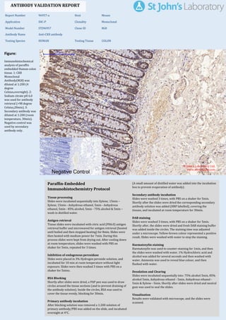
  • 1. Figure: Immunohistochemical analysis of paraffin embedded Human colon tissue. 1: CK8 Monoclonal Antibody(8G8) was diluted at 1:200 (4 degree Celsius,overnight). 2: Sodium citrate pH 6.0 was used for antibody retrieval (>98 degree Celsius,20min). 3: Secondary antibody was diluted at 1:200 (room temperature, 30min). Negative control was used by secondary antibody only. Report Number 96957-a Host Mouse Application IHC-P Clonality Monoclonal Model Number STJ96957 Clone ID 8G8 Antibody Name Anti-CK8 antibody Testing Species HUMAN Testing Tissue COLON ANTIBODY VALIDATION REPORT a. (A small amount of distilled water was added into the incubation box to prevent evaporation of antibody). 40. Secondary antibody incubation a. Slides were washed 3 times, with PBS on a shaker for 5min. Shortly after the slides were dried the corresponding secondary antibody solution was added (HRP labelled), covering the tissues, and incubated at room temperature for 30min. b. 41. DAB staining a. Slides were washed 3 times, with PBS on a shaker for 5min. b. Shortly after, the slides were dried and fresh DAB staining buffer was added inside the circles. The staining time was adjusted under a microscope. Yellow-brown colour represented a positive result. Slides were washed with water to stop the staining. c. 42. Haematoxylin staining a. Haematoxylin was used to counter-staining for 1min, and then the slides were washed with water. 1% Hydrochloric acid and alcohol was added for several seconds and then washed with water. Ammonia was used to reveal blue colour, and then flushed with water. b. 43. Desolation and Clearing i. Slides were incubated sequentially into: 75% alcohol 5min, 85% alcohol 5min, Anhydrous ethanol - 5min, Anhydrous ethanol - 5min & Xylene - 5min. Shortly after slides were dried and neutral gum was used to seal the slides. ii. 44. Visualization a. Results were validated with microscope, and the slides were scanned. Paraffin-Embedded Immunohistochemistry Protocol 34. 35. Tissue processing a. Slides were incubated sequentially into Xylene; 15min – Xylene, 15min - Anhydrous ethanol, 5min - Anhydrous ethanol, 5min - 85% alcohol, 5min - 75% alcohol & 5min – wash in distilled water. b. 36. Antigen retrieval a. Tissue slides were incubated with citric acid (PH6.0) antigen retrieval buffer and microwaved for antigen retrieval (heated until boiled and then stopped heating) for 8min. Slides were then heated with medium power for 7min. During this process slides were kept from drying out. After cooling down at room temperature, slides were washed with PBS on shaker for 5min, repeated for 3 times. b. 37. Inhibition of endogenous peroxidase a. Slides were placed in 3% Hydrogen peroxide solution, and incubated for 10 min at room temperature without light exposure. Slides were then washed 3 times with PBS on a shaker for 5mins. b. 38. BSA Blocking a. Shortly after slides were dried, a PAP pen was used to draw circles around the tissue sections (and to prevent draining of the antibody solution). Inside the circles, BSA was used to cover the tissue evenly, blocking for 30min. b. 39. Primary antibody incubation After blocking solution was removed a 1:200 solution of primary antibody/PBS was added on the slide, and incubated overnight at 4°C. St John's Laboratory Ltd. www.stjohnslabs.com
  • 2. Figure: Immunohistochemical analysis of paraffin embedded Human liver cancer tissue. 1: CK8 Monoclonal Antibody(8G8) was diluted at 1:200 (4 degree Celsius,overnight). 2: Sodium citrate pH 6.0 was used for antibody retrieval (>98 degree Celsius,20min). 3: Secondary antibody was diluted at 1:200 (room temperature, 30min). Negative control was used by secondary antibody only. Report Number 96957-b Host Mouse Application IHC-P Clonality Monoclonal Model Number STJ96957 Clone ID 8G8 Antibody Name Anti-CK8 antibody Testing Species HUMAN Testing Tissue LIVER CANCER ANTIBODY VALIDATION REPORT b. (A small amount of distilled water was added into the incubation box to prevent evaporation of antibody). 29. Secondary antibody incubation a. Slides were washed 3 times, with PBS on a shaker for 5min. Shortly after the slides were dried the corresponding secondary antibody solution was added (HRP labelled), covering the tissues, and incubated at room temperature for 30min. b. 30. DAB staining a. Slides were washed 3 times, with PBS on a shaker for 5min. b. Shortly after, the slides were dried and fresh DAB staining buffer was added inside the circles. The staining time was adjusted under a microscope. Yellow-brown colour represented a positive result. Slides were washed with water to stop the staining. c. 31. Haematoxylin staining a. Haematoxylin was used to counter-staining for 1min, and then the slides were washed with water. 1% Hydrochloric acid and alcohol was added for several seconds and then washed with water. Ammonia was used to reveal blue colour, and then flushed with water. b. 32. Desolation and Clearing i. Slides were incubated sequentially into: 75% alcohol 5min, 85% alcohol 5min, Anhydrous ethanol - 5min, Anhydrous ethanol - 5min & Xylene - 5min. Shortly after slides were dried and neutral gum was used to seal the slides. ii. 33. Visualization a. Results were validated with microscope, and the slides were scanned. Paraffin-Embedded Immunohistochemistry Protocol 23. 24. Tissue processing a. Slides were incubated sequentially into Xylene; 15min – Xylene, 15min - Anhydrous ethanol, 5min - Anhydrous ethanol, 5min - 85% alcohol, 5min - 75% alcohol & 5min – wash in distilled water. b. 25. Antigen retrieval a. Tissue slides were incubated with citric acid (PH6.0) antigen retrieval buffer and microwaved for antigen retrieval (heated until boiled and then stopped heating) for 8min. Slides were then heated with medium power for 7min. During this process slides were kept from drying out. After cooling down at room temperature, slides were washed with PBS on shaker for 5min, repeated for 3 times. b. 26. Inhibition of endogenous peroxidase a. Slides were placed in 3% Hydrogen peroxide solution, and incubated for 10 min at room temperature without light exposure. Slides were then washed 3 times with PBS on a shaker for 5mins. b. 27. BSA Blocking a. Shortly after slides were dried, a PAP pen was used to draw circles around the tissue sections (and to prevent draining of the antibody solution). Inside the circles, BSA was used to cover the tissue evenly, blocking for 30min. b. 28. Primary antibody incubation After blocking solution was removed a 1:200 solution of primary antibody/PBS was added on the slide, and incubated overnight at 4°C. St John's Laboratory Ltd. www.stjohnslabs.com
  • 3. Figure: Immunohistochemical analysis of paraffin embedded Human stomach tissue. 1: CK8 Monoclonal Antibody(8G8) was diluted at 1:200 (4 degree Celsius,overnight). 2: Sodium citrate pH 6.0 was used for antibody retrieval (>98 degree Celsius,20min). 3: Secondary antibody was diluted at 1:200 (room temperature, 30min). Negative control was used by secondary antibody only. Report Number 96957-c Host Mouse Application IHC-P Clonality Monoclonal Model Number STJ96957 Clone ID 8G8 Antibody Name Anti-CK8 antibody Testing Species HUMAN Testing Tissue STOMACH ANTIBODY VALIDATION REPORT b. (A small amount of distilled water was added into the incubation box to prevent evaporation of antibody). 18. Secondary antibody incubation a. Slides were washed 3 times, with PBS on a shaker for 5min. Shortly after the slides were dried the corresponding secondary antibody solution was added (HRP labelled), covering the tissues, and incubated at room temperature for 30min. b. 19. DAB staining a. Slides were washed 3 times, with PBS on a shaker for 5min. b. Shortly after, the slides were dried and fresh DAB staining buffer was added inside the circles. The staining time was adjusted under a microscope. Yellow-brown colour represented a positive result. Slides were washed with water to stop the staining. c. 20. Haematoxylin staining a. Haematoxylin was used to counter-staining for 1min, and then the slides were washed with water. 1% Hydrochloric acid and alcohol was added for several seconds and then washed with water. Ammonia was used to reveal blue colour, and then flushed with water. b. 21. Desolation and Clearing i. Slides were incubated sequentially into: 75% alcohol 5min, 85% alcohol 5min, Anhydrous ethanol - 5min, Anhydrous ethanol - 5min & Xylene - 5min. Shortly after slides were dried and neutral gum was used to seal the slides. ii. 22. Visualization a. Results were validated with microscope, and the slides were scanned. Paraffin-Embedded Immunohistochemistry Protocol 12. 13. Tissue processing a. Slides were incubated sequentially into Xylene; 15min – Xylene, 15min - Anhydrous ethanol, 5min - Anhydrous ethanol, 5min - 85% alcohol, 5min - 75% alcohol & 5min – wash in distilled water. b. 14. Antigen retrieval a. Tissue slides were incubated with citric acid (PH6.0) antigen retrieval buffer and microwaved for antigen retrieval (heated until boiled and then stopped heating) for 8min. Slides were then heated with medium power for 7min. During this process slides were kept from drying out. After cooling down at room temperature, slides were washed with PBS on shaker for 5min, repeated for 3 times. b. 15. Inhibition of endogenous peroxidase a. Slides were placed in 3% Hydrogen peroxide solution, and incubated for 10 min at room temperature without light exposure. Slides were then washed 3 times with PBS on a shaker for 5mins. b. 16. BSA Blocking a. Shortly after slides were dried, a PAP pen was used to draw circles around the tissue sections (and to prevent draining of the antibody solution). Inside the circles, BSA was used to cover the tissue evenly, blocking for 30min. b. 17. Primary antibody incubation After blocking solution was removed a 1:200 solution of primary antibody/PBS was added on the slide, and incubated overnight at 4°C. St John's Laboratory Ltd. www.stjohnslabs.com
  • 4. Figure: Immunohistochemical analysis of paraffin embedded Human stomach cancer tissue. 1: CK8 Monoclonal Antibody(8G8) was diluted at 1:200 (4 degree Celsius,overnight). 2: Sodium citrate pH 6.0 was used for antibody retrieval (>98 degree Celsius,20min). 3: Secondary antibody was diluted at 1:200 (room temperature, 30min). Negative control was used by secondary antibody only. Report Number 96957-d Host Mouse Application IHC-P Clonality Monoclonal Model Number STJ96957 Clone ID 8G8 Antibody Name Anti-CK8 antibody Testing Species HUMAN Testing Tissue STOMACH CANCER ANTIBODY VALIDATION REPORT b. (A small amount of distilled water was added into the incubation box to prevent evaporation of antibody). 7. Secondary antibody incubation a. Slides were washed 3 times, with PBS on a shaker for 5min. Shortly after the slides were dried the corresponding secondary antibody solution was added (HRP labelled), covering the tissues, and incubated at room temperature for 30min. b. 8. DAB staining a. Slides were washed 3 times, with PBS on a shaker for 5min. b. Shortly after, the slides were dried and fresh DAB staining buffer was added inside the circles. The staining time was adjusted under a microscope. Yellow-brown colour represented a positive result. Slides were washed with water to stop the staining. c. 9. Haematoxylin staining a. Haematoxylin was used to counter-staining for 1min, and then the slides were washed with water. 1% Hydrochloric acid and alcohol was added for several seconds and then washed with water. Ammonia was used to reveal blue colour, and then flushed with water. b. 10. Desolation and Clearing i. Slides were incubated sequentially into: 75% alcohol 5min, 85% alcohol 5min, Anhydrous ethanol - 5min, Anhydrous ethanol - 5min & Xylene - 5min. Shortly after slides were dried and neutral gum was used to seal the slides. ii. 11. Visualization a. Results were validated with microscope, and the slides were scanned. Paraffin-Embedded Immunohistochemistry Protocol 1. 2. Tissue processing a. Slides were incubated sequentially into Xylene; 15min – Xylene, 15min - Anhydrous ethanol, 5min - Anhydrous ethanol, 5min - 85% alcohol, 5min - 75% alcohol & 5min – wash in distilled water. b. 3. Antigen retrieval a. Tissue slides were incubated with citric acid (PH6.0) antigen retrieval buffer and microwaved for antigen retrieval (heated until boiled and then stopped heating) for 8min. Slides were then heated with medium power for 7min. During this process slides were kept from drying out. After cooling down at room temperature, slides were washed with PBS on shaker for 5min, repeated for 3 times. b. 4. Inhibition of endogenous peroxidase a. Slides were placed in 3% Hydrogen peroxide solution, and incubated for 10 min at room temperature without light exposure. Slides were then washed 3 times with PBS on a shaker for 5mins. b. 5. BSA Blocking a. Shortly after slides were dried, a PAP pen was used to draw circles around the tissue sections (and to prevent draining of the antibody solution). Inside the circles, BSA was used to cover the tissue evenly, blocking for 30min. b. 6. Primary antibody incubation After blocking solution was removed a 1:200 solution of primary antibody/PBS was added on the slide, and incubated overnight at 4°C. St John's Laboratory Ltd. www.stjohnslabs.com
  • 5. Figure: Immunohistochemical analysis of paraffin embedded Rat liver tissue. 1: CK8 Monoclonal Antibody(8G8) was diluted at 1:200 (4 degree Celsius,overnight). 2: Sodium citrate pH 6.0 was used for antibody retrieval (>98 degree Celsius,20min). 3: Secondary antibody was diluted at 1:200 (room temperature, 30min). Negative control was used by secondary antibody only. Report Number 96957-e Host Mouse Application IHC-P Clonality Monoclonal Model Number STJ96957 Clone ID 8G8 Antibody Name Anti-CK8 antibody Testing Species RAT Testing Tissue LIVER ANTIBODY VALIDATION REPORT c. (A small amount of distilled water was added into the incubation box to prevent evaporation of antibody). 45. Secondary antibody incubation a. Slides were washed 3 times, with PBS on a shaker for 5min. Shortly after the slides were dried the corresponding secondary antibody solution was added (HRP labelled), covering the tissues, and incubated at room temperature for 30min. b. 46. DAB staining a. Slides were washed 3 times, with PBS on a shaker for 5min. b. Shortly after, the slides were dried and fresh DAB staining buffer was added inside the circles. The staining time was adjusted under a microscope. Yellow-brown colour represented a positive result. Slides were washed with water to stop the staining. c. 47. Haematoxylin staining a. Haematoxylin was used to counter-staining for 1min, and then the slides were washed with water. 1% Hydrochloric acid and alcohol was added for several seconds and then washed with water. Ammonia was used to reveal blue colour, and then flushed with water. b. 48. Desolation and Clearing i. Slides were incubated sequentially into: 75% alcohol 5min, 85% alcohol 5min, Anhydrous ethanol - 5min, Anhydrous ethanol - 5min & Xylene - 5min. Shortly after slides were dried and neutral gum was used to seal the slides. ii. 49. Visualization a. Results were validated with microscope, and the slides were scanned. Paraffin-Embedded Immunohistochemistry Protocol 50. 51. Tissue processing a. Slides were incubated sequentially into Xylene; 15min – Xylene, 15min - Anhydrous ethanol, 5min - Anhydrous ethanol, 5min - 85% alcohol, 5min - 75% alcohol & 5min – wash in distilled water. b. 52. Antigen retrieval a. Tissue slides were incubated with citric acid (PH6.0) antigen retrieval buffer and microwaved for antigen retrieval (heated until boiled and then stopped heating) for 8min. Slides were then heated with medium power for 7min. During this process slides were kept from drying out. After cooling down at room temperature, slides were washed with PBS on shaker for 5min, repeated for 3 times. b. 53. Inhibition of endogenous peroxidase a. Slides were placed in 3% Hydrogen peroxide solution, and incubated for 10 min at room temperature without light exposure. Slides were then washed 3 times with PBS on a shaker for 5mins. b. 54. BSA Blocking a. Shortly after slides were dried, a PAP pen was used to draw circles around the tissue sections (and to prevent draining of the antibody solution). Inside the circles, BSA was used to cover the tissue evenly, blocking for 30min. b. 55. Primary antibody incubation After blocking solution was removed a 1:200 solution of primary antibody/PBS was added on the slide, and incubated overnight at 4°C. St John's Laboratory Ltd. www.stjohnslabs.com
  • 6. Figure: Immunohistochemical analysis of paraffin embedded Rat lung tissue. 1: CK8 Monoclonal Antibody(8G8) was diluted at 1:200 (4 degree Celsius,overnight). 2: Sodium citrate pH 6.0 was used for antibody retrieval (>98 degree Celsius,20min). 3: Secondary antibody was diluted at 1:200 (room temperature, 30min). Negative control was used by secondary antibody only. Report Number 96957-f Host Mouse Application IHC-P Clonality Monoclonal Model Number STJ96957 Clone ID 8G8 Antibody Name Anti-CK8 antibody Testing Species RAT Testing Tissue LUNG ANTIBODY VALIDATION REPORT b. (A small amount of distilled water was added into the incubation box to prevent evaporation of antibody). 56. Secondary antibody incubation a. Slides were washed 3 times, with PBS on a shaker for 5min. Shortly after the slides were dried the corresponding secondary antibody solution was added (HRP labelled), covering the tissues, and incubated at room temperature for 30min. b. 57. DAB staining a. Slides were washed 3 times, with PBS on a shaker for 5min. b. Shortly after, the slides were dried and fresh DAB staining buffer was added inside the circles. The staining time was adjusted under a microscope. Yellow-brown colour represented a positive result. Slides were washed with water to stop the staining. c. 58. Haematoxylin staining a. Haematoxylin was used to counter-staining for 1min, and then the slides were washed with water. 1% Hydrochloric acid and alcohol was added for several seconds and then washed with water. Ammonia was used to reveal blue colour, and then flushed with water. b. 59. Desolation and Clearing i. Slides were incubated sequentially into: 75% alcohol 5min, 85% alcohol 5min, Anhydrous ethanol - 5min, Anhydrous ethanol - 5min & Xylene - 5min. Shortly after slides were dried and neutral gum was used to seal the slides. ii. 60. Visualization a. Results were validated with microscope, and the slides were scanned. Paraffin-Embedded Immunohistochemistry Protocol 61. 62. Tissue processing a. Slides were incubated sequentially into Xylene; 15min – Xylene, 15min - Anhydrous ethanol, 5min - Anhydrous ethanol, 5min - 85% alcohol, 5min - 75% alcohol & 5min – wash in distilled water. b. 63. Antigen retrieval a. Tissue slides were incubated with citric acid (PH6.0) antigen retrieval buffer and microwaved for antigen retrieval (heated until boiled and then stopped heating) for 8min. Slides were then heated with medium power for 7min. During this process slides were kept from drying out. After cooling down at room temperature, slides were washed with PBS on shaker for 5min, repeated for 3 times. b. 64. Inhibition of endogenous peroxidase a. Slides were placed in 3% Hydrogen peroxide solution, and incubated for 10 min at room temperature without light exposure. Slides were then washed 3 times with PBS on a shaker for 5mins. b. 65. BSA Blocking a. Shortly after slides were dried, a PAP pen was used to draw circles around the tissue sections (and to prevent draining of the antibody solution). Inside the circles, BSA was used to cover the tissue evenly, blocking for 30min. b. 66. Primary antibody incubation After blocking solution was removed a 1:200 solution of primary antibody/PBS was added on the slide, and incubated overnight at 4°C. St John's Laboratory Ltd. www.stjohnslabs.com
  • 7. Figure: Immunohistochemical analysis of paraffin embedded Rat kidney tissue. 1: CK8 Monoclonal Antibody(8G8) was diluted at 1:200 (4 degree Celsius,overnight). 2: Sodium citrate pH 6.0 was used for antibody retrieval (>98 degree Celsius,20min). 3: Secondary antibody was diluted at 1:200 (room temperature, 30min). Negative control was used by secondary antibody only. Report Number 96957-g Host Mouse Application IHC-P Clonality Monoclonal Model Number STJ96957 Clone ID 8G8 Antibody Name Anti-CK8 antibody Testing Species RAT Testing Tissue KIDNEY ANTIBODY VALIDATION REPORT b. (A small amount of distilled water was added into the incubation box to prevent evaporation of antibody). 67. Secondary antibody incubation a. Slides were washed 3 times, with PBS on a shaker for 5min. Shortly after the slides were dried the corresponding secondary antibody solution was added (HRP labelled), covering the tissues, and incubated at room temperature for 30min. b. 68. DAB staining a. Slides were washed 3 times, with PBS on a shaker for 5min. b. Shortly after, the slides were dried and fresh DAB staining buffer was added inside the circles. The staining time was adjusted under a microscope. Yellow-brown colour represented a positive result. Slides were washed with water to stop the staining. c. 69. Haematoxylin staining a. Haematoxylin was used to counter-staining for 1min, and then the slides were washed with water. 1% Hydrochloric acid and alcohol was added for several seconds and then washed with water. Ammonia was used to reveal blue colour, and then flushed with water. b. 70. Desolation and Clearing i. Slides were incubated sequentially into: 75% alcohol 5min, 85% alcohol 5min, Anhydrous ethanol - 5min, Anhydrous ethanol - 5min & Xylene - 5min. Shortly after slides were dried and neutral gum was used to seal the slides. ii. 71. Visualization a. Results were validated with microscope, and the slides were scanned. Paraffin-Embedded Immunohistochemistry Protocol 72. 73. Tissue processing a. Slides were incubated sequentially into Xylene; 15min – Xylene, 15min - Anhydrous ethanol, 5min - Anhydrous ethanol, 5min - 85% alcohol, 5min - 75% alcohol & 5min – wash in distilled water. b. 74. Antigen retrieval a. Tissue slides were incubated with citric acid (PH6.0) antigen retrieval buffer and microwaved for antigen retrieval (heated until boiled and then stopped heating) for 8min. Slides were then heated with medium power for 7min. During this process slides were kept from drying out. After cooling down at room temperature, slides were washed with PBS on shaker for 5min, repeated for 3 times. b. 75. Inhibition of endogenous peroxidase a. Slides were placed in 3% Hydrogen peroxide solution, and incubated for 10 min at room temperature without light exposure. Slides were then washed 3 times with PBS on a shaker for 5mins. b. 76. BSA Blocking a. Shortly after slides were dried, a PAP pen was used to draw circles around the tissue sections (and to prevent draining of the antibody solution). Inside the circles, BSA was used to cover the tissue evenly, blocking for 30min. b. 77. Primary antibody incubation After blocking solution was removed a 1:200 solution of primary antibody/PBS was added on the slide, and incubated overnight at 4°C. St John's Laboratory Ltd. www.stjohnslabs.com
  • 8. Figure: Immunohistochemical analysis of paraffin embedded Mouse liver tissue. 1: CK8 Monoclonal Antibody(8G8) was diluted at 1:200 (4 degree Celsius,overnight). 2: Sodium citrate pH 6.0 was used for antibody retrieval (>98 degree Celsius,20min). 3: Secondary antibody was diluted at 1:200 (room temperature, 30min). Negative control was used by secondary antibody only. Report Number 96957-h Host Mouse Application IHC-P Clonality Monoclonal Model Number STJ96957 Clone ID 8G8 Antibody Name Anti-CK8 antibody Testing Species MOUSE Testing Tissue LIVER ANTIBODY VALIDATION REPORT b. (A small amount of distilled water was added into the incubation box to prevent evaporation of antibody). 78. Secondary antibody incubation a. Slides were washed 3 times, with PBS on a shaker for 5min. Shortly after the slides were dried the corresponding secondary antibody solution was added (HRP labelled), covering the tissues, and incubated at room temperature for 30min. b. 79. DAB staining a. Slides were washed 3 times, with PBS on a shaker for 5min. b. Shortly after, the slides were dried and fresh DAB staining buffer was added inside the circles. The staining time was adjusted under a microscope. Yellow-brown colour represented a positive result. Slides were washed with water to stop the staining. c. 80. Haematoxylin staining a. Haematoxylin was used to counter-staining for 1min, and then the slides were washed with water. 1% Hydrochloric acid and alcohol was added for several seconds and then washed with water. Ammonia was used to reveal blue colour, and then flushed with water. b. 81. Desolation and Clearing i. Slides were incubated sequentially into: 75% alcohol 5min, 85% alcohol 5min, Anhydrous ethanol - 5min, Anhydrous ethanol - 5min & Xylene - 5min. Shortly after slides were dried and neutral gum was used to seal the slides. ii. 82. Visualization a. Results were validated with microscope, and the slides were scanned. Paraffin-Embedded Immunohistochemistry Protocol 83. 84. Tissue processing a. Slides were incubated sequentially into Xylene; 15min – Xylene, 15min - Anhydrous ethanol, 5min - Anhydrous ethanol, 5min - 85% alcohol, 5min - 75% alcohol & 5min – wash in distilled water. b. 85. Antigen retrieval a. Tissue slides were incubated with citric acid (PH6.0) antigen retrieval buffer and microwaved for antigen retrieval (heated until boiled and then stopped heating) for 8min. Slides were then heated with medium power for 7min. During this process slides were kept from drying out. After cooling down at room temperature, slides were washed with PBS on shaker for 5min, repeated for 3 times. b. 86. Inhibition of endogenous peroxidase a. Slides were placed in 3% Hydrogen peroxide solution, and incubated for 10 min at room temperature without light exposure. Slides were then washed 3 times with PBS on a shaker for 5mins. b. 87. BSA Blocking a. Shortly after slides were dried, a PAP pen was used to draw circles around the tissue sections (and to prevent draining of the antibody solution). Inside the circles, BSA was used to cover the tissue evenly, blocking for 30min. b. 88. Primary antibody incubation After blocking solution was removed a 1:200 solution of primary antibody/PBS was added on the slide, and incubated overnight at 4°C. St John's Laboratory Ltd. www.stjohnslabs.com
  • 9. Figure: Immunohistochemical analysis of paraffin embedded Mouse lung tissue. 1: CK8 Monoclonal Antibody(8G8) was diluted at 1:200 (4 degree Celsius,overnight). 2: Sodium citrate pH 6.0 was used for antibody retrieval (>98 degree Celsius,20min). 3: Secondary antibody was diluted at 1:200 (room temperature, 30min). Negative control was used by secondary antibody only. Report Number 96957-i Host Mouse Application IHC-P Clonality Monoclonal Model Number STJ96957 Clone ID 8G8 Antibody Name Anti-CK8 antibody Testing Species MOUSE Testing Tissue LUNG ANTIBODY VALIDATION REPORT b. (A small amount of distilled water was added into the incubation box to prevent evaporation of antibody). 89. Secondary antibody incubation a. Slides were washed 3 times, with PBS on a shaker for 5min. Shortly after the slides were dried the corresponding secondary antibody solution was added (HRP labelled), covering the tissues, and incubated at room temperature for 30min. b. 90. DAB staining a. Slides were washed 3 times, with PBS on a shaker for 5min. b. Shortly after, the slides were dried and fresh DAB staining buffer was added inside the circles. The staining time was adjusted under a microscope. Yellow-brown colour represented a positive result. Slides were washed with water to stop the staining. c. 91. Haematoxylin staining a. Haematoxylin was used to counter-staining for 1min, and then the slides were washed with water. 1% Hydrochloric acid and alcohol was added for several seconds and then washed with water. Ammonia was used to reveal blue colour, and then flushed with water. b. 92. Desolation and Clearing i. Slides were incubated sequentially into: 75% alcohol 5min, 85% alcohol 5min, Anhydrous ethanol - 5min, Anhydrous ethanol - 5min & Xylene - 5min. Shortly after slides were dried and neutral gum was used to seal the slides. ii. 93. Visualization a. Results were validated with microscope, and the slides were scanned. Paraffin-Embedded Immunohistochemistry Protocol 94. 95. Tissue processing a. Slides were incubated sequentially into Xylene; 15min – Xylene, 15min - Anhydrous ethanol, 5min - Anhydrous ethanol, 5min - 85% alcohol, 5min - 75% alcohol & 5min – wash in distilled water. b. 96. Antigen retrieval a. Tissue slides were incubated with citric acid (PH6.0) antigen retrieval buffer and microwaved for antigen retrieval (heated until boiled and then stopped heating) for 8min. Slides were then heated with medium power for 7min. During this process slides were kept from drying out. After cooling down at room temperature, slides were washed with PBS on shaker for 5min, repeated for 3 times. b. 97. Inhibition of endogenous peroxidase a. Slides were placed in 3% Hydrogen peroxide solution, and incubated for 10 min at room temperature without light exposure. Slides were then washed 3 times with PBS on a shaker for 5mins. b. 98. BSA Blocking a. Shortly after slides were dried, a PAP pen was used to draw circles around the tissue sections (and to prevent draining of the antibody solution). Inside the circles, BSA was used to cover the tissue evenly, blocking for 30min. b. 99. Primary antibody incubation After blocking solution was removed a 1:200 solution of primary antibody/PBS was added on the slide, and incubated overnight at 4°C. St John's Laboratory Ltd. www.stjohnslabs.com
  • 10. Figure: Immunohistochemical analysis of paraffin embedded Mouse kidney tissue. 1: CK8 Monoclonal Antibody(8G8) was diluted at 1:200 (4 degree Celsius,overnight). 2: Sodium citrate pH 6.0 was used for antibody retrieval (>98 degree Celsius,20min). 3: Secondary antibody was diluted at 1:200 (room temperature, 30min). Negative control was used by secondary antibody only. Report Number 96957-j Host Mouse Application IHC-P Clonality Monoclonal Model Number STJ96957 Clone ID 8G8 Antibody Name Anti-CK8 antibody Testing Species MOUSE Testing Tissue KIDNEY ANTIBODY VALIDATION REPORT b. (A small amount of distilled water was added into the incubation box to prevent evaporation of antibody). 100. Secondary antibody incubation a. Slides were washed 3 times, with PBS on a shaker for 5min. Shortly after the slides were dried the corresponding secondary antibody solution was added (HRP labelled), covering the tissues, and incubated at room temperature for 30min. b. 101. DAB staining a. Slides were washed 3 times, with PBS on a shaker for 5min. b. Shortly after, the slides were dried and fresh DAB staining buffer was added inside the circles. The staining time was adjusted under a microscope. Yellow-brown colour represented a positive result. Slides were washed with water to stop the staining. c. 102. Haematoxylin staining a. Haematoxylin was used to counter-staining for 1min, and then the slides were washed with water. 1% Hydrochloric acid and alcohol was added for several seconds and then washed with water. Ammonia was used to reveal blue colour, and then flushed with water. b. 103. Desolation and Clearing i. Slides were incubated sequentially into: 75% alcohol 5min, 85% alcohol 5min, Anhydrous ethanol - 5min, Anhydrous ethanol - 5min & Xylene - 5min. Shortly after slides were dried and neutral gum was used to seal the slides. ii. 104. Visualization a. Results were validated with microscope, and the slides were scanned. Paraffin-Embedded Immunohistochemistry Protocol 105. 106. Tissue processing a. Slides were incubated sequentially into Xylene; 15min – Xylene, 15min - Anhydrous ethanol, 5min - Anhydrous ethanol, 5min - 85% alcohol, 5min - 75% alcohol & 5min – wash in distilled water. b. 107. Antigen retrieval a. Tissue slides were incubated with citric acid (PH6.0) antigen retrieval buffer and microwaved for antigen retrieval (heated until boiled and then stopped heating) for 8min. Slides were then heated with medium power for 7min. During this process slides were kept from drying out. After cooling down at room temperature, slides were washed with PBS on shaker for 5min, repeated for 3 times. b. 108. Inhibition of endogenous peroxidase a. Slides were placed in 3% Hydrogen peroxide solution, and incubated for 10 min at room temperature without light exposure. Slides were then washed 3 times with PBS on a shaker for 5mins. b. 109. BSA Blocking a. Shortly after slides were dried, a PAP pen was used to draw circles around the tissue sections (and to prevent draining of the antibody solution). Inside the circles, BSA was used to cover the tissue evenly, blocking for 30min. b. 110. Primary antibody incubation After blocking solution was removed a 1:200 solution of primary antibody/PBS was added on the slide, and incubated overnight at 4°C. St John's Laboratory Ltd. www.stjohnslabs.com