SlideShare a Scribd company logo
1 of 9
Download to read offline
ANTIBODY VALIDATION REPORT
Report Number 97382-a
Application Immunofluorescence
Model Number STJ97382
Antibody Name Anti-α-SMA antibody
Host Mouse
Clonality Monoclonal
Clone ID 6A12
Species HUMAN Tissue LIVER
Image
Description
Immunofluorescence analysis of Human
liver tissue. 1: α-SMA Monoclonal
Antibody(6A12)(red) was diluted at
1:200 (4 degree Celsius,overnight). 2:
Cy3 labled Secondary antibody was
diluted at 1:300 (room temperature,
50min).3: Picture B: DAPI(blue) 10min.
Picture A:Target. Picture B: DAPI.
Picture C: merge of A+B.
Primary Antibody Incubation
After blocking solution was removed a 1:200 primary antibody/PBS
solution was added on the slide, and incubated overnight at 4°C (a
small amount of distilled water was added into the incubation box to
prevent evaporation of antibody).
Secondary Antibody Incubation
slides were washed with PBS on a shaker for 5min, and repeated 3
times. Shortly after the slides were dried and corresponding
secondary antibody solution was added (HRP labelled), covering the
tissues, and incubated at room temperature for 50min.
DAPI Counter-Staining
slides were washed with PBS on a shaker for 5min, repeated 3 times
and then dried. DAPI staining solution was added inside the PAP
circles and incubated for 10 min at room temperature without light
exposure.
Mounting
Slides were washed with PBS on a shaker for 5min, and repeated 3
times. Shortly after slides were dried, anti-quench mountings were
used to mount slides.
Visualization
The slides were observed and placed under a NIKON inverted
fluorescence microscope (Ultra violet excitation 330-380nm,
emission 420nm; FITC green excitation 465-495nm, emission 515-
555 nm; CY3 red excitation 510-560nm, emission 590nm)
Immunofluorescence Protocol
Tissue Processing
Slides were incubated sequentially into: Xylene - 15min, Anhydrous
ethanol – 15 min, Anhydrous ethanol – 5 min, 85% alcohol – 5 min,
75% alcohol – 5 min & washed with distilled water – 5 min.
Antigen Retrieval
Tissue slides were incubated with citric acid (PH6.0) antigen
retrieval buffer, and microwaved for antigen retrieval (heated until
boiled and then stop heating) for 8min. Slides were then heated with
medium power for 7min. During this process slides are kept from
drying out. After cooling down at room temperature, slides were
washed with PBS on a shaker for 5min, and repeated 3 times.
Anti-Quench
shortly after slides were dried, a PAP pen was used to draw circles
around the tissues (to prevent draining of the antibody). Inside the
circles, anti-quench mountings were added and incubated for 5 min,
and then flushed with water for 10min.
BSA Blocking
Inside the circles, BSA was used to cover the tissue evenly, blocking
for 30min.
St John's Laboratory Ltd.
www.stjohnslabs.com
ANTIBODY VALIDATION REPORT
Report Number 97382-b
Application Immunofluorescence
Model Number STJ97382
Antibody Name Anti-α-SMA antibody
Host Mouse
Clonality Monoclonal
Clone ID 6A12
Species HUMAN Tissue LIVER
Image
Description
Immunofluorescence analysis of Human
liver tissue. 1: α-SMA Monoclonal
Antibody(6A12)(red) was diluted at
1:200 (4 degree Celsius,overnight). 2:
Cy3 labled Secondary antibody was
diluted at 1:300 (room temperature,
50min).3: Picture B: DAPI(blue) 10min.
Picture A:Target. Picture B: DAPI.
Picture C: merge of A+B.
Primary Antibody Incubation
After blocking solution was removed a 1:200 primary antibody/PBS
solution was added on the slide, and incubated overnight at 4°C (a
small amount of distilled water was added into the incubation box to
prevent evaporation of antibody).
Secondary Antibody Incubation
slides were washed with PBS on a shaker for 5min, and repeated 3
times. Shortly after the slides were dried and corresponding
secondary antibody solution was added (HRP labelled), covering the
tissues, and incubated at room temperature for 50min.
DAPI Counter-Staining
slides were washed with PBS on a shaker for 5min, repeated 3 times
and then dried. DAPI staining solution was added inside the PAP
circles and incubated for 10 min at room temperature without light
exposure.
Mounting
Slides were washed with PBS on a shaker for 5min, and repeated 3
times. Shortly after slides were dried, anti-quench mountings were
used to mount slides.
Visualization
The slides were observed and placed under a NIKON inverted
fluorescence microscope (Ultra violet excitation 330-380nm,
emission 420nm; FITC green excitation 465-495nm, emission 515-
555 nm; CY3 red excitation 510-560nm, emission 590nm)
Immunofluorescence Protocol
Tissue Processing
Slides were incubated sequentially into: Xylene - 15min, Anhydrous
ethanol – 15 min, Anhydrous ethanol – 5 min, 85% alcohol – 5 min,
75% alcohol – 5 min & washed with distilled water – 5 min.
Antigen Retrieval
Tissue slides were incubated with citric acid (PH6.0) antigen
retrieval buffer, and microwaved for antigen retrieval (heated until
boiled and then stop heating) for 8min. Slides were then heated with
medium power for 7min. During this process slides are kept from
drying out. After cooling down at room temperature, slides were
washed with PBS on a shaker for 5min, and repeated 3 times.
Anti-Quench
shortly after slides were dried, a PAP pen was used to draw circles
around the tissues (to prevent draining of the antibody). Inside the
circles, anti-quench mountings were added and incubated for 5 min,
and then flushed with water for 10min.
BSA Blocking
Inside the circles, BSA was used to cover the tissue evenly, blocking
for 30min.
St John's Laboratory Ltd.
www.stjohnslabs.com
ANTIBODY VALIDATION REPORT
Report Number 97382-c
Application Immunofluorescence
Model Number STJ97382
Antibody Name Anti-α-SMA antibody
Host Mouse
Clonality Monoclonal
Clone ID 6A12
Species HUMAN Tissue LIVER
Image
Description
Immunofluorescence analysis of Human
liver tissue. 1: α-SMA Monoclonal
Antibody(6A12)(red) was diluted at
1:200 (4 degree Celsius,overnight). 2:
Cy3 labled Secondary antibody was
diluted at 1:300 (room temperature,
50min).3: Picture B: DAPI(blue) 10min.
Picture A:Target. Picture B: DAPI.
Picture C: merge of A+B.
Primary Antibody Incubation
After blocking solution was removed a 1:200 primary antibody/PBS
solution was added on the slide, and incubated overnight at 4°C (a
small amount of distilled water was added into the incubation box to
prevent evaporation of antibody).
Secondary Antibody Incubation
slides were washed with PBS on a shaker for 5min, and repeated 3
times. Shortly after the slides were dried and corresponding
secondary antibody solution was added (HRP labelled), covering the
tissues, and incubated at room temperature for 50min.
DAPI Counter-Staining
slides were washed with PBS on a shaker for 5min, repeated 3 times
and then dried. DAPI staining solution was added inside the PAP
circles and incubated for 10 min at room temperature without light
exposure.
Mounting
Slides were washed with PBS on a shaker for 5min, and repeated 3
times. Shortly after slides were dried, anti-quench mountings were
used to mount slides.
Visualization
The slides were observed and placed under a NIKON inverted
fluorescence microscope (Ultra violet excitation 330-380nm,
emission 420nm; FITC green excitation 465-495nm, emission 515-
555 nm; CY3 red excitation 510-560nm, emission 590nm)
Immunofluorescence Protocol
Tissue Processing
Slides were incubated sequentially into: Xylene - 15min, Anhydrous
ethanol – 15 min, Anhydrous ethanol – 5 min, 85% alcohol – 5 min,
75% alcohol – 5 min & washed with distilled water – 5 min.
Antigen Retrieval
Tissue slides were incubated with citric acid (PH6.0) antigen
retrieval buffer, and microwaved for antigen retrieval (heated until
boiled and then stop heating) for 8min. Slides were then heated with
medium power for 7min. During this process slides are kept from
drying out. After cooling down at room temperature, slides were
washed with PBS on a shaker for 5min, and repeated 3 times.
Anti-Quench
shortly after slides were dried, a PAP pen was used to draw circles
around the tissues (to prevent draining of the antibody). Inside the
circles, anti-quench mountings were added and incubated for 5 min,
and then flushed with water for 10min.
BSA Blocking
Inside the circles, BSA was used to cover the tissue evenly, blocking
for 30min.
St John's Laboratory Ltd.
www.stjohnslabs.com
ANTIBODY VALIDATION REPORT
Report Number 97382-d
Application Immunofluorescence
Model Number STJ97382
Antibody Name Anti-α-SMA antibody
Host Mouse
Clonality Monoclonal
Clone ID 6A12
Species MOUSE Tissue LIVER
Image
Description
Immunofluorescence analysis of Mouse
liver tissue. 1: α-SMA Monoclonal
Antibody(6A12)(red) was diluted at
1:200 (4 degree Celsius,overnight). 2:
Cy3 labled Secondary antibody was
diluted at 1:300 (room temperature,
50min).3: Picture B: DAPI(blue) 10min.
Picture A:Target. Picture B: DAPI.
Picture C: merge of A+B.
Primary Antibody Incubation
After blocking solution was removed a 1:200 primary antibody/PBS
solution was added on the slide, and incubated overnight at 4°C (a
small amount of distilled water was added into the incubation box to
prevent evaporation of antibody).
Secondary Antibody Incubation
slides were washed with PBS on a shaker for 5min, and repeated 3
times. Shortly after the slides were dried and corresponding
secondary antibody solution was added (HRP labelled), covering the
tissues, and incubated at room temperature for 50min.
DAPI Counter-Staining
slides were washed with PBS on a shaker for 5min, repeated 3 times
and then dried. DAPI staining solution was added inside the PAP
circles and incubated for 10 min at room temperature without light
exposure.
Mounting
Slides were washed with PBS on a shaker for 5min, and repeated 3
times. Shortly after slides were dried, anti-quench mountings were
used to mount slides.
Visualization
The slides were observed and placed under a NIKON inverted
fluorescence microscope (Ultra violet excitation 330-380nm,
emission 420nm; FITC green excitation 465-495nm, emission 515-
555 nm; CY3 red excitation 510-560nm, emission 590nm)
Immunofluorescence Protocol
Tissue Processing
Slides were incubated sequentially into: Xylene - 15min, Anhydrous
ethanol – 15 min, Anhydrous ethanol – 5 min, 85% alcohol – 5 min,
75% alcohol – 5 min & washed with distilled water – 5 min.
Antigen Retrieval
Tissue slides were incubated with citric acid (PH6.0) antigen
retrieval buffer, and microwaved for antigen retrieval (heated until
boiled and then stop heating) for 8min. Slides were then heated with
medium power for 7min. During this process slides are kept from
drying out. After cooling down at room temperature, slides were
washed with PBS on a shaker for 5min, and repeated 3 times.
Anti-Quench
shortly after slides were dried, a PAP pen was used to draw circles
around the tissues (to prevent draining of the antibody). Inside the
circles, anti-quench mountings were added and incubated for 5 min,
and then flushed with water for 10min.
BSA Blocking
Inside the circles, BSA was used to cover the tissue evenly, blocking
for 30min.
St John's Laboratory Ltd.
www.stjohnslabs.com
ANTIBODY VALIDATION REPORT
Report Number 97382-e
Application Immunofluorescence
Model Number STJ97382
Antibody Name Anti-α-SMA antibody
Host Mouse
Clonality Monoclonal
Clone ID 6A12
Species MOUSE Tissue LIVER
Image
Description
Immunofluorescence analysis of Mouse
liver tissue. 1: α-SMA Monoclonal
Antibody(6A12)(red) was diluted at
1:200 (4 degree Celsius,overnight). 2:
Cy3 labled Secondary antibody was
diluted at 1:300 (room temperature,
50min).3: Picture B: DAPI(blue) 10min.
Picture A:Target. Picture B: DAPI.
Picture C: merge of A+B.
Primary Antibody Incubation
After blocking solution was removed a 1:200 primary antibody/PBS
solution was added on the slide, and incubated overnight at 4°C (a
small amount of distilled water was added into the incubation box to
prevent evaporation of antibody).
Secondary Antibody Incubation
slides were washed with PBS on a shaker for 5min, and repeated 3
times. Shortly after the slides were dried and corresponding
secondary antibody solution was added (HRP labelled), covering the
tissues, and incubated at room temperature for 50min.
DAPI Counter-Staining
slides were washed with PBS on a shaker for 5min, repeated 3 times
and then dried. DAPI staining solution was added inside the PAP
circles and incubated for 10 min at room temperature without light
exposure.
Mounting
Slides were washed with PBS on a shaker for 5min, and repeated 3
times. Shortly after slides were dried, anti-quench mountings were
used to mount slides.
Visualization
The slides were observed and placed under a NIKON inverted
fluorescence microscope (Ultra violet excitation 330-380nm,
emission 420nm; FITC green excitation 465-495nm, emission 515-
555 nm; CY3 red excitation 510-560nm, emission 590nm)
Immunofluorescence Protocol
Tissue Processing
Slides were incubated sequentially into: Xylene - 15min, Anhydrous
ethanol – 15 min, Anhydrous ethanol – 5 min, 85% alcohol – 5 min,
75% alcohol – 5 min & washed with distilled water – 5 min.
Antigen Retrieval
Tissue slides were incubated with citric acid (PH6.0) antigen
retrieval buffer, and microwaved for antigen retrieval (heated until
boiled and then stop heating) for 8min. Slides were then heated with
medium power for 7min. During this process slides are kept from
drying out. After cooling down at room temperature, slides were
washed with PBS on a shaker for 5min, and repeated 3 times.
Anti-Quench
shortly after slides were dried, a PAP pen was used to draw circles
around the tissues (to prevent draining of the antibody). Inside the
circles, anti-quench mountings were added and incubated for 5 min,
and then flushed with water for 10min.
BSA Blocking
Inside the circles, BSA was used to cover the tissue evenly, blocking
for 30min.
St John's Laboratory Ltd.
www.stjohnslabs.com
ANTIBODY VALIDATION REPORT
Report Number 97382-f
Application Immunofluorescence
Model Number STJ97382
Antibody Name Anti-α-SMA antibody
Host Mouse
Clonality Monoclonal
Clone ID 6A12
Species MOUSE Tissue LIVER
Image
Description
Immunofluorescence analysis of Mouse
liver tissue. 1: α-SMA Monoclonal
Antibody(6A12)(red) was diluted at
1:200 (4 degree Celsius,overnight). 2:
Cy3 labled Secondary antibody was
diluted at 1:300 (room temperature,
50min).3: Picture B: DAPI(blue) 10min.
Picture A:Target. Picture B: DAPI.
Picture C: merge of A+B.
Primary Antibody Incubation
After blocking solution was removed a 1:200 primary antibody/PBS
solution was added on the slide, and incubated overnight at 4°C (a
small amount of distilled water was added into the incubation box to
prevent evaporation of antibody).
Secondary Antibody Incubation
slides were washed with PBS on a shaker for 5min, and repeated 3
times. Shortly after the slides were dried and corresponding
secondary antibody solution was added (HRP labelled), covering the
tissues, and incubated at room temperature for 50min.
DAPI Counter-Staining
slides were washed with PBS on a shaker for 5min, repeated 3 times
and then dried. DAPI staining solution was added inside the PAP
circles and incubated for 10 min at room temperature without light
exposure.
Mounting
Slides were washed with PBS on a shaker for 5min, and repeated 3
times. Shortly after slides were dried, anti-quench mountings were
used to mount slides.
Visualization
The slides were observed and placed under a NIKON inverted
fluorescence microscope (Ultra violet excitation 330-380nm,
emission 420nm; FITC green excitation 465-495nm, emission 515-
555 nm; CY3 red excitation 510-560nm, emission 590nm)
Immunofluorescence Protocol
Tissue Processing
Slides were incubated sequentially into: Xylene - 15min, Anhydrous
ethanol – 15 min, Anhydrous ethanol – 5 min, 85% alcohol – 5 min,
75% alcohol – 5 min & washed with distilled water – 5 min.
Antigen Retrieval
Tissue slides were incubated with citric acid (PH6.0) antigen
retrieval buffer, and microwaved for antigen retrieval (heated until
boiled and then stop heating) for 8min. Slides were then heated with
medium power for 7min. During this process slides are kept from
drying out. After cooling down at room temperature, slides were
washed with PBS on a shaker for 5min, and repeated 3 times.
Anti-Quench
shortly after slides were dried, a PAP pen was used to draw circles
around the tissues (to prevent draining of the antibody). Inside the
circles, anti-quench mountings were added and incubated for 5 min,
and then flushed with water for 10min.
BSA Blocking
Inside the circles, BSA was used to cover the tissue evenly, blocking
for 30min.
St John's Laboratory Ltd.
www.stjohnslabs.com
ANTIBODY VALIDATION REPORT
Report Number 97382-g
Application Immunofluorescence
Model Number STJ97382
Antibody Name Anti-α-SMA antibody
Host Mouse
Clonality Monoclonal
Clone ID 6A12
Species RAT Tissue LIVER
Image
Description
Immunofluorescence analysis of Rat
liver tissue. 1: α-SMA Monoclonal
Antibody(6A12)(red) was diluted at
1:200 (4 degree Celsius,overnight). 2:
Cy3 labled Secondary antibody was
diluted at 1:300 (room temperature,
50min).3: Picture B: DAPI(blue) 10min.
Picture A:Target. Picture B: DAPI.
Picture C: merge of A+B.
Primary Antibody Incubation
After blocking solution was removed a 1:200 primary antibody/PBS
solution was added on the slide, and incubated overnight at 4°C (a
small amount of distilled water was added into the incubation box to
prevent evaporation of antibody).
Secondary Antibody Incubation
slides were washed with PBS on a shaker for 5min, and repeated 3
times. Shortly after the slides were dried and corresponding
secondary antibody solution was added (HRP labelled), covering the
tissues, and incubated at room temperature for 50min.
DAPI Counter-Staining
slides were washed with PBS on a shaker for 5min, repeated 3 times
and then dried. DAPI staining solution was added inside the PAP
circles and incubated for 10 min at room temperature without light
exposure.
Mounting
Slides were washed with PBS on a shaker for 5min, and repeated 3
times. Shortly after slides were dried, anti-quench mountings were
used to mount slides.
Visualization
The slides were observed and placed under a NIKON inverted
fluorescence microscope (Ultra violet excitation 330-380nm,
emission 420nm; FITC green excitation 465-495nm, emission 515-
555 nm; CY3 red excitation 510-560nm, emission 590nm)
Immunofluorescence Protocol
Tissue Processing
Slides were incubated sequentially into: Xylene - 15min, Anhydrous
ethanol – 15 min, Anhydrous ethanol – 5 min, 85% alcohol – 5 min,
75% alcohol – 5 min & washed with distilled water – 5 min.
Antigen Retrieval
Tissue slides were incubated with citric acid (PH6.0) antigen
retrieval buffer, and microwaved for antigen retrieval (heated until
boiled and then stop heating) for 8min. Slides were then heated with
medium power for 7min. During this process slides are kept from
drying out. After cooling down at room temperature, slides were
washed with PBS on a shaker for 5min, and repeated 3 times.
Anti-Quench
shortly after slides were dried, a PAP pen was used to draw circles
around the tissues (to prevent draining of the antibody). Inside the
circles, anti-quench mountings were added and incubated for 5 min,
and then flushed with water for 10min.
BSA Blocking
Inside the circles, BSA was used to cover the tissue evenly, blocking
for 30min.
St John's Laboratory Ltd.
www.stjohnslabs.com
ANTIBODY VALIDATION REPORT
Report Number 97382-h
Application Immunofluorescence
Model Number STJ97382
Antibody Name Anti-α-SMA antibody
Host Mouse
Clonality Monoclonal
Clone ID 6A12
Species RAT Tissue LIVER
Image
Description
Immunofluorescence analysis of Rat
liver tissue. 1: α-SMA Monoclonal
Antibody(6A12)(red) was diluted at
1:200 (4 degree Celsius,overnight). 2:
Cy3 labled Secondary antibody was
diluted at 1:300 (room temperature,
50min).3: Picture B: DAPI(blue) 10min.
Picture A:Target. Picture B: DAPI.
Picture C: merge of A+B.
Primary Antibody Incubation
After blocking solution was removed a 1:200 primary antibody/PBS
solution was added on the slide, and incubated overnight at 4°C (a
small amount of distilled water was added into the incubation box to
prevent evaporation of antibody).
Secondary Antibody Incubation
slides were washed with PBS on a shaker for 5min, and repeated 3
times. Shortly after the slides were dried and corresponding
secondary antibody solution was added (HRP labelled), covering the
tissues, and incubated at room temperature for 50min.
DAPI Counter-Staining
slides were washed with PBS on a shaker for 5min, repeated 3 times
and then dried. DAPI staining solution was added inside the PAP
circles and incubated for 10 min at room temperature without light
exposure.
Mounting
Slides were washed with PBS on a shaker for 5min, and repeated 3
times. Shortly after slides were dried, anti-quench mountings were
used to mount slides.
Visualization
The slides were observed and placed under a NIKON inverted
fluorescence microscope (Ultra violet excitation 330-380nm,
emission 420nm; FITC green excitation 465-495nm, emission 515-
555 nm; CY3 red excitation 510-560nm, emission 590nm)
Immunofluorescence Protocol
Tissue Processing
Slides were incubated sequentially into: Xylene - 15min, Anhydrous
ethanol – 15 min, Anhydrous ethanol – 5 min, 85% alcohol – 5 min,
75% alcohol – 5 min & washed with distilled water – 5 min.
Antigen Retrieval
Tissue slides were incubated with citric acid (PH6.0) antigen
retrieval buffer, and microwaved for antigen retrieval (heated until
boiled and then stop heating) for 8min. Slides were then heated with
medium power for 7min. During this process slides are kept from
drying out. After cooling down at room temperature, slides were
washed with PBS on a shaker for 5min, and repeated 3 times.
Anti-Quench
shortly after slides were dried, a PAP pen was used to draw circles
around the tissues (to prevent draining of the antibody). Inside the
circles, anti-quench mountings were added and incubated for 5 min,
and then flushed with water for 10min.
BSA Blocking
Inside the circles, BSA was used to cover the tissue evenly, blocking
for 30min.
St John's Laboratory Ltd.
www.stjohnslabs.com
ANTIBODY VALIDATION REPORT
Report Number 97382-i
Application Immunofluorescence
Model Number STJ97382
Antibody Name Anti-α-SMA antibody
Host Mouse
Clonality Monoclonal
Clone ID 6A12
Species RAT Tissue LIVER
Image
Description
Immunofluorescence analysis of Rat
liver tissue. 1: α-SMA Monoclonal
Antibody(6A12)(red) was diluted at
1:200 (4 degree Celsius,overnight). 2:
Cy3 labled Secondary antibody was
diluted at 1:300 (room temperature,
50min).3: Picture B: DAPI(blue) 10min.
Picture A:Target. Picture B: DAPI.
Picture C: merge of A+B.
Primary Antibody Incubation
After blocking solution was removed a 1:200 primary antibody/PBS
solution was added on the slide, and incubated overnight at 4°C (a
small amount of distilled water was added into the incubation box to
prevent evaporation of antibody).
Secondary Antibody Incubation
slides were washed with PBS on a shaker for 5min, and repeated 3
times. Shortly after the slides were dried and corresponding
secondary antibody solution was added (HRP labelled), covering the
tissues, and incubated at room temperature for 50min.
DAPI Counter-Staining
slides were washed with PBS on a shaker for 5min, repeated 3 times
and then dried. DAPI staining solution was added inside the PAP
circles and incubated for 10 min at room temperature without light
exposure.
Mounting
Slides were washed with PBS on a shaker for 5min, and repeated 3
times. Shortly after slides were dried, anti-quench mountings were
used to mount slides.
Visualization
The slides were observed and placed under a NIKON inverted
fluorescence microscope (Ultra violet excitation 330-380nm,
emission 420nm; FITC green excitation 465-495nm, emission 515-
555 nm; CY3 red excitation 510-560nm, emission 590nm)
Immunofluorescence Protocol
Tissue Processing
Slides were incubated sequentially into: Xylene - 15min, Anhydrous
ethanol – 15 min, Anhydrous ethanol – 5 min, 85% alcohol – 5 min,
75% alcohol – 5 min & washed with distilled water – 5 min.
Antigen Retrieval
Tissue slides were incubated with citric acid (PH6.0) antigen
retrieval buffer, and microwaved for antigen retrieval (heated until
boiled and then stop heating) for 8min. Slides were then heated with
medium power for 7min. During this process slides are kept from
drying out. After cooling down at room temperature, slides were
washed with PBS on a shaker for 5min, and repeated 3 times.
Anti-Quench
shortly after slides were dried, a PAP pen was used to draw circles
around the tissues (to prevent draining of the antibody). Inside the
circles, anti-quench mountings were added and incubated for 5 min,
and then flushed with water for 10min.
BSA Blocking
Inside the circles, BSA was used to cover the tissue evenly, blocking
for 30min.
St John's Laboratory Ltd.
www.stjohnslabs.com

More Related Content

What's hot

Immunofluorescence Antibody Validation Report for Anti-CD10 Antibody (STJ96974)
Immunofluorescence Antibody Validation Report for Anti-CD10 Antibody (STJ96974)Immunofluorescence Antibody Validation Report for Anti-CD10 Antibody (STJ96974)
Immunofluorescence Antibody Validation Report for Anti-CD10 Antibody (STJ96974)St John's Laboratory Ltd
 
Immunofluorescence Antibody Validation Report for Anti-CA IX Antibody (STJ96978)
Immunofluorescence Antibody Validation Report for Anti-CA IX Antibody (STJ96978)Immunofluorescence Antibody Validation Report for Anti-CA IX Antibody (STJ96978)
Immunofluorescence Antibody Validation Report for Anti-CA IX Antibody (STJ96978)St John's Laboratory Ltd
 
Immunofluorescence Antibody Validation Report for Anti-Caspase 9 Antibody (ST...
Immunofluorescence Antibody Validation Report for Anti-Caspase 9 Antibody (ST...Immunofluorescence Antibody Validation Report for Anti-Caspase 9 Antibody (ST...
Immunofluorescence Antibody Validation Report for Anti-Caspase 9 Antibody (ST...St John's Laboratory Ltd
 
Immunofluorescence Antibody Validation Report for Anti-HMG-1 Antibody (STJ96867)
Immunofluorescence Antibody Validation Report for Anti-HMG-1 Antibody (STJ96867)Immunofluorescence Antibody Validation Report for Anti-HMG-1 Antibody (STJ96867)
Immunofluorescence Antibody Validation Report for Anti-HMG-1 Antibody (STJ96867)St John's Laboratory Ltd
 
Immunofluorescence Antibody Validation Report for Anti-CD23 Antibody (STJ96951)
Immunofluorescence Antibody Validation Report for Anti-CD23 Antibody (STJ96951)Immunofluorescence Antibody Validation Report for Anti-CD23 Antibody (STJ96951)
Immunofluorescence Antibody Validation Report for Anti-CD23 Antibody (STJ96951)St John's Laboratory Ltd
 
Immunofluorescence Antibody Validation Report for Anti-AR Antibody (STJ97233)
Immunofluorescence Antibody Validation Report for Anti-AR Antibody (STJ97233)Immunofluorescence Antibody Validation Report for Anti-AR Antibody (STJ97233)
Immunofluorescence Antibody Validation Report for Anti-AR Antibody (STJ97233)St John's Laboratory Ltd
 
Immunofluorescence Antibody Validation Report for Anti-CDX2 Antibody (STJ96959)
Immunofluorescence Antibody Validation Report for Anti-CDX2 Antibody (STJ96959)Immunofluorescence Antibody Validation Report for Anti-CDX2 Antibody (STJ96959)
Immunofluorescence Antibody Validation Report for Anti-CDX2 Antibody (STJ96959)St John's Laboratory Ltd
 
Immunofluorescence Antibody Validation Report for Anti-PR Antibody (STJ97365)
Immunofluorescence Antibody Validation Report for Anti-PR Antibody (STJ97365)Immunofluorescence Antibody Validation Report for Anti-PR Antibody (STJ97365)
Immunofluorescence Antibody Validation Report for Anti-PR Antibody (STJ97365)St John's Laboratory Ltd
 
Immunofluorescence Antibody Validation Report for Anti-XRCC4 Antibody (STJ96988)
Immunofluorescence Antibody Validation Report for Anti-XRCC4 Antibody (STJ96988)Immunofluorescence Antibody Validation Report for Anti-XRCC4 Antibody (STJ96988)
Immunofluorescence Antibody Validation Report for Anti-XRCC4 Antibody (STJ96988)St John's Laboratory Ltd
 
Immunofluorescence Antibody Validation Report for Anti-ABCG2 Antibody (STJ97253)
Immunofluorescence Antibody Validation Report for Anti-ABCG2 Antibody (STJ97253)Immunofluorescence Antibody Validation Report for Anti-ABCG2 Antibody (STJ97253)
Immunofluorescence Antibody Validation Report for Anti-ABCG2 Antibody (STJ97253)St John's Laboratory Ltd
 
Immunofluorescence Antibody Validation Report for Anti-IL8 Antibody (STJ96518)
Immunofluorescence Antibody Validation Report for Anti-IL8 Antibody (STJ96518)Immunofluorescence Antibody Validation Report for Anti-IL8 Antibody (STJ96518)
Immunofluorescence Antibody Validation Report for Anti-IL8 Antibody (STJ96518)St John's Laboratory Ltd
 
Immunofluorescence Antibody Validation Report for Anti-Luciferase Antibody (S...
Immunofluorescence Antibody Validation Report for Anti-Luciferase Antibody (S...Immunofluorescence Antibody Validation Report for Anti-Luciferase Antibody (S...
Immunofluorescence Antibody Validation Report for Anti-Luciferase Antibody (S...St John's Laboratory Ltd
 
Immunofluorescence Antibody Validation Report for Anti-Galectin-3 Antibody (S...
Immunofluorescence Antibody Validation Report for Anti-Galectin-3 Antibody (S...Immunofluorescence Antibody Validation Report for Anti-Galectin-3 Antibody (S...
Immunofluorescence Antibody Validation Report for Anti-Galectin-3 Antibody (S...St John's Laboratory Ltd
 
Immunofluorescence Antibody Validation Report for Anti-OPG Antibody (STJ94831)
Immunofluorescence Antibody Validation Report for Anti-OPG Antibody (STJ94831)Immunofluorescence Antibody Validation Report for Anti-OPG Antibody (STJ94831)
Immunofluorescence Antibody Validation Report for Anti-OPG Antibody (STJ94831)St John's Laboratory Ltd
 
Immunofluorescence Antibody Validation Report for Anti-Cytokeratin 6 Antibody...
Immunofluorescence Antibody Validation Report for Anti-Cytokeratin 6 Antibody...Immunofluorescence Antibody Validation Report for Anti-Cytokeratin 6 Antibody...
Immunofluorescence Antibody Validation Report for Anti-Cytokeratin 6 Antibody...St John's Laboratory Ltd
 
Immunofluorescence Antibody Validation Report for Anti-CK7 Antibody (STJ96956)
Immunofluorescence Antibody Validation Report for Anti-CK7 Antibody (STJ96956)Immunofluorescence Antibody Validation Report for Anti-CK7 Antibody (STJ96956)
Immunofluorescence Antibody Validation Report for Anti-CK7 Antibody (STJ96956)St John's Laboratory Ltd
 
Immunofluorescence Antibody Validation Report for Anti-ABCB5 Antibody (STJ96976)
Immunofluorescence Antibody Validation Report for Anti-ABCB5 Antibody (STJ96976)Immunofluorescence Antibody Validation Report for Anti-ABCB5 Antibody (STJ96976)
Immunofluorescence Antibody Validation Report for Anti-ABCB5 Antibody (STJ96976)St John's Laboratory Ltd
 
Immunofluorescence Antibody Validation Report for Anti-Collagen IV Antibody (...
Immunofluorescence Antibody Validation Report for Anti-Collagen IV Antibody (...Immunofluorescence Antibody Validation Report for Anti-Collagen IV Antibody (...
Immunofluorescence Antibody Validation Report for Anti-Collagen IV Antibody (...St John's Laboratory Ltd
 
Immunofluorescence Antibody Validation Report for Anti-Catenin-β Antibody (ST...
Immunofluorescence Antibody Validation Report for Anti-Catenin-β Antibody (ST...Immunofluorescence Antibody Validation Report for Anti-Catenin-β Antibody (ST...
Immunofluorescence Antibody Validation Report for Anti-Catenin-β Antibody (ST...St John's Laboratory Ltd
 
Immunofluorescence Antibody Validation Report for Anti-CYCS Antibody (STJ97419)
Immunofluorescence Antibody Validation Report for Anti-CYCS Antibody (STJ97419)Immunofluorescence Antibody Validation Report for Anti-CYCS Antibody (STJ97419)
Immunofluorescence Antibody Validation Report for Anti-CYCS Antibody (STJ97419)St John's Laboratory Ltd
 

What's hot (20)

Immunofluorescence Antibody Validation Report for Anti-CD10 Antibody (STJ96974)
Immunofluorescence Antibody Validation Report for Anti-CD10 Antibody (STJ96974)Immunofluorescence Antibody Validation Report for Anti-CD10 Antibody (STJ96974)
Immunofluorescence Antibody Validation Report for Anti-CD10 Antibody (STJ96974)
 
Immunofluorescence Antibody Validation Report for Anti-CA IX Antibody (STJ96978)
Immunofluorescence Antibody Validation Report for Anti-CA IX Antibody (STJ96978)Immunofluorescence Antibody Validation Report for Anti-CA IX Antibody (STJ96978)
Immunofluorescence Antibody Validation Report for Anti-CA IX Antibody (STJ96978)
 
Immunofluorescence Antibody Validation Report for Anti-Caspase 9 Antibody (ST...
Immunofluorescence Antibody Validation Report for Anti-Caspase 9 Antibody (ST...Immunofluorescence Antibody Validation Report for Anti-Caspase 9 Antibody (ST...
Immunofluorescence Antibody Validation Report for Anti-Caspase 9 Antibody (ST...
 
Immunofluorescence Antibody Validation Report for Anti-HMG-1 Antibody (STJ96867)
Immunofluorescence Antibody Validation Report for Anti-HMG-1 Antibody (STJ96867)Immunofluorescence Antibody Validation Report for Anti-HMG-1 Antibody (STJ96867)
Immunofluorescence Antibody Validation Report for Anti-HMG-1 Antibody (STJ96867)
 
Immunofluorescence Antibody Validation Report for Anti-CD23 Antibody (STJ96951)
Immunofluorescence Antibody Validation Report for Anti-CD23 Antibody (STJ96951)Immunofluorescence Antibody Validation Report for Anti-CD23 Antibody (STJ96951)
Immunofluorescence Antibody Validation Report for Anti-CD23 Antibody (STJ96951)
 
Immunofluorescence Antibody Validation Report for Anti-AR Antibody (STJ97233)
Immunofluorescence Antibody Validation Report for Anti-AR Antibody (STJ97233)Immunofluorescence Antibody Validation Report for Anti-AR Antibody (STJ97233)
Immunofluorescence Antibody Validation Report for Anti-AR Antibody (STJ97233)
 
Immunofluorescence Antibody Validation Report for Anti-CDX2 Antibody (STJ96959)
Immunofluorescence Antibody Validation Report for Anti-CDX2 Antibody (STJ96959)Immunofluorescence Antibody Validation Report for Anti-CDX2 Antibody (STJ96959)
Immunofluorescence Antibody Validation Report for Anti-CDX2 Antibody (STJ96959)
 
Immunofluorescence Antibody Validation Report for Anti-PR Antibody (STJ97365)
Immunofluorescence Antibody Validation Report for Anti-PR Antibody (STJ97365)Immunofluorescence Antibody Validation Report for Anti-PR Antibody (STJ97365)
Immunofluorescence Antibody Validation Report for Anti-PR Antibody (STJ97365)
 
Immunofluorescence Antibody Validation Report for Anti-XRCC4 Antibody (STJ96988)
Immunofluorescence Antibody Validation Report for Anti-XRCC4 Antibody (STJ96988)Immunofluorescence Antibody Validation Report for Anti-XRCC4 Antibody (STJ96988)
Immunofluorescence Antibody Validation Report for Anti-XRCC4 Antibody (STJ96988)
 
Immunofluorescence Antibody Validation Report for Anti-ABCG2 Antibody (STJ97253)
Immunofluorescence Antibody Validation Report for Anti-ABCG2 Antibody (STJ97253)Immunofluorescence Antibody Validation Report for Anti-ABCG2 Antibody (STJ97253)
Immunofluorescence Antibody Validation Report for Anti-ABCG2 Antibody (STJ97253)
 
Immunofluorescence Antibody Validation Report for Anti-IL8 Antibody (STJ96518)
Immunofluorescence Antibody Validation Report for Anti-IL8 Antibody (STJ96518)Immunofluorescence Antibody Validation Report for Anti-IL8 Antibody (STJ96518)
Immunofluorescence Antibody Validation Report for Anti-IL8 Antibody (STJ96518)
 
Immunofluorescence Antibody Validation Report for Anti-Luciferase Antibody (S...
Immunofluorescence Antibody Validation Report for Anti-Luciferase Antibody (S...Immunofluorescence Antibody Validation Report for Anti-Luciferase Antibody (S...
Immunofluorescence Antibody Validation Report for Anti-Luciferase Antibody (S...
 
Immunofluorescence Antibody Validation Report for Anti-Galectin-3 Antibody (S...
Immunofluorescence Antibody Validation Report for Anti-Galectin-3 Antibody (S...Immunofluorescence Antibody Validation Report for Anti-Galectin-3 Antibody (S...
Immunofluorescence Antibody Validation Report for Anti-Galectin-3 Antibody (S...
 
Immunofluorescence Antibody Validation Report for Anti-OPG Antibody (STJ94831)
Immunofluorescence Antibody Validation Report for Anti-OPG Antibody (STJ94831)Immunofluorescence Antibody Validation Report for Anti-OPG Antibody (STJ94831)
Immunofluorescence Antibody Validation Report for Anti-OPG Antibody (STJ94831)
 
Immunofluorescence Antibody Validation Report for Anti-Cytokeratin 6 Antibody...
Immunofluorescence Antibody Validation Report for Anti-Cytokeratin 6 Antibody...Immunofluorescence Antibody Validation Report for Anti-Cytokeratin 6 Antibody...
Immunofluorescence Antibody Validation Report for Anti-Cytokeratin 6 Antibody...
 
Immunofluorescence Antibody Validation Report for Anti-CK7 Antibody (STJ96956)
Immunofluorescence Antibody Validation Report for Anti-CK7 Antibody (STJ96956)Immunofluorescence Antibody Validation Report for Anti-CK7 Antibody (STJ96956)
Immunofluorescence Antibody Validation Report for Anti-CK7 Antibody (STJ96956)
 
Immunofluorescence Antibody Validation Report for Anti-ABCB5 Antibody (STJ96976)
Immunofluorescence Antibody Validation Report for Anti-ABCB5 Antibody (STJ96976)Immunofluorescence Antibody Validation Report for Anti-ABCB5 Antibody (STJ96976)
Immunofluorescence Antibody Validation Report for Anti-ABCB5 Antibody (STJ96976)
 
Immunofluorescence Antibody Validation Report for Anti-Collagen IV Antibody (...
Immunofluorescence Antibody Validation Report for Anti-Collagen IV Antibody (...Immunofluorescence Antibody Validation Report for Anti-Collagen IV Antibody (...
Immunofluorescence Antibody Validation Report for Anti-Collagen IV Antibody (...
 
Immunofluorescence Antibody Validation Report for Anti-Catenin-β Antibody (ST...
Immunofluorescence Antibody Validation Report for Anti-Catenin-β Antibody (ST...Immunofluorescence Antibody Validation Report for Anti-Catenin-β Antibody (ST...
Immunofluorescence Antibody Validation Report for Anti-Catenin-β Antibody (ST...
 
Immunofluorescence Antibody Validation Report for Anti-CYCS Antibody (STJ97419)
Immunofluorescence Antibody Validation Report for Anti-CYCS Antibody (STJ97419)Immunofluorescence Antibody Validation Report for Anti-CYCS Antibody (STJ97419)
Immunofluorescence Antibody Validation Report for Anti-CYCS Antibody (STJ97419)
 

Viewers also liked

Immunofluorescence Antibody Validation Report for Anti-NFkB p65 Antibody (STJ...
Immunofluorescence Antibody Validation Report for Anti-NFkB p65 Antibody (STJ...Immunofluorescence Antibody Validation Report for Anti-NFkB p65 Antibody (STJ...
Immunofluorescence Antibody Validation Report for Anti-NFkB p65 Antibody (STJ...St John's Laboratory Ltd
 
Immunohistochemistry Antibody Validation Report for Anti-α-SMA Antibody (STJ9...
Immunohistochemistry Antibody Validation Report for Anti-α-SMA Antibody (STJ9...Immunohistochemistry Antibody Validation Report for Anti-α-SMA Antibody (STJ9...
Immunohistochemistry Antibody Validation Report for Anti-α-SMA Antibody (STJ9...St John's Laboratory Ltd
 
Immunohistochemistry Antibody Validation Report for Anti-NFkB p65 Antibody (S...
Immunohistochemistry Antibody Validation Report for Anti-NFkB p65 Antibody (S...Immunohistochemistry Antibody Validation Report for Anti-NFkB p65 Antibody (S...
Immunohistochemistry Antibody Validation Report for Anti-NFkB p65 Antibody (S...St John's Laboratory Ltd
 
Immunohistochemistry Antibody Validation Report for Anti-Fibronectin Antibody...
Immunohistochemistry Antibody Validation Report for Anti-Fibronectin Antibody...Immunohistochemistry Antibody Validation Report for Anti-Fibronectin Antibody...
Immunohistochemistry Antibody Validation Report for Anti-Fibronectin Antibody...St John's Laboratory Ltd
 
Immunofluorescence Antibody Validation Report for Anti-Active Caspase-3 Antib...
Immunofluorescence Antibody Validation Report for Anti-Active Caspase-3 Antib...Immunofluorescence Antibody Validation Report for Anti-Active Caspase-3 Antib...
Immunofluorescence Antibody Validation Report for Anti-Active Caspase-3 Antib...St John's Laboratory Ltd
 
Western Blot Antibody Customer Review for Anti-Bad Antibody (STJ91797)
Western Blot Antibody Customer Review for Anti-Bad Antibody (STJ91797)Western Blot Antibody Customer Review for Anti-Bad Antibody (STJ91797)
Western Blot Antibody Customer Review for Anti-Bad Antibody (STJ91797)St John's Laboratory Ltd
 
Customer Review For AXIN2 Polyclonal Antibody (STJ26209)
Customer Review For AXIN2 Polyclonal Antibody (STJ26209)Customer Review For AXIN2 Polyclonal Antibody (STJ26209)
Customer Review For AXIN2 Polyclonal Antibody (STJ26209)St John's Laboratory Ltd
 
Immunofluorescence Antibody Validation Report for Anti-Mcl-1 Antibody (STJ96525)
Immunofluorescence Antibody Validation Report for Anti-Mcl-1 Antibody (STJ96525)Immunofluorescence Antibody Validation Report for Anti-Mcl-1 Antibody (STJ96525)
Immunofluorescence Antibody Validation Report for Anti-Mcl-1 Antibody (STJ96525)St John's Laboratory Ltd
 
Immunofluorescence Antibody Validation Report for Anti-Bax Antibody (STJ91820)
Immunofluorescence Antibody Validation Report for Anti-Bax Antibody (STJ91820)Immunofluorescence Antibody Validation Report for Anti-Bax Antibody (STJ91820)
Immunofluorescence Antibody Validation Report for Anti-Bax Antibody (STJ91820)St John's Laboratory Ltd
 
Immunohistochemistry Antibody Validation Report for Anti-Bax Antibody (STJ91820)
Immunohistochemistry Antibody Validation Report for Anti-Bax Antibody (STJ91820)Immunohistochemistry Antibody Validation Report for Anti-Bax Antibody (STJ91820)
Immunohistochemistry Antibody Validation Report for Anti-Bax Antibody (STJ91820)St John's Laboratory Ltd
 
Immunofluorescence Antibody Validation Report for Anti-α skeletal muscle acti...
Immunofluorescence Antibody Validation Report for Anti-α skeletal muscle acti...Immunofluorescence Antibody Validation Report for Anti-α skeletal muscle acti...
Immunofluorescence Antibody Validation Report for Anti-α skeletal muscle acti...St John's Laboratory Ltd
 
Immunofluorescence Antibody Validation Report for Anti-Cystatin C Antibody (S...
Immunofluorescence Antibody Validation Report for Anti-Cystatin C Antibody (S...Immunofluorescence Antibody Validation Report for Anti-Cystatin C Antibody (S...
Immunofluorescence Antibody Validation Report for Anti-Cystatin C Antibody (S...St John's Laboratory Ltd
 
Immunofluorescence Antibody Validation Report for Anti-MICU1 Antibody (STJ97447)
Immunofluorescence Antibody Validation Report for Anti-MICU1 Antibody (STJ97447)Immunofluorescence Antibody Validation Report for Anti-MICU1 Antibody (STJ97447)
Immunofluorescence Antibody Validation Report for Anti-MICU1 Antibody (STJ97447)St John's Laboratory Ltd
 
Immunofluorescence Antibody Validation Report for Anti-HSP90β Antibody (STJ97...
Immunofluorescence Antibody Validation Report for Anti-HSP90β Antibody (STJ97...Immunofluorescence Antibody Validation Report for Anti-HSP90β Antibody (STJ97...
Immunofluorescence Antibody Validation Report for Anti-HSP90β Antibody (STJ97...St John's Laboratory Ltd
 
Immunofluorescence Antibody Validation Report for Anti-β-tubulin (HRP)Antibo...
Immunofluorescence Antibody Validation Report for Anti-β-tubulin  (HRP)Antibo...Immunofluorescence Antibody Validation Report for Anti-β-tubulin  (HRP)Antibo...
Immunofluorescence Antibody Validation Report for Anti-β-tubulin (HRP)Antibo...St John's Laboratory Ltd
 
Immunofluorescence Antibody Validation Report for Anti-HDAC1 Antibody (STJ97722)
Immunofluorescence Antibody Validation Report for Anti-HDAC1 Antibody (STJ97722)Immunofluorescence Antibody Validation Report for Anti-HDAC1 Antibody (STJ97722)
Immunofluorescence Antibody Validation Report for Anti-HDAC1 Antibody (STJ97722)St John's Laboratory Ltd
 
Immunofluorescence Antibody Validation Report for Anti-CD15 Antibody (STJ97007)
Immunofluorescence Antibody Validation Report for Anti-CD15 Antibody (STJ97007)Immunofluorescence Antibody Validation Report for Anti-CD15 Antibody (STJ97007)
Immunofluorescence Antibody Validation Report for Anti-CD15 Antibody (STJ97007)St John's Laboratory Ltd
 

Viewers also liked (17)

Immunofluorescence Antibody Validation Report for Anti-NFkB p65 Antibody (STJ...
Immunofluorescence Antibody Validation Report for Anti-NFkB p65 Antibody (STJ...Immunofluorescence Antibody Validation Report for Anti-NFkB p65 Antibody (STJ...
Immunofluorescence Antibody Validation Report for Anti-NFkB p65 Antibody (STJ...
 
Immunohistochemistry Antibody Validation Report for Anti-α-SMA Antibody (STJ9...
Immunohistochemistry Antibody Validation Report for Anti-α-SMA Antibody (STJ9...Immunohistochemistry Antibody Validation Report for Anti-α-SMA Antibody (STJ9...
Immunohistochemistry Antibody Validation Report for Anti-α-SMA Antibody (STJ9...
 
Immunohistochemistry Antibody Validation Report for Anti-NFkB p65 Antibody (S...
Immunohistochemistry Antibody Validation Report for Anti-NFkB p65 Antibody (S...Immunohistochemistry Antibody Validation Report for Anti-NFkB p65 Antibody (S...
Immunohistochemistry Antibody Validation Report for Anti-NFkB p65 Antibody (S...
 
Immunohistochemistry Antibody Validation Report for Anti-Fibronectin Antibody...
Immunohistochemistry Antibody Validation Report for Anti-Fibronectin Antibody...Immunohistochemistry Antibody Validation Report for Anti-Fibronectin Antibody...
Immunohistochemistry Antibody Validation Report for Anti-Fibronectin Antibody...
 
Immunofluorescence Antibody Validation Report for Anti-Active Caspase-3 Antib...
Immunofluorescence Antibody Validation Report for Anti-Active Caspase-3 Antib...Immunofluorescence Antibody Validation Report for Anti-Active Caspase-3 Antib...
Immunofluorescence Antibody Validation Report for Anti-Active Caspase-3 Antib...
 
Western Blot Antibody Customer Review for Anti-Bad Antibody (STJ91797)
Western Blot Antibody Customer Review for Anti-Bad Antibody (STJ91797)Western Blot Antibody Customer Review for Anti-Bad Antibody (STJ91797)
Western Blot Antibody Customer Review for Anti-Bad Antibody (STJ91797)
 
Customer Review For AXIN2 Polyclonal Antibody (STJ26209)
Customer Review For AXIN2 Polyclonal Antibody (STJ26209)Customer Review For AXIN2 Polyclonal Antibody (STJ26209)
Customer Review For AXIN2 Polyclonal Antibody (STJ26209)
 
Immunofluorescence Antibody Validation Report for Anti-Mcl-1 Antibody (STJ96525)
Immunofluorescence Antibody Validation Report for Anti-Mcl-1 Antibody (STJ96525)Immunofluorescence Antibody Validation Report for Anti-Mcl-1 Antibody (STJ96525)
Immunofluorescence Antibody Validation Report for Anti-Mcl-1 Antibody (STJ96525)
 
Immunofluorescence Antibody Validation Report for Anti-Bax Antibody (STJ91820)
Immunofluorescence Antibody Validation Report for Anti-Bax Antibody (STJ91820)Immunofluorescence Antibody Validation Report for Anti-Bax Antibody (STJ91820)
Immunofluorescence Antibody Validation Report for Anti-Bax Antibody (STJ91820)
 
Immunohistochemistry Antibody Validation Report for Anti-Bax Antibody (STJ91820)
Immunohistochemistry Antibody Validation Report for Anti-Bax Antibody (STJ91820)Immunohistochemistry Antibody Validation Report for Anti-Bax Antibody (STJ91820)
Immunohistochemistry Antibody Validation Report for Anti-Bax Antibody (STJ91820)
 
Immunofluorescence Antibody Validation Report for Anti-α skeletal muscle acti...
Immunofluorescence Antibody Validation Report for Anti-α skeletal muscle acti...Immunofluorescence Antibody Validation Report for Anti-α skeletal muscle acti...
Immunofluorescence Antibody Validation Report for Anti-α skeletal muscle acti...
 
Immunofluorescence Antibody Validation Report for Anti-Cystatin C Antibody (S...
Immunofluorescence Antibody Validation Report for Anti-Cystatin C Antibody (S...Immunofluorescence Antibody Validation Report for Anti-Cystatin C Antibody (S...
Immunofluorescence Antibody Validation Report for Anti-Cystatin C Antibody (S...
 
Immunofluorescence Antibody Validation Report for Anti-MICU1 Antibody (STJ97447)
Immunofluorescence Antibody Validation Report for Anti-MICU1 Antibody (STJ97447)Immunofluorescence Antibody Validation Report for Anti-MICU1 Antibody (STJ97447)
Immunofluorescence Antibody Validation Report for Anti-MICU1 Antibody (STJ97447)
 
Immunofluorescence Antibody Validation Report for Anti-HSP90β Antibody (STJ97...
Immunofluorescence Antibody Validation Report for Anti-HSP90β Antibody (STJ97...Immunofluorescence Antibody Validation Report for Anti-HSP90β Antibody (STJ97...
Immunofluorescence Antibody Validation Report for Anti-HSP90β Antibody (STJ97...
 
Immunofluorescence Antibody Validation Report for Anti-β-tubulin (HRP)Antibo...
Immunofluorescence Antibody Validation Report for Anti-β-tubulin  (HRP)Antibo...Immunofluorescence Antibody Validation Report for Anti-β-tubulin  (HRP)Antibo...
Immunofluorescence Antibody Validation Report for Anti-β-tubulin (HRP)Antibo...
 
Immunofluorescence Antibody Validation Report for Anti-HDAC1 Antibody (STJ97722)
Immunofluorescence Antibody Validation Report for Anti-HDAC1 Antibody (STJ97722)Immunofluorescence Antibody Validation Report for Anti-HDAC1 Antibody (STJ97722)
Immunofluorescence Antibody Validation Report for Anti-HDAC1 Antibody (STJ97722)
 
Immunofluorescence Antibody Validation Report for Anti-CD15 Antibody (STJ97007)
Immunofluorescence Antibody Validation Report for Anti-CD15 Antibody (STJ97007)Immunofluorescence Antibody Validation Report for Anti-CD15 Antibody (STJ97007)
Immunofluorescence Antibody Validation Report for Anti-CD15 Antibody (STJ97007)
 

Similar to Immunofluorescence Antibody Validation Report for Anti-α-SMA Antibody (STJ97382)

Immunofluorescence Antibody Validation Report for Anti-Smad2 Antibody (STJ95694)
Immunofluorescence Antibody Validation Report for Anti-Smad2 Antibody (STJ95694)Immunofluorescence Antibody Validation Report for Anti-Smad2 Antibody (STJ95694)
Immunofluorescence Antibody Validation Report for Anti-Smad2 Antibody (STJ95694)St John's Laboratory Ltd
 
Immunofluorescence Antibody Validation Report for Anti-Smad3 Antibody (STJ95699)
Immunofluorescence Antibody Validation Report for Anti-Smad3 Antibody (STJ95699)Immunofluorescence Antibody Validation Report for Anti-Smad3 Antibody (STJ95699)
Immunofluorescence Antibody Validation Report for Anti-Smad3 Antibody (STJ95699)St John's Laboratory Ltd
 
Immunofluorescence Antibody Validation Report for Anti-ATG5 Antibody (STJ98903)
Immunofluorescence Antibody Validation Report for Anti-ATG5 Antibody (STJ98903)Immunofluorescence Antibody Validation Report for Anti-ATG5 Antibody (STJ98903)
Immunofluorescence Antibody Validation Report for Anti-ATG5 Antibody (STJ98903)St John's Laboratory Ltd
 
Immunofluorescence Antibody Validation Report for Anti-GAPDH Antibody (STJ96931)
Immunofluorescence Antibody Validation Report for Anti-GAPDH Antibody (STJ96931)Immunofluorescence Antibody Validation Report for Anti-GAPDH Antibody (STJ96931)
Immunofluorescence Antibody Validation Report for Anti-GAPDH Antibody (STJ96931)St John's Laboratory Ltd
 
Immunofluorescence Antibody Validation Report for Anti-GFAP Antibody (STJ96961)
Immunofluorescence Antibody Validation Report for Anti-GFAP Antibody (STJ96961)Immunofluorescence Antibody Validation Report for Anti-GFAP Antibody (STJ96961)
Immunofluorescence Antibody Validation Report for Anti-GFAP Antibody (STJ96961)St John's Laboratory Ltd
 
Immunofluorescence Antibody Validation Report for Anti-TIMP-1 Antibody (STJ96...
Immunofluorescence Antibody Validation Report for Anti-TIMP-1 Antibody (STJ96...Immunofluorescence Antibody Validation Report for Anti-TIMP-1 Antibody (STJ96...
Immunofluorescence Antibody Validation Report for Anti-TIMP-1 Antibody (STJ96...St John's Laboratory Ltd
 
Immunofluorescence Antibody Validation Report for Anti-NSE Antibody (STJ96968)
Immunofluorescence Antibody Validation Report for Anti-NSE Antibody (STJ96968)Immunofluorescence Antibody Validation Report for Anti-NSE Antibody (STJ96968)
Immunofluorescence Antibody Validation Report for Anti-NSE Antibody (STJ96968)St John's Laboratory Ltd
 
Immunofluorescence Antibody Validation Report for Anti-Amyloid-β Antibody (ST...
Immunofluorescence Antibody Validation Report for Anti-Amyloid-β Antibody (ST...Immunofluorescence Antibody Validation Report for Anti-Amyloid-β Antibody (ST...
Immunofluorescence Antibody Validation Report for Anti-Amyloid-β Antibody (ST...St John's Laboratory Ltd
 
Immunofluorescence Antibody Validation Report for Anti-HP-1α Antibody (STJ98895)
Immunofluorescence Antibody Validation Report for Anti-HP-1α Antibody (STJ98895)Immunofluorescence Antibody Validation Report for Anti-HP-1α Antibody (STJ98895)
Immunofluorescence Antibody Validation Report for Anti-HP-1α Antibody (STJ98895)St John's Laboratory Ltd
 
Immunofluorescence Antibody Validation Report for Anti-Lamin A/C Antibody (ST...
Immunofluorescence Antibody Validation Report for Anti-Lamin A/C Antibody (ST...Immunofluorescence Antibody Validation Report for Anti-Lamin A/C Antibody (ST...
Immunofluorescence Antibody Validation Report for Anti-Lamin A/C Antibody (ST...St John's Laboratory Ltd
 
Immunofluorescence Antibody Validation Report for Anti-SIRT1 Antibody (STJ95667)
Immunofluorescence Antibody Validation Report for Anti-SIRT1 Antibody (STJ95667)Immunofluorescence Antibody Validation Report for Anti-SIRT1 Antibody (STJ95667)
Immunofluorescence Antibody Validation Report for Anti-SIRT1 Antibody (STJ95667)St John's Laboratory Ltd
 
Immunofluorescence Antibody Validation Report for Anti-Caspase-7 Antibody (ST...
Immunofluorescence Antibody Validation Report for Anti-Caspase-7 Antibody (ST...Immunofluorescence Antibody Validation Report for Anti-Caspase-7 Antibody (ST...
Immunofluorescence Antibody Validation Report for Anti-Caspase-7 Antibody (ST...St John's Laboratory Ltd
 
Immunofluorescence Antibody Validation Report for Anti-Ub Antibody (STJ97236)
Immunofluorescence Antibody Validation Report for Anti-Ub Antibody (STJ97236)Immunofluorescence Antibody Validation Report for Anti-Ub Antibody (STJ97236)
Immunofluorescence Antibody Validation Report for Anti-Ub Antibody (STJ97236)St John's Laboratory Ltd
 
Immunofluorescence Antibody Validation Report for Anti-alpha Lactalbumin Anti...
Immunofluorescence Antibody Validation Report for Anti-alpha Lactalbumin Anti...Immunofluorescence Antibody Validation Report for Anti-alpha Lactalbumin Anti...
Immunofluorescence Antibody Validation Report for Anti-alpha Lactalbumin Anti...St John's Laboratory Ltd
 
Immunofluorescence Antibody Validation Report for Anti-VE-Cadherin Antibody (...
Immunofluorescence Antibody Validation Report for Anti-VE-Cadherin Antibody (...Immunofluorescence Antibody Validation Report for Anti-VE-Cadherin Antibody (...
Immunofluorescence Antibody Validation Report for Anti-VE-Cadherin Antibody (...St John's Laboratory Ltd
 
Immunofluorescence Antibody Validation Report for Anti-CYCS Antibody (STJ98953)
Immunofluorescence Antibody Validation Report for Anti-CYCS Antibody (STJ98953)Immunofluorescence Antibody Validation Report for Anti-CYCS Antibody (STJ98953)
Immunofluorescence Antibody Validation Report for Anti-CYCS Antibody (STJ98953)St John's Laboratory Ltd
 
Immunofluorescence Antibody Validation Report for Anti-PPAR Delta Antibody (S...
Immunofluorescence Antibody Validation Report for Anti-PPAR Delta Antibody (S...Immunofluorescence Antibody Validation Report for Anti-PPAR Delta Antibody (S...
Immunofluorescence Antibody Validation Report for Anti-PPAR Delta Antibody (S...St John's Laboratory Ltd
 
Immunofluorescence Antibody Validation Report for Anti-α-tubulin Antibody (ST...
Immunofluorescence Antibody Validation Report for Anti-α-tubulin Antibody (ST...Immunofluorescence Antibody Validation Report for Anti-α-tubulin Antibody (ST...
Immunofluorescence Antibody Validation Report for Anti-α-tubulin Antibody (ST...St John's Laboratory Ltd
 
Immunofluorescence Antibody Validation Report for Anti-Survivin Antibody (STJ...
Immunofluorescence Antibody Validation Report for Anti-Survivin Antibody (STJ...Immunofluorescence Antibody Validation Report for Anti-Survivin Antibody (STJ...
Immunofluorescence Antibody Validation Report for Anti-Survivin Antibody (STJ...St John's Laboratory Ltd
 
Immunofluorescence Antibody Validation Report for Anti-Smad4 Antibody (STJ95702)
Immunofluorescence Antibody Validation Report for Anti-Smad4 Antibody (STJ95702)Immunofluorescence Antibody Validation Report for Anti-Smad4 Antibody (STJ95702)
Immunofluorescence Antibody Validation Report for Anti-Smad4 Antibody (STJ95702)St John's Laboratory Ltd
 

Similar to Immunofluorescence Antibody Validation Report for Anti-α-SMA Antibody (STJ97382) (20)

Immunofluorescence Antibody Validation Report for Anti-Smad2 Antibody (STJ95694)
Immunofluorescence Antibody Validation Report for Anti-Smad2 Antibody (STJ95694)Immunofluorescence Antibody Validation Report for Anti-Smad2 Antibody (STJ95694)
Immunofluorescence Antibody Validation Report for Anti-Smad2 Antibody (STJ95694)
 
Immunofluorescence Antibody Validation Report for Anti-Smad3 Antibody (STJ95699)
Immunofluorescence Antibody Validation Report for Anti-Smad3 Antibody (STJ95699)Immunofluorescence Antibody Validation Report for Anti-Smad3 Antibody (STJ95699)
Immunofluorescence Antibody Validation Report for Anti-Smad3 Antibody (STJ95699)
 
Immunofluorescence Antibody Validation Report for Anti-ATG5 Antibody (STJ98903)
Immunofluorescence Antibody Validation Report for Anti-ATG5 Antibody (STJ98903)Immunofluorescence Antibody Validation Report for Anti-ATG5 Antibody (STJ98903)
Immunofluorescence Antibody Validation Report for Anti-ATG5 Antibody (STJ98903)
 
Immunofluorescence Antibody Validation Report for Anti-GAPDH Antibody (STJ96931)
Immunofluorescence Antibody Validation Report for Anti-GAPDH Antibody (STJ96931)Immunofluorescence Antibody Validation Report for Anti-GAPDH Antibody (STJ96931)
Immunofluorescence Antibody Validation Report for Anti-GAPDH Antibody (STJ96931)
 
Immunofluorescence Antibody Validation Report for Anti-GFAP Antibody (STJ96961)
Immunofluorescence Antibody Validation Report for Anti-GFAP Antibody (STJ96961)Immunofluorescence Antibody Validation Report for Anti-GFAP Antibody (STJ96961)
Immunofluorescence Antibody Validation Report for Anti-GFAP Antibody (STJ96961)
 
Immunofluorescence Antibody Validation Report for Anti-TIMP-1 Antibody (STJ96...
Immunofluorescence Antibody Validation Report for Anti-TIMP-1 Antibody (STJ96...Immunofluorescence Antibody Validation Report for Anti-TIMP-1 Antibody (STJ96...
Immunofluorescence Antibody Validation Report for Anti-TIMP-1 Antibody (STJ96...
 
Immunofluorescence Antibody Validation Report for Anti-NSE Antibody (STJ96968)
Immunofluorescence Antibody Validation Report for Anti-NSE Antibody (STJ96968)Immunofluorescence Antibody Validation Report for Anti-NSE Antibody (STJ96968)
Immunofluorescence Antibody Validation Report for Anti-NSE Antibody (STJ96968)
 
Immunofluorescence Antibody Validation Report for Anti-Amyloid-β Antibody (ST...
Immunofluorescence Antibody Validation Report for Anti-Amyloid-β Antibody (ST...Immunofluorescence Antibody Validation Report for Anti-Amyloid-β Antibody (ST...
Immunofluorescence Antibody Validation Report for Anti-Amyloid-β Antibody (ST...
 
Immunofluorescence Antibody Validation Report for Anti-HP-1α Antibody (STJ98895)
Immunofluorescence Antibody Validation Report for Anti-HP-1α Antibody (STJ98895)Immunofluorescence Antibody Validation Report for Anti-HP-1α Antibody (STJ98895)
Immunofluorescence Antibody Validation Report for Anti-HP-1α Antibody (STJ98895)
 
Immunofluorescence Antibody Validation Report for Anti-Lamin A/C Antibody (ST...
Immunofluorescence Antibody Validation Report for Anti-Lamin A/C Antibody (ST...Immunofluorescence Antibody Validation Report for Anti-Lamin A/C Antibody (ST...
Immunofluorescence Antibody Validation Report for Anti-Lamin A/C Antibody (ST...
 
Immunofluorescence Antibody Validation Report for Anti-SIRT1 Antibody (STJ95667)
Immunofluorescence Antibody Validation Report for Anti-SIRT1 Antibody (STJ95667)Immunofluorescence Antibody Validation Report for Anti-SIRT1 Antibody (STJ95667)
Immunofluorescence Antibody Validation Report for Anti-SIRT1 Antibody (STJ95667)
 
Immunofluorescence Antibody Validation Report for Anti-Caspase-7 Antibody (ST...
Immunofluorescence Antibody Validation Report for Anti-Caspase-7 Antibody (ST...Immunofluorescence Antibody Validation Report for Anti-Caspase-7 Antibody (ST...
Immunofluorescence Antibody Validation Report for Anti-Caspase-7 Antibody (ST...
 
Immunofluorescence Antibody Validation Report for Anti-Ub Antibody (STJ97236)
Immunofluorescence Antibody Validation Report for Anti-Ub Antibody (STJ97236)Immunofluorescence Antibody Validation Report for Anti-Ub Antibody (STJ97236)
Immunofluorescence Antibody Validation Report for Anti-Ub Antibody (STJ97236)
 
Immunofluorescence Antibody Validation Report for Anti-alpha Lactalbumin Anti...
Immunofluorescence Antibody Validation Report for Anti-alpha Lactalbumin Anti...Immunofluorescence Antibody Validation Report for Anti-alpha Lactalbumin Anti...
Immunofluorescence Antibody Validation Report for Anti-alpha Lactalbumin Anti...
 
Immunofluorescence Antibody Validation Report for Anti-VE-Cadherin Antibody (...
Immunofluorescence Antibody Validation Report for Anti-VE-Cadherin Antibody (...Immunofluorescence Antibody Validation Report for Anti-VE-Cadherin Antibody (...
Immunofluorescence Antibody Validation Report for Anti-VE-Cadherin Antibody (...
 
Immunofluorescence Antibody Validation Report for Anti-CYCS Antibody (STJ98953)
Immunofluorescence Antibody Validation Report for Anti-CYCS Antibody (STJ98953)Immunofluorescence Antibody Validation Report for Anti-CYCS Antibody (STJ98953)
Immunofluorescence Antibody Validation Report for Anti-CYCS Antibody (STJ98953)
 
Immunofluorescence Antibody Validation Report for Anti-PPAR Delta Antibody (S...
Immunofluorescence Antibody Validation Report for Anti-PPAR Delta Antibody (S...Immunofluorescence Antibody Validation Report for Anti-PPAR Delta Antibody (S...
Immunofluorescence Antibody Validation Report for Anti-PPAR Delta Antibody (S...
 
Immunofluorescence Antibody Validation Report for Anti-α-tubulin Antibody (ST...
Immunofluorescence Antibody Validation Report for Anti-α-tubulin Antibody (ST...Immunofluorescence Antibody Validation Report for Anti-α-tubulin Antibody (ST...
Immunofluorescence Antibody Validation Report for Anti-α-tubulin Antibody (ST...
 
Immunofluorescence Antibody Validation Report for Anti-Survivin Antibody (STJ...
Immunofluorescence Antibody Validation Report for Anti-Survivin Antibody (STJ...Immunofluorescence Antibody Validation Report for Anti-Survivin Antibody (STJ...
Immunofluorescence Antibody Validation Report for Anti-Survivin Antibody (STJ...
 
Immunofluorescence Antibody Validation Report for Anti-Smad4 Antibody (STJ95702)
Immunofluorescence Antibody Validation Report for Anti-Smad4 Antibody (STJ95702)Immunofluorescence Antibody Validation Report for Anti-Smad4 Antibody (STJ95702)
Immunofluorescence Antibody Validation Report for Anti-Smad4 Antibody (STJ95702)
 

More from St John's Laboratory Ltd

Immunohistochemistry Customer Review Anti-Smo antibody (STJ95710)
Immunohistochemistry Customer Review Anti-Smo antibody (STJ95710)Immunohistochemistry Customer Review Anti-Smo antibody (STJ95710)
Immunohistochemistry Customer Review Anti-Smo antibody (STJ95710)St John's Laboratory Ltd
 
Western Blot Customer Review Anti Glucocorticoid Receptor antibody (STJ97101)
Western Blot Customer Review Anti Glucocorticoid Receptor antibody (STJ97101)Western Blot Customer Review Anti Glucocorticoid Receptor antibody (STJ97101)
Western Blot Customer Review Anti Glucocorticoid Receptor antibody (STJ97101)St John's Laboratory Ltd
 
Western Blot Customer Review Anti-Phospho-Cofilin (S3) Antibody (STJ90230)
Western Blot Customer Review Anti-Phospho-Cofilin (S3) Antibody (STJ90230)Western Blot Customer Review Anti-Phospho-Cofilin (S3) Antibody (STJ90230)
Western Blot Customer Review Anti-Phospho-Cofilin (S3) Antibody (STJ90230)St John's Laboratory Ltd
 
Western Blot Customer Review Anti-WAVE2 Antibody (STJ96263)
Western Blot Customer Review Anti-WAVE2 Antibody (STJ96263)Western Blot Customer Review Anti-WAVE2 Antibody (STJ96263)
Western Blot Customer Review Anti-WAVE2 Antibody (STJ96263)St John's Laboratory Ltd
 
Western Blot Customer Review Anti-Endophilin I Antibody (STJ92924)
Western Blot Customer Review Anti-Endophilin I Antibody (STJ92924)Western Blot Customer Review Anti-Endophilin I Antibody (STJ92924)
Western Blot Customer Review Anti-Endophilin I Antibody (STJ92924)St John's Laboratory Ltd
 
Western Blot Customer Review Anti-β-tubulin Antibody (STJ97037)
Western Blot Customer Review Anti-β-tubulin Antibody (STJ97037)Western Blot Customer Review Anti-β-tubulin Antibody (STJ97037)
Western Blot Customer Review Anti-β-tubulin Antibody (STJ97037)St John's Laboratory Ltd
 
Immunohistochemistry Antibody Validation Report for Anti-Caspase-8 Antibody (...
Immunohistochemistry Antibody Validation Report for Anti-Caspase-8 Antibody (...Immunohistochemistry Antibody Validation Report for Anti-Caspase-8 Antibody (...
Immunohistochemistry Antibody Validation Report for Anti-Caspase-8 Antibody (...St John's Laboratory Ltd
 
Immunohistochemistry Antibody Validation Report for Anti-Gamma Tubulin Antibo...
Immunohistochemistry Antibody Validation Report for Anti-Gamma Tubulin Antibo...Immunohistochemistry Antibody Validation Report for Anti-Gamma Tubulin Antibo...
Immunohistochemistry Antibody Validation Report for Anti-Gamma Tubulin Antibo...St John's Laboratory Ltd
 
Immunohistochemistry Antibody Validation Report for Anti-Epsilon Tubulin Anti...
Immunohistochemistry Antibody Validation Report for Anti-Epsilon Tubulin Anti...Immunohistochemistry Antibody Validation Report for Anti-Epsilon Tubulin Anti...
Immunohistochemistry Antibody Validation Report for Anti-Epsilon Tubulin Anti...St John's Laboratory Ltd
 
Immunohistochemistry Antibody Validation Report for Anti-Luciferase Antibody ...
Immunohistochemistry Antibody Validation Report for Anti-Luciferase Antibody ...Immunohistochemistry Antibody Validation Report for Anti-Luciferase Antibody ...
Immunohistochemistry Antibody Validation Report for Anti-Luciferase Antibody ...St John's Laboratory Ltd
 
Immunohistochemistry Antibody Validation Report for Anti-LC3A Antibody (STJ97...
Immunohistochemistry Antibody Validation Report for Anti-LC3A Antibody (STJ97...Immunohistochemistry Antibody Validation Report for Anti-LC3A Antibody (STJ97...
Immunohistochemistry Antibody Validation Report for Anti-LC3A Antibody (STJ97...St John's Laboratory Ltd
 
Immunohistochemistry Antibody Validation Report for Anti-CHOP Antibody (STJ97...
Immunohistochemistry Antibody Validation Report for Anti-CHOP Antibody (STJ97...Immunohistochemistry Antibody Validation Report for Anti-CHOP Antibody (STJ97...
Immunohistochemistry Antibody Validation Report for Anti-CHOP Antibody (STJ97...St John's Laboratory Ltd
 
Immunohistochemistry Antibody Validation Report for Anti-phospho-MLKL (S358) ...
Immunohistochemistry Antibody Validation Report for Anti-phospho-MLKL (S358) ...Immunohistochemistry Antibody Validation Report for Anti-phospho-MLKL (S358) ...
Immunohistochemistry Antibody Validation Report for Anti-phospho-MLKL (S358) ...St John's Laboratory Ltd
 
Immunohistochemistry Antibody Validation Report for Anti-ERK1 Antibody (STJ97...
Immunohistochemistry Antibody Validation Report for Anti-ERK1 Antibody (STJ97...Immunohistochemistry Antibody Validation Report for Anti-ERK1 Antibody (STJ97...
Immunohistochemistry Antibody Validation Report for Anti-ERK1 Antibody (STJ97...St John's Laboratory Ltd
 
Immunohistochemistry Antibody Validation Report for Anti-PDGFRα Antibody (STJ...
Immunohistochemistry Antibody Validation Report for Anti-PDGFRα Antibody (STJ...Immunohistochemistry Antibody Validation Report for Anti-PDGFRα Antibody (STJ...
Immunohistochemistry Antibody Validation Report for Anti-PDGFRα Antibody (STJ...St John's Laboratory Ltd
 
Immunohistochemistry Antibody Validation Report for Anti-Caspase-1 Antibody (...
Immunohistochemistry Antibody Validation Report for Anti-Caspase-1 Antibody (...Immunohistochemistry Antibody Validation Report for Anti-Caspase-1 Antibody (...
Immunohistochemistry Antibody Validation Report for Anti-Caspase-1 Antibody (...St John's Laboratory Ltd
 
Immunohistochemistry Antibody Validation Report for Anti-Amyloid-β Antibody (...
Immunohistochemistry Antibody Validation Report for Anti-Amyloid-β Antibody (...Immunohistochemistry Antibody Validation Report for Anti-Amyloid-β Antibody (...
Immunohistochemistry Antibody Validation Report for Anti-Amyloid-β Antibody (...St John's Laboratory Ltd
 
Immunohistochemistry Antibody Validation Report for Anti-Bcl-2 Antibody (STJ9...
Immunohistochemistry Antibody Validation Report for Anti-Bcl-2 Antibody (STJ9...Immunohistochemistry Antibody Validation Report for Anti-Bcl-2 Antibody (STJ9...
Immunohistochemistry Antibody Validation Report for Anti-Bcl-2 Antibody (STJ9...St John's Laboratory Ltd
 
Immunohistochemistry Antibody Validation Report for Anti-α-SMA Antibody (STJ9...
Immunohistochemistry Antibody Validation Report for Anti-α-SMA Antibody (STJ9...Immunohistochemistry Antibody Validation Report for Anti-α-SMA Antibody (STJ9...
Immunohistochemistry Antibody Validation Report for Anti-α-SMA Antibody (STJ9...St John's Laboratory Ltd
 

More from St John's Laboratory Ltd (20)

Immunohistochemistry Customer Review Anti-Smo antibody (STJ95710)
Immunohistochemistry Customer Review Anti-Smo antibody (STJ95710)Immunohistochemistry Customer Review Anti-Smo antibody (STJ95710)
Immunohistochemistry Customer Review Anti-Smo antibody (STJ95710)
 
Western Blot Customer Review Anti Glucocorticoid Receptor antibody (STJ97101)
Western Blot Customer Review Anti Glucocorticoid Receptor antibody (STJ97101)Western Blot Customer Review Anti Glucocorticoid Receptor antibody (STJ97101)
Western Blot Customer Review Anti Glucocorticoid Receptor antibody (STJ97101)
 
Western Blot Customer Review Anti-Phospho-Cofilin (S3) Antibody (STJ90230)
Western Blot Customer Review Anti-Phospho-Cofilin (S3) Antibody (STJ90230)Western Blot Customer Review Anti-Phospho-Cofilin (S3) Antibody (STJ90230)
Western Blot Customer Review Anti-Phospho-Cofilin (S3) Antibody (STJ90230)
 
Scientist of the Month - May Byron Baron
Scientist of the Month - May Byron BaronScientist of the Month - May Byron Baron
Scientist of the Month - May Byron Baron
 
Western Blot Customer Review Anti-WAVE2 Antibody (STJ96263)
Western Blot Customer Review Anti-WAVE2 Antibody (STJ96263)Western Blot Customer Review Anti-WAVE2 Antibody (STJ96263)
Western Blot Customer Review Anti-WAVE2 Antibody (STJ96263)
 
Western Blot Customer Review Anti-Endophilin I Antibody (STJ92924)
Western Blot Customer Review Anti-Endophilin I Antibody (STJ92924)Western Blot Customer Review Anti-Endophilin I Antibody (STJ92924)
Western Blot Customer Review Anti-Endophilin I Antibody (STJ92924)
 
Western Blot Customer Review Anti-β-tubulin Antibody (STJ97037)
Western Blot Customer Review Anti-β-tubulin Antibody (STJ97037)Western Blot Customer Review Anti-β-tubulin Antibody (STJ97037)
Western Blot Customer Review Anti-β-tubulin Antibody (STJ97037)
 
Immunohistochemistry Antibody Validation Report for Anti-Caspase-8 Antibody (...
Immunohistochemistry Antibody Validation Report for Anti-Caspase-8 Antibody (...Immunohistochemistry Antibody Validation Report for Anti-Caspase-8 Antibody (...
Immunohistochemistry Antibody Validation Report for Anti-Caspase-8 Antibody (...
 
Immunohistochemistry Antibody Validation Report for Anti-Gamma Tubulin Antibo...
Immunohistochemistry Antibody Validation Report for Anti-Gamma Tubulin Antibo...Immunohistochemistry Antibody Validation Report for Anti-Gamma Tubulin Antibo...
Immunohistochemistry Antibody Validation Report for Anti-Gamma Tubulin Antibo...
 
Immunohistochemistry Antibody Validation Report for Anti-Epsilon Tubulin Anti...
Immunohistochemistry Antibody Validation Report for Anti-Epsilon Tubulin Anti...Immunohistochemistry Antibody Validation Report for Anti-Epsilon Tubulin Anti...
Immunohistochemistry Antibody Validation Report for Anti-Epsilon Tubulin Anti...
 
Immunohistochemistry Antibody Validation Report for Anti-Luciferase Antibody ...
Immunohistochemistry Antibody Validation Report for Anti-Luciferase Antibody ...Immunohistochemistry Antibody Validation Report for Anti-Luciferase Antibody ...
Immunohistochemistry Antibody Validation Report for Anti-Luciferase Antibody ...
 
Immunohistochemistry Antibody Validation Report for Anti-LC3A Antibody (STJ97...
Immunohistochemistry Antibody Validation Report for Anti-LC3A Antibody (STJ97...Immunohistochemistry Antibody Validation Report for Anti-LC3A Antibody (STJ97...
Immunohistochemistry Antibody Validation Report for Anti-LC3A Antibody (STJ97...
 
Immunohistochemistry Antibody Validation Report for Anti-CHOP Antibody (STJ97...
Immunohistochemistry Antibody Validation Report for Anti-CHOP Antibody (STJ97...Immunohistochemistry Antibody Validation Report for Anti-CHOP Antibody (STJ97...
Immunohistochemistry Antibody Validation Report for Anti-CHOP Antibody (STJ97...
 
Immunohistochemistry Antibody Validation Report for Anti-phospho-MLKL (S358) ...
Immunohistochemistry Antibody Validation Report for Anti-phospho-MLKL (S358) ...Immunohistochemistry Antibody Validation Report for Anti-phospho-MLKL (S358) ...
Immunohistochemistry Antibody Validation Report for Anti-phospho-MLKL (S358) ...
 
Immunohistochemistry Antibody Validation Report for Anti-ERK1 Antibody (STJ97...
Immunohistochemistry Antibody Validation Report for Anti-ERK1 Antibody (STJ97...Immunohistochemistry Antibody Validation Report for Anti-ERK1 Antibody (STJ97...
Immunohistochemistry Antibody Validation Report for Anti-ERK1 Antibody (STJ97...
 
Immunohistochemistry Antibody Validation Report for Anti-PDGFRα Antibody (STJ...
Immunohistochemistry Antibody Validation Report for Anti-PDGFRα Antibody (STJ...Immunohistochemistry Antibody Validation Report for Anti-PDGFRα Antibody (STJ...
Immunohistochemistry Antibody Validation Report for Anti-PDGFRα Antibody (STJ...
 
Immunohistochemistry Antibody Validation Report for Anti-Caspase-1 Antibody (...
Immunohistochemistry Antibody Validation Report for Anti-Caspase-1 Antibody (...Immunohistochemistry Antibody Validation Report for Anti-Caspase-1 Antibody (...
Immunohistochemistry Antibody Validation Report for Anti-Caspase-1 Antibody (...
 
Immunohistochemistry Antibody Validation Report for Anti-Amyloid-β Antibody (...
Immunohistochemistry Antibody Validation Report for Anti-Amyloid-β Antibody (...Immunohistochemistry Antibody Validation Report for Anti-Amyloid-β Antibody (...
Immunohistochemistry Antibody Validation Report for Anti-Amyloid-β Antibody (...
 
Immunohistochemistry Antibody Validation Report for Anti-Bcl-2 Antibody (STJ9...
Immunohistochemistry Antibody Validation Report for Anti-Bcl-2 Antibody (STJ9...Immunohistochemistry Antibody Validation Report for Anti-Bcl-2 Antibody (STJ9...
Immunohistochemistry Antibody Validation Report for Anti-Bcl-2 Antibody (STJ9...
 
Immunohistochemistry Antibody Validation Report for Anti-α-SMA Antibody (STJ9...
Immunohistochemistry Antibody Validation Report for Anti-α-SMA Antibody (STJ9...Immunohistochemistry Antibody Validation Report for Anti-α-SMA Antibody (STJ9...
Immunohistochemistry Antibody Validation Report for Anti-α-SMA Antibody (STJ9...
 

Recently uploaded

Spermiogenesis or Spermateleosis or metamorphosis of spermatid
Spermiogenesis or Spermateleosis or metamorphosis of spermatidSpermiogenesis or Spermateleosis or metamorphosis of spermatid
Spermiogenesis or Spermateleosis or metamorphosis of spermatidSarthak Sekhar Mondal
 
Cultivation of KODO MILLET . made by Ghanshyam pptx
Cultivation of KODO MILLET . made by Ghanshyam pptxCultivation of KODO MILLET . made by Ghanshyam pptx
Cultivation of KODO MILLET . made by Ghanshyam pptxpradhanghanshyam7136
 
Nightside clouds and disequilibrium chemistry on the hot Jupiter WASP-43b
Nightside clouds and disequilibrium chemistry on the hot Jupiter WASP-43bNightside clouds and disequilibrium chemistry on the hot Jupiter WASP-43b
Nightside clouds and disequilibrium chemistry on the hot Jupiter WASP-43bSérgio Sacani
 
Raman spectroscopy.pptx M Pharm, M Sc, Advanced Spectral Analysis
Raman spectroscopy.pptx M Pharm, M Sc, Advanced Spectral AnalysisRaman spectroscopy.pptx M Pharm, M Sc, Advanced Spectral Analysis
Raman spectroscopy.pptx M Pharm, M Sc, Advanced Spectral AnalysisDiwakar Mishra
 
Call Us ≽ 9953322196 ≼ Call Girls In Mukherjee Nagar(Delhi) |
Call Us ≽ 9953322196 ≼ Call Girls In Mukherjee Nagar(Delhi) |Call Us ≽ 9953322196 ≼ Call Girls In Mukherjee Nagar(Delhi) |
Call Us ≽ 9953322196 ≼ Call Girls In Mukherjee Nagar(Delhi) |aasikanpl
 
Work, Energy and Power for class 10 ICSE Physics
Work, Energy and Power for class 10 ICSE PhysicsWork, Energy and Power for class 10 ICSE Physics
Work, Energy and Power for class 10 ICSE Physicsvishikhakeshava1
 
Botany 4th semester file By Sumit Kumar yadav.pdf
Botany 4th semester file By Sumit Kumar yadav.pdfBotany 4th semester file By Sumit Kumar yadav.pdf
Botany 4th semester file By Sumit Kumar yadav.pdfSumit Kumar yadav
 
Analytical Profile of Coleus Forskohlii | Forskolin .pptx
Analytical Profile of Coleus Forskohlii | Forskolin .pptxAnalytical Profile of Coleus Forskohlii | Forskolin .pptx
Analytical Profile of Coleus Forskohlii | Forskolin .pptxSwapnil Therkar
 
Natural Polymer Based Nanomaterials
Natural Polymer Based NanomaterialsNatural Polymer Based Nanomaterials
Natural Polymer Based NanomaterialsAArockiyaNisha
 
Biopesticide (2).pptx .This slides helps to know the different types of biop...
Biopesticide (2).pptx  .This slides helps to know the different types of biop...Biopesticide (2).pptx  .This slides helps to know the different types of biop...
Biopesticide (2).pptx .This slides helps to know the different types of biop...RohitNehra6
 
STERILITY TESTING OF PHARMACEUTICALS ppt by DR.C.P.PRINCE
STERILITY TESTING OF PHARMACEUTICALS ppt by DR.C.P.PRINCESTERILITY TESTING OF PHARMACEUTICALS ppt by DR.C.P.PRINCE
STERILITY TESTING OF PHARMACEUTICALS ppt by DR.C.P.PRINCEPRINCE C P
 
Formation of low mass protostars and their circumstellar disks
Formation of low mass protostars and their circumstellar disksFormation of low mass protostars and their circumstellar disks
Formation of low mass protostars and their circumstellar disksSérgio Sacani
 
Boyles law module in the grade 10 science
Boyles law module in the grade 10 scienceBoyles law module in the grade 10 science
Boyles law module in the grade 10 sciencefloriejanemacaya1
 
Isotopic evidence of long-lived volcanism on Io
Isotopic evidence of long-lived volcanism on IoIsotopic evidence of long-lived volcanism on Io
Isotopic evidence of long-lived volcanism on IoSérgio Sacani
 
Recombinant DNA technology (Immunological screening)
Recombinant DNA technology (Immunological screening)Recombinant DNA technology (Immunological screening)
Recombinant DNA technology (Immunological screening)PraveenaKalaiselvan1
 
Lucknow 💋 Russian Call Girls Lucknow Finest Escorts Service 8923113531 Availa...
Lucknow 💋 Russian Call Girls Lucknow Finest Escorts Service 8923113531 Availa...Lucknow 💋 Russian Call Girls Lucknow Finest Escorts Service 8923113531 Availa...
Lucknow 💋 Russian Call Girls Lucknow Finest Escorts Service 8923113531 Availa...anilsa9823
 
Analytical Profile of Coleus Forskohlii | Forskolin .pdf
Analytical Profile of Coleus Forskohlii | Forskolin .pdfAnalytical Profile of Coleus Forskohlii | Forskolin .pdf
Analytical Profile of Coleus Forskohlii | Forskolin .pdfSwapnil Therkar
 
Stunning ➥8448380779▻ Call Girls In Panchshil Enclave Delhi NCR
Stunning ➥8448380779▻ Call Girls In Panchshil Enclave Delhi NCRStunning ➥8448380779▻ Call Girls In Panchshil Enclave Delhi NCR
Stunning ➥8448380779▻ Call Girls In Panchshil Enclave Delhi NCRDelhi Call girls
 

Recently uploaded (20)

Spermiogenesis or Spermateleosis or metamorphosis of spermatid
Spermiogenesis or Spermateleosis or metamorphosis of spermatidSpermiogenesis or Spermateleosis or metamorphosis of spermatid
Spermiogenesis or Spermateleosis or metamorphosis of spermatid
 
Cultivation of KODO MILLET . made by Ghanshyam pptx
Cultivation of KODO MILLET . made by Ghanshyam pptxCultivation of KODO MILLET . made by Ghanshyam pptx
Cultivation of KODO MILLET . made by Ghanshyam pptx
 
Nightside clouds and disequilibrium chemistry on the hot Jupiter WASP-43b
Nightside clouds and disequilibrium chemistry on the hot Jupiter WASP-43bNightside clouds and disequilibrium chemistry on the hot Jupiter WASP-43b
Nightside clouds and disequilibrium chemistry on the hot Jupiter WASP-43b
 
Raman spectroscopy.pptx M Pharm, M Sc, Advanced Spectral Analysis
Raman spectroscopy.pptx M Pharm, M Sc, Advanced Spectral AnalysisRaman spectroscopy.pptx M Pharm, M Sc, Advanced Spectral Analysis
Raman spectroscopy.pptx M Pharm, M Sc, Advanced Spectral Analysis
 
Call Us ≽ 9953322196 ≼ Call Girls In Mukherjee Nagar(Delhi) |
Call Us ≽ 9953322196 ≼ Call Girls In Mukherjee Nagar(Delhi) |Call Us ≽ 9953322196 ≼ Call Girls In Mukherjee Nagar(Delhi) |
Call Us ≽ 9953322196 ≼ Call Girls In Mukherjee Nagar(Delhi) |
 
Work, Energy and Power for class 10 ICSE Physics
Work, Energy and Power for class 10 ICSE PhysicsWork, Energy and Power for class 10 ICSE Physics
Work, Energy and Power for class 10 ICSE Physics
 
Botany 4th semester file By Sumit Kumar yadav.pdf
Botany 4th semester file By Sumit Kumar yadav.pdfBotany 4th semester file By Sumit Kumar yadav.pdf
Botany 4th semester file By Sumit Kumar yadav.pdf
 
Analytical Profile of Coleus Forskohlii | Forskolin .pptx
Analytical Profile of Coleus Forskohlii | Forskolin .pptxAnalytical Profile of Coleus Forskohlii | Forskolin .pptx
Analytical Profile of Coleus Forskohlii | Forskolin .pptx
 
Natural Polymer Based Nanomaterials
Natural Polymer Based NanomaterialsNatural Polymer Based Nanomaterials
Natural Polymer Based Nanomaterials
 
Biopesticide (2).pptx .This slides helps to know the different types of biop...
Biopesticide (2).pptx  .This slides helps to know the different types of biop...Biopesticide (2).pptx  .This slides helps to know the different types of biop...
Biopesticide (2).pptx .This slides helps to know the different types of biop...
 
STERILITY TESTING OF PHARMACEUTICALS ppt by DR.C.P.PRINCE
STERILITY TESTING OF PHARMACEUTICALS ppt by DR.C.P.PRINCESTERILITY TESTING OF PHARMACEUTICALS ppt by DR.C.P.PRINCE
STERILITY TESTING OF PHARMACEUTICALS ppt by DR.C.P.PRINCE
 
Formation of low mass protostars and their circumstellar disks
Formation of low mass protostars and their circumstellar disksFormation of low mass protostars and their circumstellar disks
Formation of low mass protostars and their circumstellar disks
 
Boyles law module in the grade 10 science
Boyles law module in the grade 10 scienceBoyles law module in the grade 10 science
Boyles law module in the grade 10 science
 
Isotopic evidence of long-lived volcanism on Io
Isotopic evidence of long-lived volcanism on IoIsotopic evidence of long-lived volcanism on Io
Isotopic evidence of long-lived volcanism on Io
 
Recombinant DNA technology (Immunological screening)
Recombinant DNA technology (Immunological screening)Recombinant DNA technology (Immunological screening)
Recombinant DNA technology (Immunological screening)
 
CELL -Structural and Functional unit of life.pdf
CELL -Structural and Functional unit of life.pdfCELL -Structural and Functional unit of life.pdf
CELL -Structural and Functional unit of life.pdf
 
Lucknow 💋 Russian Call Girls Lucknow Finest Escorts Service 8923113531 Availa...
Lucknow 💋 Russian Call Girls Lucknow Finest Escorts Service 8923113531 Availa...Lucknow 💋 Russian Call Girls Lucknow Finest Escorts Service 8923113531 Availa...
Lucknow 💋 Russian Call Girls Lucknow Finest Escorts Service 8923113531 Availa...
 
9953056974 Young Call Girls In Mahavir enclave Indian Quality Escort service
9953056974 Young Call Girls In Mahavir enclave Indian Quality Escort service9953056974 Young Call Girls In Mahavir enclave Indian Quality Escort service
9953056974 Young Call Girls In Mahavir enclave Indian Quality Escort service
 
Analytical Profile of Coleus Forskohlii | Forskolin .pdf
Analytical Profile of Coleus Forskohlii | Forskolin .pdfAnalytical Profile of Coleus Forskohlii | Forskolin .pdf
Analytical Profile of Coleus Forskohlii | Forskolin .pdf
 
Stunning ➥8448380779▻ Call Girls In Panchshil Enclave Delhi NCR
Stunning ➥8448380779▻ Call Girls In Panchshil Enclave Delhi NCRStunning ➥8448380779▻ Call Girls In Panchshil Enclave Delhi NCR
Stunning ➥8448380779▻ Call Girls In Panchshil Enclave Delhi NCR
 

Immunofluorescence Antibody Validation Report for Anti-α-SMA Antibody (STJ97382)

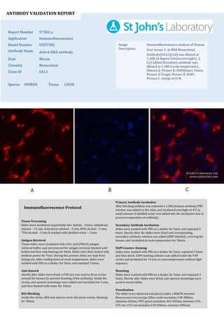
  • 1. ANTIBODY VALIDATION REPORT Report Number 97382-a Application Immunofluorescence Model Number STJ97382 Antibody Name Anti-α-SMA antibody Host Mouse Clonality Monoclonal Clone ID 6A12 Species HUMAN Tissue LIVER Image Description Immunofluorescence analysis of Human liver tissue. 1: α-SMA Monoclonal Antibody(6A12)(red) was diluted at 1:200 (4 degree Celsius,overnight). 2: Cy3 labled Secondary antibody was diluted at 1:300 (room temperature, 50min).3: Picture B: DAPI(blue) 10min. Picture A:Target. Picture B: DAPI. Picture C: merge of A+B. Primary Antibody Incubation After blocking solution was removed a 1:200 primary antibody/PBS solution was added on the slide, and incubated overnight at 4°C (a small amount of distilled water was added into the incubation box to prevent evaporation of antibody). Secondary Antibody Incubation slides were washed with PBS on a shaker for 5min, and repeated 3 times. Shortly after the slides were dried and corresponding secondary antibody solution was added (HRP labelled), covering the tissues, and incubated at room temperature for 50min. DAPI Counter-Staining slides were washed with PBS on a shaker for 5min, repeated 3 times and then dried. DAPI staining solution was added inside the PAP circles and incubated for 10 min at room temperature without light exposure. Mounting Slides were washed with PBS on a shaker for 5min, and repeated 3 times. Shortly after slides were dried, anti-quench mountings were used to mount slides. Visualization The slides were observed and placed under a NIKON inverted fluorescence microscope (Ultra violet excitation 330-380nm, emission 420nm; FITC green excitation 465-495nm, emission 515- 555 nm; CY3 red excitation 510-560nm, emission 590nm) Immunofluorescence Protocol Tissue Processing Slides were incubated sequentially into: Xylene - 15min, Anhydrous ethanol – 15 min, Anhydrous ethanol – 5 min, 85% alcohol – 5 min, 75% alcohol – 5 min & washed with distilled water – 5 min. Antigen Retrieval Tissue slides were incubated with citric acid (PH6.0) antigen retrieval buffer, and microwaved for antigen retrieval (heated until boiled and then stop heating) for 8min. Slides were then heated with medium power for 7min. During this process slides are kept from drying out. After cooling down at room temperature, slides were washed with PBS on a shaker for 5min, and repeated 3 times. Anti-Quench shortly after slides were dried, a PAP pen was used to draw circles around the tissues (to prevent draining of the antibody). Inside the circles, anti-quench mountings were added and incubated for 5 min, and then flushed with water for 10min. BSA Blocking Inside the circles, BSA was used to cover the tissue evenly, blocking for 30min. St John's Laboratory Ltd. www.stjohnslabs.com
  • 2. ANTIBODY VALIDATION REPORT Report Number 97382-b Application Immunofluorescence Model Number STJ97382 Antibody Name Anti-α-SMA antibody Host Mouse Clonality Monoclonal Clone ID 6A12 Species HUMAN Tissue LIVER Image Description Immunofluorescence analysis of Human liver tissue. 1: α-SMA Monoclonal Antibody(6A12)(red) was diluted at 1:200 (4 degree Celsius,overnight). 2: Cy3 labled Secondary antibody was diluted at 1:300 (room temperature, 50min).3: Picture B: DAPI(blue) 10min. Picture A:Target. Picture B: DAPI. Picture C: merge of A+B. Primary Antibody Incubation After blocking solution was removed a 1:200 primary antibody/PBS solution was added on the slide, and incubated overnight at 4°C (a small amount of distilled water was added into the incubation box to prevent evaporation of antibody). Secondary Antibody Incubation slides were washed with PBS on a shaker for 5min, and repeated 3 times. Shortly after the slides were dried and corresponding secondary antibody solution was added (HRP labelled), covering the tissues, and incubated at room temperature for 50min. DAPI Counter-Staining slides were washed with PBS on a shaker for 5min, repeated 3 times and then dried. DAPI staining solution was added inside the PAP circles and incubated for 10 min at room temperature without light exposure. Mounting Slides were washed with PBS on a shaker for 5min, and repeated 3 times. Shortly after slides were dried, anti-quench mountings were used to mount slides. Visualization The slides were observed and placed under a NIKON inverted fluorescence microscope (Ultra violet excitation 330-380nm, emission 420nm; FITC green excitation 465-495nm, emission 515- 555 nm; CY3 red excitation 510-560nm, emission 590nm) Immunofluorescence Protocol Tissue Processing Slides were incubated sequentially into: Xylene - 15min, Anhydrous ethanol – 15 min, Anhydrous ethanol – 5 min, 85% alcohol – 5 min, 75% alcohol – 5 min & washed with distilled water – 5 min. Antigen Retrieval Tissue slides were incubated with citric acid (PH6.0) antigen retrieval buffer, and microwaved for antigen retrieval (heated until boiled and then stop heating) for 8min. Slides were then heated with medium power for 7min. During this process slides are kept from drying out. After cooling down at room temperature, slides were washed with PBS on a shaker for 5min, and repeated 3 times. Anti-Quench shortly after slides were dried, a PAP pen was used to draw circles around the tissues (to prevent draining of the antibody). Inside the circles, anti-quench mountings were added and incubated for 5 min, and then flushed with water for 10min. BSA Blocking Inside the circles, BSA was used to cover the tissue evenly, blocking for 30min. St John's Laboratory Ltd. www.stjohnslabs.com
  • 3. ANTIBODY VALIDATION REPORT Report Number 97382-c Application Immunofluorescence Model Number STJ97382 Antibody Name Anti-α-SMA antibody Host Mouse Clonality Monoclonal Clone ID 6A12 Species HUMAN Tissue LIVER Image Description Immunofluorescence analysis of Human liver tissue. 1: α-SMA Monoclonal Antibody(6A12)(red) was diluted at 1:200 (4 degree Celsius,overnight). 2: Cy3 labled Secondary antibody was diluted at 1:300 (room temperature, 50min).3: Picture B: DAPI(blue) 10min. Picture A:Target. Picture B: DAPI. Picture C: merge of A+B. Primary Antibody Incubation After blocking solution was removed a 1:200 primary antibody/PBS solution was added on the slide, and incubated overnight at 4°C (a small amount of distilled water was added into the incubation box to prevent evaporation of antibody). Secondary Antibody Incubation slides were washed with PBS on a shaker for 5min, and repeated 3 times. Shortly after the slides were dried and corresponding secondary antibody solution was added (HRP labelled), covering the tissues, and incubated at room temperature for 50min. DAPI Counter-Staining slides were washed with PBS on a shaker for 5min, repeated 3 times and then dried. DAPI staining solution was added inside the PAP circles and incubated for 10 min at room temperature without light exposure. Mounting Slides were washed with PBS on a shaker for 5min, and repeated 3 times. Shortly after slides were dried, anti-quench mountings were used to mount slides. Visualization The slides were observed and placed under a NIKON inverted fluorescence microscope (Ultra violet excitation 330-380nm, emission 420nm; FITC green excitation 465-495nm, emission 515- 555 nm; CY3 red excitation 510-560nm, emission 590nm) Immunofluorescence Protocol Tissue Processing Slides were incubated sequentially into: Xylene - 15min, Anhydrous ethanol – 15 min, Anhydrous ethanol – 5 min, 85% alcohol – 5 min, 75% alcohol – 5 min & washed with distilled water – 5 min. Antigen Retrieval Tissue slides were incubated with citric acid (PH6.0) antigen retrieval buffer, and microwaved for antigen retrieval (heated until boiled and then stop heating) for 8min. Slides were then heated with medium power for 7min. During this process slides are kept from drying out. After cooling down at room temperature, slides were washed with PBS on a shaker for 5min, and repeated 3 times. Anti-Quench shortly after slides were dried, a PAP pen was used to draw circles around the tissues (to prevent draining of the antibody). Inside the circles, anti-quench mountings were added and incubated for 5 min, and then flushed with water for 10min. BSA Blocking Inside the circles, BSA was used to cover the tissue evenly, blocking for 30min. St John's Laboratory Ltd. www.stjohnslabs.com
  • 4. ANTIBODY VALIDATION REPORT Report Number 97382-d Application Immunofluorescence Model Number STJ97382 Antibody Name Anti-α-SMA antibody Host Mouse Clonality Monoclonal Clone ID 6A12 Species MOUSE Tissue LIVER Image Description Immunofluorescence analysis of Mouse liver tissue. 1: α-SMA Monoclonal Antibody(6A12)(red) was diluted at 1:200 (4 degree Celsius,overnight). 2: Cy3 labled Secondary antibody was diluted at 1:300 (room temperature, 50min).3: Picture B: DAPI(blue) 10min. Picture A:Target. Picture B: DAPI. Picture C: merge of A+B. Primary Antibody Incubation After blocking solution was removed a 1:200 primary antibody/PBS solution was added on the slide, and incubated overnight at 4°C (a small amount of distilled water was added into the incubation box to prevent evaporation of antibody). Secondary Antibody Incubation slides were washed with PBS on a shaker for 5min, and repeated 3 times. Shortly after the slides were dried and corresponding secondary antibody solution was added (HRP labelled), covering the tissues, and incubated at room temperature for 50min. DAPI Counter-Staining slides were washed with PBS on a shaker for 5min, repeated 3 times and then dried. DAPI staining solution was added inside the PAP circles and incubated for 10 min at room temperature without light exposure. Mounting Slides were washed with PBS on a shaker for 5min, and repeated 3 times. Shortly after slides were dried, anti-quench mountings were used to mount slides. Visualization The slides were observed and placed under a NIKON inverted fluorescence microscope (Ultra violet excitation 330-380nm, emission 420nm; FITC green excitation 465-495nm, emission 515- 555 nm; CY3 red excitation 510-560nm, emission 590nm) Immunofluorescence Protocol Tissue Processing Slides were incubated sequentially into: Xylene - 15min, Anhydrous ethanol – 15 min, Anhydrous ethanol – 5 min, 85% alcohol – 5 min, 75% alcohol – 5 min & washed with distilled water – 5 min. Antigen Retrieval Tissue slides were incubated with citric acid (PH6.0) antigen retrieval buffer, and microwaved for antigen retrieval (heated until boiled and then stop heating) for 8min. Slides were then heated with medium power for 7min. During this process slides are kept from drying out. After cooling down at room temperature, slides were washed with PBS on a shaker for 5min, and repeated 3 times. Anti-Quench shortly after slides were dried, a PAP pen was used to draw circles around the tissues (to prevent draining of the antibody). Inside the circles, anti-quench mountings were added and incubated for 5 min, and then flushed with water for 10min. BSA Blocking Inside the circles, BSA was used to cover the tissue evenly, blocking for 30min. St John's Laboratory Ltd. www.stjohnslabs.com
  • 5. ANTIBODY VALIDATION REPORT Report Number 97382-e Application Immunofluorescence Model Number STJ97382 Antibody Name Anti-α-SMA antibody Host Mouse Clonality Monoclonal Clone ID 6A12 Species MOUSE Tissue LIVER Image Description Immunofluorescence analysis of Mouse liver tissue. 1: α-SMA Monoclonal Antibody(6A12)(red) was diluted at 1:200 (4 degree Celsius,overnight). 2: Cy3 labled Secondary antibody was diluted at 1:300 (room temperature, 50min).3: Picture B: DAPI(blue) 10min. Picture A:Target. Picture B: DAPI. Picture C: merge of A+B. Primary Antibody Incubation After blocking solution was removed a 1:200 primary antibody/PBS solution was added on the slide, and incubated overnight at 4°C (a small amount of distilled water was added into the incubation box to prevent evaporation of antibody). Secondary Antibody Incubation slides were washed with PBS on a shaker for 5min, and repeated 3 times. Shortly after the slides were dried and corresponding secondary antibody solution was added (HRP labelled), covering the tissues, and incubated at room temperature for 50min. DAPI Counter-Staining slides were washed with PBS on a shaker for 5min, repeated 3 times and then dried. DAPI staining solution was added inside the PAP circles and incubated for 10 min at room temperature without light exposure. Mounting Slides were washed with PBS on a shaker for 5min, and repeated 3 times. Shortly after slides were dried, anti-quench mountings were used to mount slides. Visualization The slides were observed and placed under a NIKON inverted fluorescence microscope (Ultra violet excitation 330-380nm, emission 420nm; FITC green excitation 465-495nm, emission 515- 555 nm; CY3 red excitation 510-560nm, emission 590nm) Immunofluorescence Protocol Tissue Processing Slides were incubated sequentially into: Xylene - 15min, Anhydrous ethanol – 15 min, Anhydrous ethanol – 5 min, 85% alcohol – 5 min, 75% alcohol – 5 min & washed with distilled water – 5 min. Antigen Retrieval Tissue slides were incubated with citric acid (PH6.0) antigen retrieval buffer, and microwaved for antigen retrieval (heated until boiled and then stop heating) for 8min. Slides were then heated with medium power for 7min. During this process slides are kept from drying out. After cooling down at room temperature, slides were washed with PBS on a shaker for 5min, and repeated 3 times. Anti-Quench shortly after slides were dried, a PAP pen was used to draw circles around the tissues (to prevent draining of the antibody). Inside the circles, anti-quench mountings were added and incubated for 5 min, and then flushed with water for 10min. BSA Blocking Inside the circles, BSA was used to cover the tissue evenly, blocking for 30min. St John's Laboratory Ltd. www.stjohnslabs.com
  • 6. ANTIBODY VALIDATION REPORT Report Number 97382-f Application Immunofluorescence Model Number STJ97382 Antibody Name Anti-α-SMA antibody Host Mouse Clonality Monoclonal Clone ID 6A12 Species MOUSE Tissue LIVER Image Description Immunofluorescence analysis of Mouse liver tissue. 1: α-SMA Monoclonal Antibody(6A12)(red) was diluted at 1:200 (4 degree Celsius,overnight). 2: Cy3 labled Secondary antibody was diluted at 1:300 (room temperature, 50min).3: Picture B: DAPI(blue) 10min. Picture A:Target. Picture B: DAPI. Picture C: merge of A+B. Primary Antibody Incubation After blocking solution was removed a 1:200 primary antibody/PBS solution was added on the slide, and incubated overnight at 4°C (a small amount of distilled water was added into the incubation box to prevent evaporation of antibody). Secondary Antibody Incubation slides were washed with PBS on a shaker for 5min, and repeated 3 times. Shortly after the slides were dried and corresponding secondary antibody solution was added (HRP labelled), covering the tissues, and incubated at room temperature for 50min. DAPI Counter-Staining slides were washed with PBS on a shaker for 5min, repeated 3 times and then dried. DAPI staining solution was added inside the PAP circles and incubated for 10 min at room temperature without light exposure. Mounting Slides were washed with PBS on a shaker for 5min, and repeated 3 times. Shortly after slides were dried, anti-quench mountings were used to mount slides. Visualization The slides were observed and placed under a NIKON inverted fluorescence microscope (Ultra violet excitation 330-380nm, emission 420nm; FITC green excitation 465-495nm, emission 515- 555 nm; CY3 red excitation 510-560nm, emission 590nm) Immunofluorescence Protocol Tissue Processing Slides were incubated sequentially into: Xylene - 15min, Anhydrous ethanol – 15 min, Anhydrous ethanol – 5 min, 85% alcohol – 5 min, 75% alcohol – 5 min & washed with distilled water – 5 min. Antigen Retrieval Tissue slides were incubated with citric acid (PH6.0) antigen retrieval buffer, and microwaved for antigen retrieval (heated until boiled and then stop heating) for 8min. Slides were then heated with medium power for 7min. During this process slides are kept from drying out. After cooling down at room temperature, slides were washed with PBS on a shaker for 5min, and repeated 3 times. Anti-Quench shortly after slides were dried, a PAP pen was used to draw circles around the tissues (to prevent draining of the antibody). Inside the circles, anti-quench mountings were added and incubated for 5 min, and then flushed with water for 10min. BSA Blocking Inside the circles, BSA was used to cover the tissue evenly, blocking for 30min. St John's Laboratory Ltd. www.stjohnslabs.com
  • 7. ANTIBODY VALIDATION REPORT Report Number 97382-g Application Immunofluorescence Model Number STJ97382 Antibody Name Anti-α-SMA antibody Host Mouse Clonality Monoclonal Clone ID 6A12 Species RAT Tissue LIVER Image Description Immunofluorescence analysis of Rat liver tissue. 1: α-SMA Monoclonal Antibody(6A12)(red) was diluted at 1:200 (4 degree Celsius,overnight). 2: Cy3 labled Secondary antibody was diluted at 1:300 (room temperature, 50min).3: Picture B: DAPI(blue) 10min. Picture A:Target. Picture B: DAPI. Picture C: merge of A+B. Primary Antibody Incubation After blocking solution was removed a 1:200 primary antibody/PBS solution was added on the slide, and incubated overnight at 4°C (a small amount of distilled water was added into the incubation box to prevent evaporation of antibody). Secondary Antibody Incubation slides were washed with PBS on a shaker for 5min, and repeated 3 times. Shortly after the slides were dried and corresponding secondary antibody solution was added (HRP labelled), covering the tissues, and incubated at room temperature for 50min. DAPI Counter-Staining slides were washed with PBS on a shaker for 5min, repeated 3 times and then dried. DAPI staining solution was added inside the PAP circles and incubated for 10 min at room temperature without light exposure. Mounting Slides were washed with PBS on a shaker for 5min, and repeated 3 times. Shortly after slides were dried, anti-quench mountings were used to mount slides. Visualization The slides were observed and placed under a NIKON inverted fluorescence microscope (Ultra violet excitation 330-380nm, emission 420nm; FITC green excitation 465-495nm, emission 515- 555 nm; CY3 red excitation 510-560nm, emission 590nm) Immunofluorescence Protocol Tissue Processing Slides were incubated sequentially into: Xylene - 15min, Anhydrous ethanol – 15 min, Anhydrous ethanol – 5 min, 85% alcohol – 5 min, 75% alcohol – 5 min & washed with distilled water – 5 min. Antigen Retrieval Tissue slides were incubated with citric acid (PH6.0) antigen retrieval buffer, and microwaved for antigen retrieval (heated until boiled and then stop heating) for 8min. Slides were then heated with medium power for 7min. During this process slides are kept from drying out. After cooling down at room temperature, slides were washed with PBS on a shaker for 5min, and repeated 3 times. Anti-Quench shortly after slides were dried, a PAP pen was used to draw circles around the tissues (to prevent draining of the antibody). Inside the circles, anti-quench mountings were added and incubated for 5 min, and then flushed with water for 10min. BSA Blocking Inside the circles, BSA was used to cover the tissue evenly, blocking for 30min. St John's Laboratory Ltd. www.stjohnslabs.com
  • 8. ANTIBODY VALIDATION REPORT Report Number 97382-h Application Immunofluorescence Model Number STJ97382 Antibody Name Anti-α-SMA antibody Host Mouse Clonality Monoclonal Clone ID 6A12 Species RAT Tissue LIVER Image Description Immunofluorescence analysis of Rat liver tissue. 1: α-SMA Monoclonal Antibody(6A12)(red) was diluted at 1:200 (4 degree Celsius,overnight). 2: Cy3 labled Secondary antibody was diluted at 1:300 (room temperature, 50min).3: Picture B: DAPI(blue) 10min. Picture A:Target. Picture B: DAPI. Picture C: merge of A+B. Primary Antibody Incubation After blocking solution was removed a 1:200 primary antibody/PBS solution was added on the slide, and incubated overnight at 4°C (a small amount of distilled water was added into the incubation box to prevent evaporation of antibody). Secondary Antibody Incubation slides were washed with PBS on a shaker for 5min, and repeated 3 times. Shortly after the slides were dried and corresponding secondary antibody solution was added (HRP labelled), covering the tissues, and incubated at room temperature for 50min. DAPI Counter-Staining slides were washed with PBS on a shaker for 5min, repeated 3 times and then dried. DAPI staining solution was added inside the PAP circles and incubated for 10 min at room temperature without light exposure. Mounting Slides were washed with PBS on a shaker for 5min, and repeated 3 times. Shortly after slides were dried, anti-quench mountings were used to mount slides. Visualization The slides were observed and placed under a NIKON inverted fluorescence microscope (Ultra violet excitation 330-380nm, emission 420nm; FITC green excitation 465-495nm, emission 515- 555 nm; CY3 red excitation 510-560nm, emission 590nm) Immunofluorescence Protocol Tissue Processing Slides were incubated sequentially into: Xylene - 15min, Anhydrous ethanol – 15 min, Anhydrous ethanol – 5 min, 85% alcohol – 5 min, 75% alcohol – 5 min & washed with distilled water – 5 min. Antigen Retrieval Tissue slides were incubated with citric acid (PH6.0) antigen retrieval buffer, and microwaved for antigen retrieval (heated until boiled and then stop heating) for 8min. Slides were then heated with medium power for 7min. During this process slides are kept from drying out. After cooling down at room temperature, slides were washed with PBS on a shaker for 5min, and repeated 3 times. Anti-Quench shortly after slides were dried, a PAP pen was used to draw circles around the tissues (to prevent draining of the antibody). Inside the circles, anti-quench mountings were added and incubated for 5 min, and then flushed with water for 10min. BSA Blocking Inside the circles, BSA was used to cover the tissue evenly, blocking for 30min. St John's Laboratory Ltd. www.stjohnslabs.com
  • 9. ANTIBODY VALIDATION REPORT Report Number 97382-i Application Immunofluorescence Model Number STJ97382 Antibody Name Anti-α-SMA antibody Host Mouse Clonality Monoclonal Clone ID 6A12 Species RAT Tissue LIVER Image Description Immunofluorescence analysis of Rat liver tissue. 1: α-SMA Monoclonal Antibody(6A12)(red) was diluted at 1:200 (4 degree Celsius,overnight). 2: Cy3 labled Secondary antibody was diluted at 1:300 (room temperature, 50min).3: Picture B: DAPI(blue) 10min. Picture A:Target. Picture B: DAPI. Picture C: merge of A+B. Primary Antibody Incubation After blocking solution was removed a 1:200 primary antibody/PBS solution was added on the slide, and incubated overnight at 4°C (a small amount of distilled water was added into the incubation box to prevent evaporation of antibody). Secondary Antibody Incubation slides were washed with PBS on a shaker for 5min, and repeated 3 times. Shortly after the slides were dried and corresponding secondary antibody solution was added (HRP labelled), covering the tissues, and incubated at room temperature for 50min. DAPI Counter-Staining slides were washed with PBS on a shaker for 5min, repeated 3 times and then dried. DAPI staining solution was added inside the PAP circles and incubated for 10 min at room temperature without light exposure. Mounting Slides were washed with PBS on a shaker for 5min, and repeated 3 times. Shortly after slides were dried, anti-quench mountings were used to mount slides. Visualization The slides were observed and placed under a NIKON inverted fluorescence microscope (Ultra violet excitation 330-380nm, emission 420nm; FITC green excitation 465-495nm, emission 515- 555 nm; CY3 red excitation 510-560nm, emission 590nm) Immunofluorescence Protocol Tissue Processing Slides were incubated sequentially into: Xylene - 15min, Anhydrous ethanol – 15 min, Anhydrous ethanol – 5 min, 85% alcohol – 5 min, 75% alcohol – 5 min & washed with distilled water – 5 min. Antigen Retrieval Tissue slides were incubated with citric acid (PH6.0) antigen retrieval buffer, and microwaved for antigen retrieval (heated until boiled and then stop heating) for 8min. Slides were then heated with medium power for 7min. During this process slides are kept from drying out. After cooling down at room temperature, slides were washed with PBS on a shaker for 5min, and repeated 3 times. Anti-Quench shortly after slides were dried, a PAP pen was used to draw circles around the tissues (to prevent draining of the antibody). Inside the circles, anti-quench mountings were added and incubated for 5 min, and then flushed with water for 10min. BSA Blocking Inside the circles, BSA was used to cover the tissue evenly, blocking for 30min. St John's Laboratory Ltd. www.stjohnslabs.com