1. The Department of Human anatomy
The Department of Human anatomy
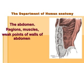
The abdomen.
The abdomen.
Regions, muscles,
Regions, muscles,
weak points of walls of
weak points of walls of
abdomen
abdomen
2. PLAN
PLAN
Abdominal regions.
Abdominal regions.
Walls of abdomen.
Walls of abdomen.
Muscles of abdomen: classification.
Muscles of abdomen: classification.
Superficial and internal inguinal
Superficial and internal inguinal
rings. Abdominal press.
rings. Abdominal press.
Fasciae of abdomen.
Fasciae of abdomen.
Weak points of walls of abdomen.
Weak points of walls of abdomen.
3. Abdomen is the
Abdomen is the
region of body
region of body
lying between
lying between
the diaphragm
the diaphragm
above and pelvis
above and pelvis
below. It contains
below. It contains
the contents of
the contents of
digestive and
digestive and
urinary systems.
urinary systems.
4. Two horizontal and
Two horizontal and
two vertical lines
two vertical lines
divide the abdomen
divide the abdomen
into nine "regions."
into nine "regions."
6. I. Epigastrium:
I. Epigastrium:
1 (regio epigastrica)
1 (regio epigastrica)
2. (regio hypocondrica dexter)
2. (regio hypocondrica dexter)
3. (regio hypochondrica sinistra)
3. (regio hypochondrica sinistra)
ІІ. Mesogastrium:
ІІ. Mesogastrium:
4. (regio umbilicalis)
4. (regio umbilicalis)
5. (regio lateralis dextra)
5. (regio lateralis dextra)
6. (regio lateralis sinistra)
6. (regio lateralis sinistra)
ІІІ. Hypogastrium:
ІІІ. Hypogastrium:
7. (regio pubica)
7. (regio pubica)
8. (regio inguinalis dextra)
8. (regio inguinalis dextra)
9. (regio inguinalis sinister)
9. (regio inguinalis sinister)
("hypo" means "below" and "epi" means
("hypo" means "below" and "epi" means
"above", "chond" means "cartilage"
"above", "chond" means "cartilage"
(in this case, the cartilage of the rib)
(in this case, the cartilage of the rib)
and "gast" means stomach).
and "gast" means stomach).
7. Walls of abdomen:
Walls of abdomen:
Superior Wall:
Superior Wall: The superior wall of abdomen is formed by the
The superior wall of abdomen is formed by the
diaphragm. It is a strong muscular sheath that has a vital role in
diaphragm. It is a strong muscular sheath that has a vital role in
respiratory system.
respiratory system.
Inferior Wall:
Inferior Wall: the conditional line to the entry of the pelvic
the conditional line to the entry of the pelvic
cavity.
cavity.
Anterior Wall:
Anterior Wall: The anterior wall of abdomen is formed above by
The anterior wall of abdomen is formed above by
the lower part of thoracic cage. Below it is formed by Rectus
the lower part of thoracic cage. Below it is formed by Rectus
abdominis, External oblique, Internal oblique and Transversus
abdominis, External oblique, Internal oblique and Transversus
abdominis muscles along with their fasciae.
abdominis muscles along with their fasciae.
Posterior Wall:
Posterior Wall: The posterior wall of abdomen is formed in the
The posterior wall of abdomen is formed in the
midline by the five lumbar vertebrae and their intervertebral
midline by the five lumbar vertebrae and their intervertebral
discs. Laterally, it is formed by the twelfth rib, psoas muscle,
discs. Laterally, it is formed by the twelfth rib, psoas muscle,
quadratus lumborum muscle and the border of bony pelvis. The
quadratus lumborum muscle and the border of bony pelvis. The
aponeurosis of origin of transversus abdominis muscle is also
aponeurosis of origin of transversus abdominis muscle is also
involved in the formation of posterior wall of the abdomen.
involved in the formation of posterior wall of the abdomen.
8. The layers of the abdominal wall are (from
The layers of the abdominal wall are (from
superficial to deep):
superficial to deep):
Skin
Skin
Fascia
Fascia
Camper's fascia - fatty superficial layer.
Camper's fascia - fatty superficial layer.
Scarpa's fascia - deep fibrous layer.
Scarpa's fascia - deep fibrous layer.
Muscle
Muscle
Rectus abdominis
Rectus abdominis
External oblique muscle
External oblique muscle
Internal oblique muscle
Internal oblique muscle
Transverse abdominal muscle
Transverse abdominal muscle
Fascia transversalis
Fascia transversalis
Peritoneum
Peritoneum
9. Abdominal muscles
The muscles
of anterior wall
The muscles
of posterior wall
The muscles
of laterar wall
Pyramidalis
Obliquus externus
abdominis
Rectus Abdominus
Obliquus internus
abdominis
Transversus abdominis
Quadratus lumborum
10. The muscles
of anterior wall
Rectus Abdominus
Origin
5, 6, 7 costal cartilages,
5, 6, 7 costal cartilages,
xiphoid process
xiphoid process
Insertion
Insertion
Pubic crest and pubic
symphysis
Action
Action
Flexes trunk, aids forced
Flexes trunk, aids forced
expiration and raise intra-
expiration and raise intra-
abdominal pressure
abdominal pressure
11. Pyramidalis
Origin
Pubic crest
Insertion
Lower linea alba
Action
Action
Is a tensor of the linea
Is a tensor of the linea
alba
alba
Reinforces lower rectus
Reinforces lower rectus
sheath
sheath
12. The muscles of lateral wall
Obliquus externus Abdominis
Origin
Anterior angles of lower eight
ribs
Insertion
Insertion
Outer anterior half of iliac crest,
Outer anterior half of iliac crest,
inguinal lig, pubic tubercle and
inguinal lig, pubic tubercle and
crest, and aponeurosis of
crest, and aponeurosis of
anterior rectus sheath
anterior rectus sheath
Action
Supports abdominal wall,
assists forced expiration, aids
raising intraabdominal pressure
and, with muscles of opposite
side, abducts and rotates trunk
13. Obliquus internus
abdominis
Origin
Lumbar fascia, ant. two thirds
of iliac crest and lateral two
thirds of inguinal ligament
Insertion
Insertion
Costal margin, aponeurosis of
Costal margin, aponeurosis of
rectus sheath (ant. and post. ),
rectus sheath (ant. and post. ),
conjoint tendon to pubic crest
conjoint tendon to pubic crest
and pectineal line
and pectineal line
Action
Action
Supports abdominal wall,
Supports abdominal wall,
assists forced respiration, aids
assists forced respiration, aids
raising intraabdominal pressure
raising intraabdominal pressure
& , with muscles of other side ,
& , with muscles of other side ,
abducts and rotates trunk.
abducts and rotates trunk.
Conjoint tendon supports
Conjoint tendon supports
posterior wall of inguinal canal
posterior wall of inguinal canal
14. Transversus abdominis
Origin
Costal margin , lumbar fascia,
ant two thirds of iliac crest and
lateral half of inguinal ligament
Insertion
Insertion
Aponeurosis of posterior and
Aponeurosis of posterior and
anterior rectus sheath and
anterior rectus sheath and
conjoint tendon to pubic crest
conjoint tendon to pubic crest
and pectineal line
and pectineal line
Action
Action
Supports abdominal wall, aids
Supports abdominal wall, aids
forced expiration and raising
forced expiration and raising
intraabdominal pressure.
intraabdominal pressure.
Conjoint tendon supports
Conjoint tendon supports
posterior wall of inguinal canal
posterior wall of inguinal canal
15. The muscles of posterior
wall
Quadratus lumborum
Origin
Inferior border of 12th rib
Insertion
Transverse processes of
L1-4, iliolumbar ligament
and post. 1/3 of iliac crest
Action
Action
Fixes 12th rib during
Fixes 12th rib during
respiration and lateral
respiration and lateral
Flexes of trunk
Flexes of trunk
16. Rectus sheath above and below the umbilical chord
Rectus sheath above and below the umbilical chord
1 – (m. rectus abdominis);
1 – (m. rectus abdominis);
2 – (m. obliguus externus abdominis);
2 – (m. obliguus externus abdominis);
3 – (m. obliguus internus abdominis);
3 – (m. obliguus internus abdominis);
4 – (m. transversus abdominis);
4 – (m. transversus abdominis);
а) (aponeurosis m. obliguus externus abdominis);
а) (aponeurosis m. obliguus externus abdominis);
b) (lamina anterior m. obliguus internus abdominis);
b) (lamina anterior m. obliguus internus abdominis);
c) (lamina posterior m. obliguus internus abdominis);
c) (lamina posterior m. obliguus internus abdominis);
d) (aponeurosis m. transverses abdominis);
d) (aponeurosis m. transverses abdominis);
e) (fascia transversalis).
e) (fascia transversalis).
17. weak points of walls of abdomen
weak points of walls of abdomen
1) canalis inquinalis
1) canalis inquinalis
2) linea alba
2) linea alba
3) anulus umbilicalis
3) anulus umbilicalis
4) trigonum lumbale Petiti
4) trigonum lumbale Petiti
5) spatium tendinosum lumbale
5) spatium tendinosum lumbale
6) trigonum hypochondriacum
6) trigonum hypochondriacum
7) linea semilunaris Spigelii
7) linea semilunaris Spigelii
8) trigonum sternocostale dext. et sin.
8) trigonum sternocostale dext. et sin.
trigonum costolumbalis dext. et sin.
trigonum costolumbalis dext. et sin.
9) canalis obturatorius
9) canalis obturatorius
10) Foramen supra- et infrapiriformis
10) Foramen supra- et infrapiriformis
11) canalis femoralis
11) canalis femoralis
18.
19. Inguinal canal is an oblique
Inguinal canal is an oblique
passage through the lower part
passage through the lower part
of anterior abdominal wall. It is
of anterior abdominal wall. It is
present in both males and
present in both males and
females.
females.
20. Functions of inguinal canal:
Functions of inguinal canal:
In males, the inguinal canal allows structures of
In males, the inguinal canal allows structures of
the spermatic cord to pass to and from the testis
the spermatic cord to pass to and from the testis
to the abdomen. This allows the testes to leave
to the abdomen. This allows the testes to leave
the abdominal cavity. The importance of this
the abdominal cavity. The importance of this
phenomenon can be appreciated by reminding
phenomenon can be appreciated by reminding
the fact that spermatogenesis takes place only if
the fact that spermatogenesis takes place only if
the testes leave the abdominal cavity to go to a
the testes leave the abdominal cavity to go to a
cooler environment in the scrotum.
cooler environment in the scrotum.
In the females, the inguinal canal is smaller as
In the females, the inguinal canal is smaller as
compared to males. It transmits the round
compared to males. It transmits the round
ligament of uterus to pass from the uterus to
ligament of uterus to pass from the uterus to
labium majus.
labium majus.
In both males and females, the inguinal canal also
In both males and females, the inguinal canal also
transmits the ilioinguinal nerve.
transmits the ilioinguinal nerve.
21. Structure of inguinal canal:
Structure of inguinal canal:
Inguinal canal is about 4-5 cm long in adults. It
Inguinal canal is about 4-5 cm long in adults. It
extends from
extends from deep
deep inguinal ring
inguinal ring, downward and
, downward and
medially, to the
medially, to the superficial inguinal ring
superficial inguinal ring. The deep
. The deep
inguinal ring is an oval opening in fascia
inguinal ring is an oval opening in fascia
transversalis, while the superficial inguinal ring is a
transversalis, while the superficial inguinal ring is a
triangular defect in the aponeuorsis of external
triangular defect in the aponeuorsis of external
oblique muscle.
oblique muscle.
The inguinal canal lies immediately above and
The inguinal canal lies immediately above and
parallel to the inguinal ligament. However, in
parallel to the inguinal ligament. However, in
infants, the deep inguinal ring lies almost posterior
infants, the deep inguinal ring lies almost posterior
to the superficial, so that the canal is shorter. As a
to the superficial, so that the canal is shorter. As a
result of growth in following years, the deep
result of growth in following years, the deep
inguinal ring moves laterally and the canal
inguinal ring moves laterally and the canal
elongates.
elongates.
22. Walls of inguinal canal:
Walls of inguinal canal:
23. Anterior wall:
Anterior wall: The
The
anterior wall is formed
anterior wall is formed
along its entire length
along its entire length
by the aponeuorsis of
by the aponeuorsis of
external oblique
external oblique
muscle. In its lateral
muscle. In its lateral
part, it is reinforced by
part, it is reinforced by
the origin of internal
the origin of internal
oblique from the
oblique from the
inguinal ligament.
inguinal ligament.
24. Posterior wall:
Posterior wall: The
The
posterior wall of inguinal
posterior wall of inguinal
canal is formed along its
canal is formed along its
entire length by the
entire length by the
fascia transversalis.
fascia transversalis.
25. Floor of inguinal canal
Floor of inguinal canal
(inferior wall):
(inferior wall): The floor of
The floor of
the canal is formed by the
the canal is formed by the
inguinal ligament. This
inguinal ligament. This
ligament is in fact the rolled-
ligament is in fact the rolled-
under inferior edge of
under inferior edge of
aponeuorsis of external
aponeuorsis of external
oblique muscle. The medial
oblique muscle. The medial
part of this wall is formed by
part of this wall is formed by
the lacunar ligament.
the lacunar ligament.
Roof of inguinal canal
Roof of inguinal canal
(superior wall):
(superior wall): The roof of
The roof of
the canal is formed by the
the canal is formed by the
lowest fibers of internal
lowest fibers of internal
oblique and transversus
oblique and transversus
abdominis muscles.
abdominis muscles.
26. Internal surface of anterior abdominal
Internal surface of anterior abdominal
wall
wall
27.
28. Inguinal canal as a potential weakness in
Inguinal canal as a potential weakness in
abdominal wall:
abdominal wall:
The presence of inguinal canal in lower part
The presence of inguinal canal in lower part
of anterior abdominal wall presents a
of anterior abdominal wall presents a
potential weakness the structure of the wall.
potential weakness the structure of the wall.
The canal is oblique in its form. This causes
The canal is oblique in its form. This causes
the weaker parts of it to lie at some distance
the weaker parts of it to lie at some distance
apart, thus reducing chances of any type of
apart, thus reducing chances of any type of
damage.
damage.
29. The weak inguinal canal can be exploited
The weak inguinal canal can be exploited
during forceful activities like coughing,
during forceful activities like coughing,
sneezing, straining, parturition, defecation
sneezing, straining, parturition, defecation
etc. To prevent this, the arching lowest
etc. To prevent this, the arching lowest
fibers of internal oblique and transversus
fibers of internal oblique and transversus
abdominis muscles contract and flatten out
abdominis muscles contract and flatten out
the arched roof so that it is lowered
the arched roof so that it is lowered
towards the floor. In this condition, the
towards the floor. In this condition, the
canal is virtually closed.
canal is virtually closed.
30. An
An inguinal hernia
inguinal hernia
is a protrusion
is a protrusion
of abdominal-
of abdominal-
cavity contents
cavity contents
through the inguinal
through the inguinal
canal. They are very
canal. They are very
common (lifetime
common (lifetime
risk 27% for men,
risk 27% for men,
3% for women), and
3% for women), and
their repair is one of
their repair is one of
the most frequently
the most frequently
performed surgical
performed surgical
operations.
operations.
31. There are two types of
There are two types of
inguinal hernia,
inguinal hernia, direct
direct and
and indirect
indirect, which are
, which are
defined by their relationship to the inferior
defined by their relationship to the inferior
epigastric vessels. Direct inguinal hernias occur
epigastric vessels. Direct inguinal hernias occur
medial to the inferior epigastric vessels when
medial to the inferior epigastric vessels when
abdominal contents herniate through a weak spot
abdominal contents herniate through a weak spot
in the fascia of the posterior wall of the inguinal
in the fascia of the posterior wall of the inguinal
canal, which is formed by the transversalis fascia.
canal, which is formed by the transversalis fascia.
Indirect inguinal hernias occur when abdominal
Indirect inguinal hernias occur when abdominal
contents protrude through the deep inguinal ring,
contents protrude through the deep inguinal ring,
lateral to the inferior epigastric vessels; this may
lateral to the inferior epigastric vessels; this may
be caused by failure of embryonic closure of
be caused by failure of embryonic closure of
the processus vaginalis.
the processus vaginalis.
32. Inguinal hernias, in turn, belong
Inguinal hernias, in turn, belong
to groin hernias, which also
to groin hernias, which also
includes femoral hernias. A femoral hernia
includes femoral hernias. A femoral hernia
is not via the inguinal canal, but via the
is not via the inguinal canal, but via the
femoral canal, which normally allows
femoral canal, which normally allows
passage of the common femoral artery and
passage of the common femoral artery and
vein from the pelvis to the leg.
vein from the pelvis to the leg.
In Amyand's hernia, the content of the
In Amyand's hernia, the content of the
hernial sac is the vermiform appendix.
hernial sac is the vermiform appendix.
In Littre's hernia, the content of the hernial
In Littre's hernia, the content of the hernial
sac contains a Meckel's Diverticulum.
sac contains a Meckel's Diverticulum.