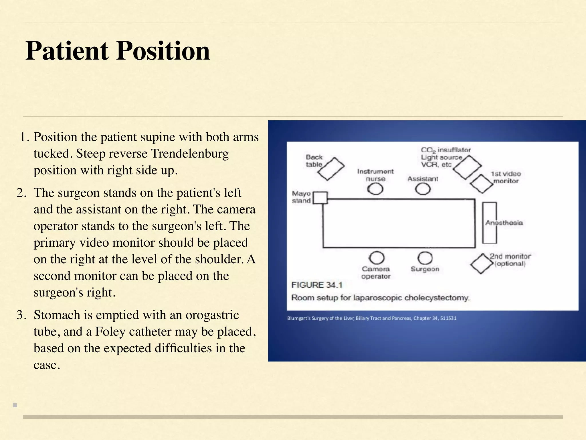
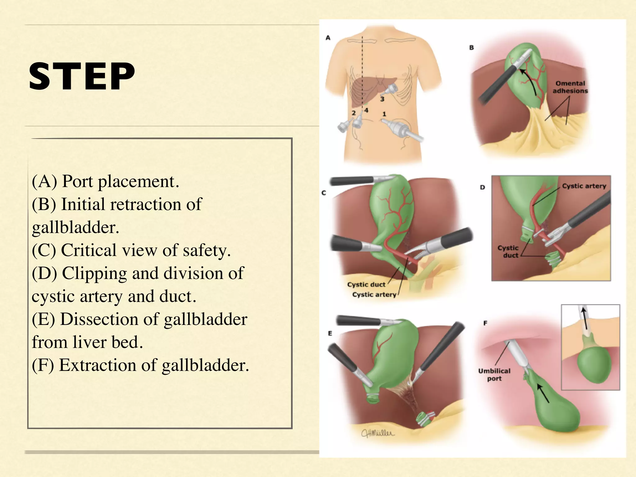
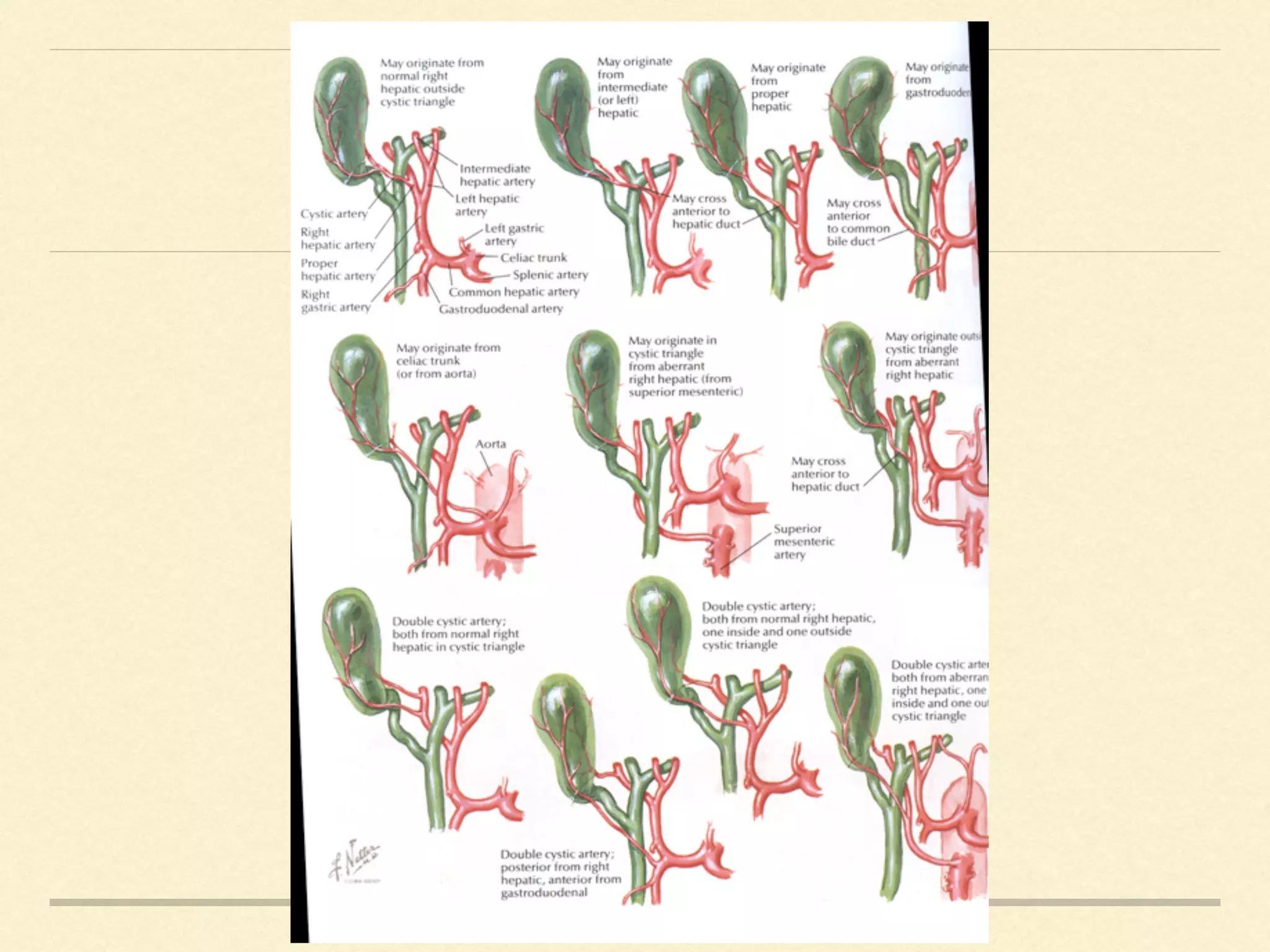
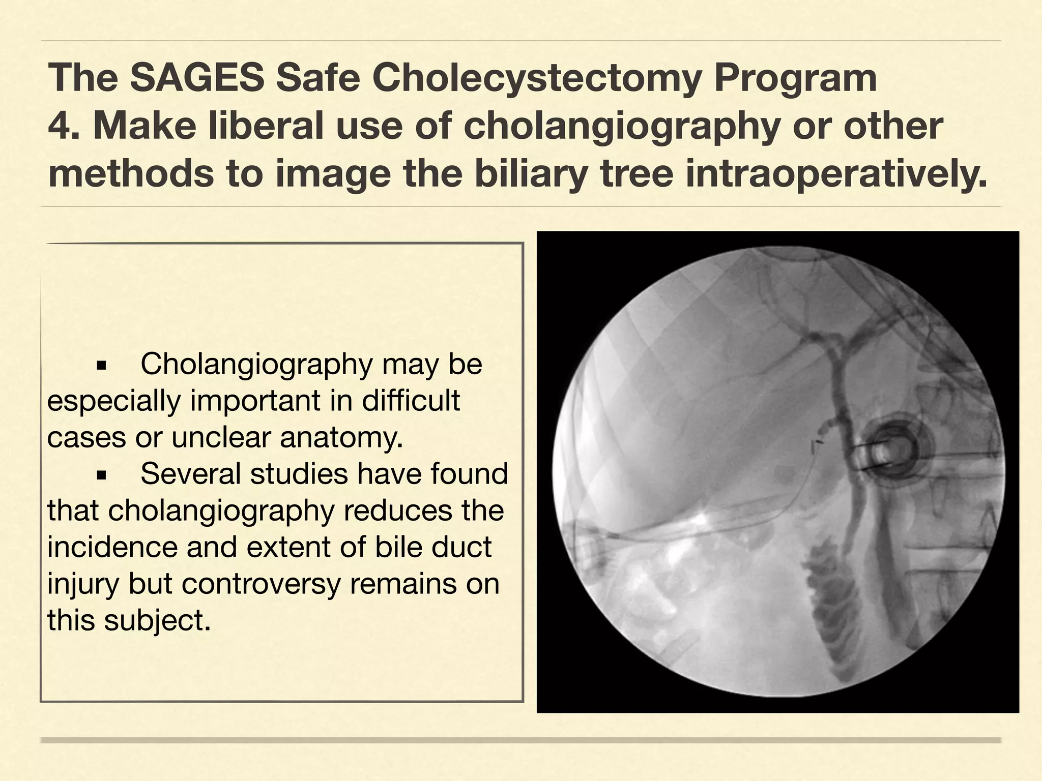
TIP & TECHNIQUE FOR SAFE
LAPAROSCOPIC CHOLECYSTECTOM
Y

Kamales Prasitvarakul ,MD. FRCS
T

Division of Minimally Invasive Surgery center
Department of Surgery
Hatyai Hospital
Songkla Thailand
Patient Position
 

1
.	
Position the patient supine with both arms
tucked. Steep reverse Trendelenburg
position with right side up
.

2
.	
The surgeon stands on the patient's left
and the assistant on the right. The camera
operator stands to the surgeon's left. The
primary video monitor should be placed
on the right at the level of the shoulder. A
second monitor can be placed on the
surgeon's right
.

3
.	
Stomach is emptied with an orogastric
tube, and a Foley catheter may be placed,
based on the expected dif
fi
culties in the
case.


Abdominal assess and Trocar Placement
 

Abdominal Access
 

1. Veress needle
 

2. The open Hasson’s techniqu
e

3. Direct trocar placement without prior pneumoperitoneum
.

Trocar Placement
 

Typically, use four trocar
s

a
.	
Umbilical 12-mm optical entry trocar
.

b
.	
Epigastric 5-mm trocar is placed based on the liver edge
,just to the right of the falciform ligament.
 

c. Two additional 5-mm trocars laterally on the right side,
one in midclavicular line and another in anterior axillary
line.


**Three additional trocars are typically placed under
direct vision**


STEP
(A) Port placement
.

(B) Initial retraction of
gallbladder
.

(C) Critical view of safety
.

(D) Clipping and division of
cystic artery and duct
.

(E) Dissection of gallbladder
from liver bed
.

(F) Extraction of gallbladder
.
The SAGES Safe Cholecystectomy Program
1. Use the Critical View of Safety (CVS) method
Three criteria are required to
achieve the CVS: 

A. The hepatocystic triangle
is cleared of fat and
fi
brous
tissue. The hepatocystic
triangle is de
fi
ned as the
triangle formed by the cystic
duct, the common hepatic
duct, and inferior edge of the
liver. The common bile duct
and common hepatic duct do
not have to be exposed.
The SAGES Safe Cholecystectomy Program
1. Use the Critical View of Safety (CVS) method
B. The lower one third of the
gallbladder is separated
from the liver to expose the
cystic plate. The cystic plate
is also known as liver bed of
the gallbladder and lies in the
gallbladder fossa.

C. Two and only two
structures should be seen
entering the gallbladder.
Visualization of the doublet view
(anterior)
Visualization of the doublet view
(posterior)
The SAGES Safe Cholecystectomy Program
2. Intraoperative Time Out CVS
• Consider an Intraoperative Time Out during
laparoscopic cholecystectomy prior to clipping, cutting
or transecting any ductal structures. 

• The Intraoperative Time Out should consist of a stop
point in the operation to con
fi
rm that the CVS has been
achieved utilizing the Doublet View.
The SAGES Safe Cholecystectomy Program
3. Understand the potential for aberrant anatomy
in all cases.
• Aberrant anatomy may include
a short cystic duct, aberrant
hepatic ducts, or a right hepatic
artery that crosses anterior to the
common bile duct . These are
some but not all common
variants. •
The SAGES Safe Cholecystectomy Program
4. Make liberal use of cholangiography or other
methods to image the biliary tree intraoperatively.
	 ▪	 Cholangiography may be
especially important in di
ffi
cult
cases or unclear anatomy.

	 ▪	 Several studies have found
that cholangiography reduces the
incidence and extent of bile duct
injury but controversy remains on
this subject.
The SAGES Safe Cholecystectomy Program
5. Recognize when the dissection is approaching a zone of signi
fi
cant risk and halt
the dissection before entering the zone. Finish the operation by a safe method
other than cholecystectomy if conditions around the gallbladder are too
dangerous.
	 ▪	 Severe in
fl
ammation in the porta hepatis and neck of the gallbladder, the CVS can
be di
ffi
cult to achieve. The surgical judgment that a zone of  signi
fi
cant risk  is being
approached can be made when there is failure to obtain adequate exposure of the
anatomy of the hepatocystic triangle  or when the dissection is not progressing due to
bleeding, in
fl
ammation or
fi
brosis.

	 ▪	 Consider laparoscopic subtotal cholecystectomy or Semi top-down technique
or cholecystostomy tube placement, and/or conversion to an open procedure based
on the judgment of the attending surgeon.

6. Get help from another surgeon when the dissection or conditions are di
ffi
cult.
THANK YOU

Tip and technique for safe LC

  • 1.
    TIP & TECHNIQUEFOR SAFE LAPAROSCOPIC CHOLECYSTECTOM Y Kamales Prasitvarakul ,MD. FRCS T Division of Minimally Invasive Surgery center Department of Surgery Hatyai Hospital Songkla Thailand
  • 2.
    Patient Position 1 . Positionthe patient supine with both arms tucked. Steep reverse Trendelenburg position with right side up . 2 . The surgeon stands on the patient's left and the assistant on the right. The camera operator stands to the surgeon's left. The primary video monitor should be placed on the right at the level of the shoulder. A second monitor can be placed on the surgeon's right . 3 . Stomach is emptied with an orogastric tube, and a Foley catheter may be placed, based on the expected dif fi culties in the case. 

  • 3.
    Abdominal assess andTrocar Placement Abdominal Access 1. Veress needle 2. The open Hasson’s techniqu e 3. Direct trocar placement without prior pneumoperitoneum . Trocar Placement Typically, use four trocar s a . Umbilical 12-mm optical entry trocar . b . Epigastric 5-mm trocar is placed based on the liver edge ,just to the right of the falciform ligament. c. Two additional 5-mm trocars laterally on the right side, one in midclavicular line and another in anterior axillary line. **Three additional trocars are typically placed under direct vision** 

  • 4.
    STEP (A) Port placement . (B)Initial retraction of gallbladder . (C) Critical view of safety . (D) Clipping and division of cystic artery and duct . (E) Dissection of gallbladder from liver bed . (F) Extraction of gallbladder .
  • 6.
    The SAGES SafeCholecystectomy Program 1. Use the Critical View of Safety (CVS) method Three criteria are required to achieve the CVS: A. The hepatocystic triangle is cleared of fat and fi brous tissue. The hepatocystic triangle is de fi ned as the triangle formed by the cystic duct, the common hepatic duct, and inferior edge of the liver. The common bile duct and common hepatic duct do not have to be exposed.
  • 7.
    The SAGES SafeCholecystectomy Program 1. Use the Critical View of Safety (CVS) method B. The lower one third of the gallbladder is separated from the liver to expose the cystic plate. The cystic plate is also known as liver bed of the gallbladder and lies in the gallbladder fossa. C. Two and only two structures should be seen entering the gallbladder. Visualization of the doublet view (anterior) Visualization of the doublet view (posterior)
  • 8.
    The SAGES SafeCholecystectomy Program 2. Intraoperative Time Out CVS • Consider an Intraoperative Time Out during laparoscopic cholecystectomy prior to clipping, cutting or transecting any ductal structures. • The Intraoperative Time Out should consist of a stop point in the operation to con fi rm that the CVS has been achieved utilizing the Doublet View.
  • 9.
    The SAGES SafeCholecystectomy Program 3. Understand the potential for aberrant anatomy in all cases. • Aberrant anatomy may include a short cystic duct, aberrant hepatic ducts, or a right hepatic artery that crosses anterior to the common bile duct . These are some but not all common variants. •
  • 11.
    The SAGES SafeCholecystectomy Program 4. Make liberal use of cholangiography or other methods to image the biliary tree intraoperatively. ▪ Cholangiography may be especially important in di ffi cult cases or unclear anatomy. ▪ Several studies have found that cholangiography reduces the incidence and extent of bile duct injury but controversy remains on this subject.
  • 12.
    The SAGES SafeCholecystectomy Program 5. Recognize when the dissection is approaching a zone of signi fi cant risk and halt the dissection before entering the zone. Finish the operation by a safe method other than cholecystectomy if conditions around the gallbladder are too dangerous. ▪ Severe in fl ammation in the porta hepatis and neck of the gallbladder, the CVS can be di ffi cult to achieve. The surgical judgment that a zone of  signi fi cant risk  is being approached can be made when there is failure to obtain adequate exposure of the anatomy of the hepatocystic triangle  or when the dissection is not progressing due to bleeding, in fl ammation or fi brosis. ▪ Consider laparoscopic subtotal cholecystectomy or Semi top-down technique or cholecystostomy tube placement, and/or conversion to an open procedure based on the judgment of the attending surgeon. 6. Get help from another surgeon when the dissection or conditions are di ffi cult.
  • 13.