
Recommended
More Related Content
Similar to Sarcoidosis PK (2) (1).pptx
Similar to Sarcoidosis PK (2) (1).pptx (20)
More from ssuser227d6b
More from ssuser227d6b (16)
Recently uploaded
Recently uploaded (20)
Sarcoidosis PK (2) (1).pptx
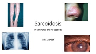
- 1. Sarcoidosis in 6 minutes and 40 seconds Matt Dickson
- 2. Definition • Chronic granulomatous disorder • Multiorgan involvement • First case described by Sir Jonathan Hutchinson 1878
- 3. Epidemiology • Incidence of 5-10 per 100,000 • Bimodal age distribution • Highest incidence: • Northern Europe (Scandinavia) • Irish • African Americans • West Indians
- 4. Aetiology • Unknown • HLA-DRB1*1101 associated with disease susceptibility • HLA-DRB1*0301 associated with acute and remitting disease
- 6. Pathophysiology • ACE levels • Lymphopenia • Delayed type hypersensitivity reactions
- 7. Presentation • Variable – acute vs chronic • Thoracic involvement ~90% • Respiratory symptoms • Constitutional symptoms • Lofgren's syndrome: Fever Bilateral Hilar Lymphadenopathy Erythema nodosum Arthralgia • Asymptomatic
- 10. Other investigations • FBC – anaemia, leucopenia • Hypercalaemia • Immunoglobulins • ACE • ECG/Echo • PFTs • Bronchoscopy • EBUS
- 11. Management • Treatment not recommended for : • Asymptomatic stage I disease • Asymptomatic stage II or III disease with mildly abnormal lung function and stable disease • Oral corticosteroids first line • 0.5mg/kg/day prednisolone for ~ 4 weeks, reduce to maintenance dose (5-20mg OD) for period of 6 months to 2 years • ICS not of significant benefit • In treatment failure/life threatening – pulsed IVMP
- 12. Management • Second-line agents • Methotrexate • Azathioprine • Mycophenolate • Leflunomide • Third-line agents • Biologics e.g infliximab • Lung transplantation
- 13. Prognosis • Remission rates can correlate with the Scadding classification • Lofgren's syndrome or Stage I – 80-90% will resolve • Poor prognosis with chronic disease: • Lupus pernio • Nasal mucosa involvement • Chronic uveiitis • Chronic hypercalcaemia • Nephrocalcinosis • Neural involvement • >40 • Black population
- 14. Extrapulmonary manifestations • Hypercalcaemia • Dysregulated calcitriol production • Increased intestinal absorption • Mild – dietary advice, reduce sun exposure • Significant - steroids
- 15. • Skin • Papules and plaques • Erythema nodosum • Lupus pernio Extrapulmonary manifestations
- 16. Extrapulmonary manifestations • Heart • Conduction defects • Palpitations • Syncope • Wall motion abnormalities
- 17. Extrapulmonary manifestations • Eyes • Uveiitis • Episcleritis • Scleritis • Glaucoma • Conjunctivitis • Kidney • Obstructive uropathy • Nephrocalcinosis • Glomerulonephritis
- 18. Extrapulmonary manifestations • CNS • MSK • GI • Others
- 19. Conclusion • Multisystem disorder • Non-caseous granulomas • Thoracic involvement most common • Acute vs Chronic • Bilateral hilar lymphadenopathy → fibrosis
- 20. Conclusion • Mainstay of treatment is steroids • Variable prognosis • Disease requiring MDT input
Editor's Notes
- I’m going to try and cover sarcoidosis in less than 7 minutes, and hopefully I’ve picked out the most relevant bits we’ll all need to remember in clinic and for exams.
- Sarcoidosis is a multisystem disorder that is characterized by noncaseous epithelioid cell granulomas, which may affect almost any organ, most commonly affecting the lungs, skin and eyes. The first case was described by Sir Jonathan Hutchinson in 1878, who coined the term "lupus vulgaris multiplex non-ulcerans".
- The disease has an incidence worldwide of about 5-10 per 100,000, prevalence of 1 in 10,000 in UK. with a bimodal age distribution, with two peaks in the thid and fifth decade of life. Highest incidence populations include people from Northern Europe, in particular Scandavians, and also the Irish, African Americans and West Indians. The disease is typically more aggressive in black populations. Chronic uveitis is more common in black populations, lupus pernio in Peurto Ricans, EN in Europeans.
- Result of abnormal immunological response to a benign environmental trigger or antigen; various genetic predispositions described and familial cases are described
- Non-caseating granulomas with multinucleated giant cells in the centre CD4 T cell activation and expansion is triggered, with T cells proliferation and release of mediators attracting inflammatory cells with macrophage activation and aggregation – this leads to granuloma formation and cytokines maintain the granuloma Granulomata cause increased local fibroblast stimulation and can lead to eventual fibrosis of tissues
- Metabolic activity of macrophages causes raised ACE levels in serum, lung tissue and BAL fluid –serum ACE increased in 80% Increased T cell activity causes B – lymphocyte stimulation leading to raised serum immunoglobulins and immune complexes Delayed type IV hypersensitivity reactions are depressed in sarcoid – the migration of activated lymphocytes to active compartment (lungs) of inflammation leads to lymphopenia. Seen as reduced response to tuberculin skin test eg
- Presentation can be variable, and effect almost any organ. It can present acutely, or symptoms develop insidiously. 80% of those presenting with acute symptoms will go into remission, while in 20%, the disease will take a chronic course 90% have thoracic involvement, with spontaneous remission in 2/3, 1/3 have a chronic course Clinical features include cough, SOB, costochondritis, CP, weight loss, arthralgia and low grade fever Acute sarcoidosis can present as Lofgren's syndrome, which generally has a self limiting course. It consists of fever, bilateral hilar lymphadenopathy, EN and arthralgia Up to 50% can be asymptomatic, with bilateral hilar lymphadenopathy picked up incidentally
- Acute sarcoidosis on chest radiograph is typically characterised by symmetrical, bilateral hilar and mediastinal lymphadenopathy. When effecting the parenchyma, classic changes include mid and upper zone predominant nodular or reticulonodular opacities and upper zone predominant fibrosis. However the chest radiograph is normal in 20% of cases. Staging (Scadding classication) can be used as can correlate with prognosis
- Classical parenchymal changes include micronodules in subpleural and bronchovascular distribution, fissural nodularity (beading) and bronchial distortion. Can see irregular linear opacities, ground glass in bronchovascular distribution and nodular/ill-defined shadows. In long standing disease, calcified/egg shell calcification of nodes can be present. A minority can have UIP pattern fibrosis.
- The usual battery of bloods should be sent ~ 20% will be anaemic, ~40% leucopenic Hypercalaemia can be a feature Immunoglobulin and ACE can be raised in active disease (in ~80%) ECG to check for cardiac involvement PFTs can be normal, obstructive, restrictive or simply show reduce gas transfer Bronchoscopy/EBUS may not be necessary. Endo/transbronchial bx or TBNA can have a sensitivity of up to 90%. BAL shows a lymphocytosis generally of between 15-25%, and a CD4:CD8 ratio on BAL of >4 can be highly specific for sarcoid.
- Surveillance only for asymptomatic stage I, or asymptomatic stage II or III stable disease with mildly abnormal lung function Symptomatic or higher stage disease – 0.5mg/kg prednisolone for 4 weeks, reducing to maintenance dose for period of 6 months to 2 years ICS of no significant benefit Consider IVMP if treatment failure or in life threatening disease
- Other treatment options should be considered if disease progression on steroids, intolerable side effects, or an inability to taper below 10-15mg OD. Options include methotrexate, azathioprine, MMF and leflunomide. Third line agents are the biologics, TNF alpha inhibitors, most commonly infliximab, but others include etanercept, adalimumab and golimimumab. Last resort would be referral for lung transplantation, but granulomata can recur in transplanted lung
- Dysregulated production of calcitriol by activated macrophages and granulomas causes increased calcium absorption from the intestines More common in Caucasians and in men. If mildly elevated, limit dietary intake, avoid sun, increase fluid intake. Otherwise steroids, reducing to low dose once calcium controlled (which should occur within 2 weeks). Alternatively, hydroxychloroquine can be used
- Skin involvement is found in 25% of patients, is more common in women Can present as papules or plaques, can arise in old scars or tattoo sites EN is a panniculitis effecting subcutaneous fat, presenting as papules, nodules or plaques, usually on shins. There is no utility is biopsy of these. Lupus pernio – bluish lesion present on nose, cheeks and ears, associated with chronic, more aggressive disease. Should be treated with systemic steroids/methotrexate
- 5% - more commonly presents with conduction defects on ECG. Myocardial granulomata commonly occur in the interventricular septum. Aneurysms and pericarditis can occur. Echocardiography may demonstrate a restrictive cardiomyopathy. Generally, check ECG every 6 months in clinic, echocardiogram in those with symptoms/ECG findings +/- cardiac MR. Steroids again are mainstay of treatment
- 25% of patients, more common in women and Afro-carribeans Can present with any of uveitis, episcleritis, scleritis, glaucoma, conjunctivitis and retinal involvement. Topical/systemic steroids can be used 35% will have renal involvement, most commonly due to effects of hypercalcaemia, presenting with obstructive uropathy, nephrolithiasis, nephrocalcinosis. Can also get a sarcoid nephropathy due to granuloma formation in renal tissue.
- Can effect central nervous system in up to 15% of patients, most commonly presents with facial nerve palsy or optic neuritis. Can present with psych features. Should be investigated with MRI and CSF sampling. Steroids often fail, and second line immunosuppression may be needed Arthralgia is common but arthritis unusual, but can effect both large and small joints Can get granulomatous formation within the liver, with hepatomegaly, portal fibrosis and cirrhosis Others organs that can be affected included spleen, pharyx/larynx, parotids, breasts, ovaries and testes
- Sarcoidosis is a multisystem disorder that is characterized by noncaseous epithelioid cell granulomas, which may affect almost any organ. Thoracic involvement is the most common. It can present acutely, or symptoms develop insidiously, and presentations can be variable, from being completely asymptomatic with bilateral hilar lymphadenopathy, to hypoxic and breathless with pulmonary fibrosis.
- The mainstay of treatment is steroids, with second line agents being considered in treatment failure, poor tolerance of steroids or inability to wean to less than 10-15mg OD. Prognosis for thoracic disease is variable 80% of those presenting with acute symptoms will go into remission, while in 20%, the disease will take a chronic course. It is important to remember that treatment is not required for all. Disease that requires MDT input and surveillance for extra-thoracic involvement