3. • late 19th and early 20th centuries - identification of
spiral microorganisms in the stomachs of animals and
humans.
• 1982/83 - Barry Marshall and Robin Warren won 2005
Nobel prize for the discovery of Helicobacter pylori (H.
pylori).
• H. pylori lead to a variety of upper gastrointestinal
disorders, such as chronic gastritis and peptic ulcer,
and is a class 1 carcinogen in gastric cancer.
Fig.1: Barry Marshall and Robin
Warren.
3
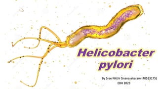
4. Morphology
• Spiral.
• Gram-negative.
• Length of 2.5 – 5.0 μm.
• Width of 0.5 – 1.0 μm.
• 2-6 unipolar flagella (approximate length of 3 μm
and thickness of 2.5 nm) with a distinctive bulb at
its end.
4
Fig.2: Structure of
H. pylori
bacterium.
5. • Surface consists of lipopolysaccharides (LPS) urease, and adhesins.
5
growth requirements.
• Microaerophile.
• Optimal growth conditions:
• O2: 2 – 5%
• CO2: 5 – 10%
• High humidity
• 34 – 40 °C optimum (37°C)
• Neutralophile: 5.5 to 8.0 pH (will survive brief exposure to pHs of < 4)
6. • Fastidious microbe. Ex:- requires supplements
such as blood, serum, yeast extract, lysed human
erythrocytes, IsoVitaleX, hemin, cyclodextrin,
cholesterol, etc.
• Isolated from gastric biopsy specimens.
• Colonies appear between 3 – 14 days.
• H. pylori form small (~1-mm), translucent, smooth
colonies.
• Urease, catalase, and oxidase positive.
6
Fig.3: H. pylori
colonies on blood
agar.
7. • The prevalence of H. pylori is >80% in developing countries.
• The prevalence of H. pylori in developed countries is <40%.
• Prevalence of H. pylori inversely correlates with socioeconomic status.
• Infection mainly occurs during childhood and persists lifelong if untreated.
• Human to human transmission:
7
• oral-oral,
• fecal-oral,
• and/or iatrogenic routes.
8. • After entering the host stomach, four steps are critical for disease pathogenesis:-
8
Fig.4: major
steps in H.
pylori
infection and
pathogenesis
.
9. 9
• pH of the gastric mucosa = 4 –
6.5 pH.
• Urease enzyme activity.
• Converts urea into ammonia and
carbamate.
• Ammonia produced increases the
pH.
• Decreases the viscosity.
Step 1: Survival in the acidic stomach.
Fig.5: urease activity in H. pylori.
10. • Action of flagella helps to reach the basal layer
(pH~7).
• Chemotaxis towards gastric epithelium.
• Mucinase, protease, and phospholipase – mucus
disruption.
10
Step 2: Flagella movement toward epithelial cells.
Fig.6: H. pylori
movement toward
epithelium cells.
11. Step 3: Adhesion to host cells via adhesin-receptor interaction.
• OMPs are the main adhesins of H.
pylori:
• Pilus surface adhesin CagL – binds to
β1 integrins.
11
• Blood group antigen-binding adhesion
(BabA) – binds to Lewis b antigen.
• Sialic acid-binding adhesion (SabA) –
binds Lewis x and a antigen.
• AlpA and B – binds to laminin.
Fig.8: interaction between host cells receptors and adhesins
of H. pylori.
12. Step 4: Toxin release and host tissue damage.
• Cytotoxin-associated
gene A (CagA).
• Delivery into host cells –
type IV secretion system
(T4SS).
• Tyrosine phosphorylation
of CagA.
12
Fig.9: signaling
pathways triggered
by CagA.
13. • Vacuolating cytotoxin A (VacA) – oligomeric structure – immunomodulator.
• Delivery - type V secretion system.
• Membrane embedded vacA acts as an anion selection channel.
• Leakage of nutrients by disruption of tight junction barriers.
• Endocytosed vacA leads to the vacuolation of host cells.
• Cell apoptosis via the mitochondrial-dependent pathway.
13
Fig.10:
functions
of VacA.
14. • Bacterial contact with TLRs
(Ex:TLR4) on monocytes and
other APCs leads to production
of proinflammatory cytokines. Ex:
TNF-α (tumour necrosis factor-
α), IL-1β and IL-8.
• Proinflammatory cytokines act as
chemoattractants.
• Granulocytic infiltration.
14
Fig.11: innate immune
response to H. pylori.
15. 15
• Th1-predominant host
immune response –
further elevates pro-
inflammatory cytokines
IL-12, IL-18 and TNF-α.
• Th17 – inflammatory
response – gastric
cancer.
• Production of IgA and
IgG. Fig.12: adaptive immune response
to H. pylori.
16. • Evasion of recognition by pattern recognition receptors – LPS.
• Inhibition of phagocytic killing – VacA and etc.
• Inhibition of killing by reactive oxygen and nitrogen species – arginase, catalase, and
superoxide dismutase.
• Apoptosis of macrophages and monocytes – VacA.
• Inhibition of antigen presentation– VacA and CagA.
• Apoptosis of gastric epithelial cells – VacA and etc.
• Inhibition of T cell function – CagA and VacA.
16
17. 17
• Laboratory identification methods:
• Culturing, staining or urease activity
of gastric biopsy specimens.
• Detection of serum IgG.
• urea breath test.
• urinary excretion of (15N) ammonia.
• stool antigen assays.
Fig.13: rapid urease test kit.
18. • Treatment is two-pronged:
18
• Drugs to decrease stomach acid: bismuth subsalicylate (Pepto-Bismol).
• Antibiotics to kill the bacteria: metronidazole and either tetracycline or amoxicillin;
clarithromycin, ranitidine, and bismuth citrate; or clarithromycin, amoxicillin, and
lansoprazole.
Fig.14: three regimens of H.
pylori treatment kit.
19. Algood, H., & Cover, T. (2006). Innate immune recognition of H. pylori [Image]. Research gate
https://www.researchgate.net/figure/Innate-immune-recognition-of-H-pylori-Innate-immune-recognition-of-H-pylori-leads-
to_fig3_6754710
B Van Der Weyden, M., M Armstrong, R., & T Gregory, A. (2005). Marshall and Warren, 1984. [Image]. Med J Aust.
https://www.mja.com.au/journal/2005/183/11/2005-nobel-prize-physiology-or-medicine#0_i1091639
Bonsor, D., & Sundberg, E. (2019). Roles of Adhesion to Epithelial Cells in Gastric Colonization by Helicobacter
pylori [Image]. Springer link https://link.springer.com/chapter/10.1007/5584_2019_359
Dreamstime (n.d). Helicobacter pylori cell structures and anatomy [Image]. https://www.dreamstime.com/basic-rgb-
image186817330
Dunn, B., Cohen, H., & Blaser, M. (1997). Helicobacter pylori. Clinical Microbiology Reviews, 10(4), 720-741.
https://doi.org/10.1128/cmr.10.4.720
Huang, Y., Wang, Q., Cheng, D., Xu, W., & Lu, N. (2016). Adhesion and Invasion of Gastric Mucosa Epithelial Cells by
Helicobacter pylori. Frontiers In Cellular And Infection Microbiology, 6. https://doi.org/10.3389/fcimb.2016.00159
19
20. Kao, C., & Sheu, B. (2016). Schematic diagram of Helicobacter pylori infection and pathogenesis. [Image]. Research gate
https://www.researchgate.net/figure/Schematic-diagram-of-Helicobacter-pylori-infection-and-pathogenesis-The-urease-
activity_fig1_301234135
Kao, C., Sheu, B., & Wu, J. (2016). Helicobacter pylori infection: An overview of bacterial virulence factors and
pathogenesis. Biomedical Journal, 39(1), 14-23. https://doi.org/10.1016/j.bj.2015.06.002
Karkhah, A., Ebrahimpour, S., Rostamtabar, M., Koppolu, V., Darvish, S., & Vasigala, V. et al. (2019). H. pylori evasion
mechanisms from immune responses [Image]. Science direct
https://www.sciencedirect.com/science/article/pii/S0944501318308358#!
Khan, S., Karim, A., & Iqbal, S. (2009). Role of urease in the colonization of gastric mucosa by Helicobacter pylori [Image].
Research gate https://www.researchgate.net/figure/Role-of-urease-in-the-colonization-of-gastric-mucosa-by-Helicobacter-
pylori-taken-from_fig1_38096135
Kusters, J., van Vliet, A., & Kuipers, E. (2006). Pathogenesis of Helicobacter pylori Infection. Clinical Microbiology
Reviews, 19(3), 449-490. https://doi.org/10.1128/cmr.00054-05
Lina, T. (2014). Immune evasion strategies used by Helicobacter pylori. World Journal Of Gastroenterology, 20(36), 12753.
https://doi.org/10.3748/wjg.v20.i36.12753
20
21. Palframan, S., Kwok, T., & Gabriel, K. (2012). VacA, a multi-functional toxin [Image]. Frontiers
https://www.frontiersin.org/articles/10.3389/fcimb.2012.00092/full
Pokhrel, P. (2015). 3 day culture of helicobacter pylori on blood agar [Image]. Microbiology notes
https://microbiologynotes.com/cultural-characteristics-of-helicobacter-pylori/
Roesler, B. M., (Ed.). (2014). Trends in Helicobacter pylori Infection. IntechOpen. https://doi.org/10.5772/57053
Siupsinskiene, N. (2017). Negative and (b) positive rapid urease test results for the diagnosis of Helicobacter
pylori [Image]. Research gate https://www.researchgate.net/figure/a-Negative-and-b-positive-rapid-urease-test-results-for-
the-diagnosis-of-Helicobacter_fig1_315473838
Wellona pharma, (n.d) H Pylori Kit. [image] wellona pharma https://wellonapharma.com/product/finished/h-pylori-kit
Wen, S., & Moss, S. (2009). The pathogenesis of H. pylori infection [Image]. Science direct
https://www.sciencedirect.com/science/article/abs/pii/S0304383508009099?via%3Dihub
Willey, J., Prescott, L., Sherwood, L., & Woolverton, C. (2014). Prescott's microbiology (9th ed., p. 904). McGraw-Hill.
Zhang, R. (2016). Role of Helicobacter pylori infection in pathogenesis of gastric carcinoma. World Journal Of
Gastrointestinal Pathophysiology, 7(1), 97. https://doi.org/10.4291/wjgp.v7.i1.97
21