
Recommended
More Related Content
Similar to Learning Objectives Covered1. Explain Respiratory Failure and th.docx
Similar to Learning Objectives Covered1. Explain Respiratory Failure and th.docx (20)
More from smile790243
More from smile790243 (20)
Recently uploaded
Recently uploaded (20)
Learning Objectives Covered1. Explain Respiratory Failure and th.docx
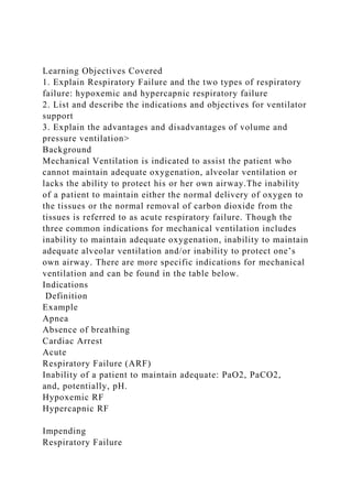
- 1. Learning Objectives Covered 1. Explain Respiratory Failure and the two types of respiratory failure: hypoxemic and hypercapnic respiratory failure 2. List and describe the indications and objectives for ventilator support 3. Explain the advantages and disadvantages of volume and pressure ventilation> Background Mechanical Ventilation is indicated to assist the patient who cannot maintain adequate oxygenation, alveolar ventilation or lacks the ability to protect his or her own airway.The inability of a patient to maintain either the normal delivery of oxygen to the tissues or the normal removal of carbon dioxide from the tissues is referred to as acute respiratory failure. Though the three common indications for mechanical ventilation includes inability to maintain adequate oxygenation, inability to maintain adequate alveolar ventilation and/or inability to protect one’s own airway. There are more specific indications for mechanical ventilation and can be found in the table below. Indications Definition Example Apnea Absence of breathing Cardiac Arrest Acute Respiratory Failure (ARF) Inability of a patient to maintain adequate: PaO2, PaCO2, and, potentially, pH. Hypoxemic RF Hypercapnic RF Impending Respiratory Failure
- 2. Respiratory failure is immi-nent in spite of therapies. Commonly defined as: Pt is barely maintaining (or gradually deteriorating) normal blood gases but with significant WOB. Neuromuscular Disease (N-M) Status Asthmaticus Chronic Respiratory Failure Repeated failures after attempts to liberate from the ventilator (extubations, Trach Collar trials, etc.) SEVERE: Obesity Hypoventilation Syndrome COPD Pulmonary Fibrosis Prophylactic Ventilatory Support Clinical indication = high risk of respiratory failure. Ventilatory support is instituted to ↓ WOB,minimize O2consumption and hypoxemia, reduce cardiopulmonary stress, and/or control airway with sedation. Brain injury Heart muscle Injury Major surgery Shock (prolonged) Smoke injury Trauma (some)
- 3. Hyperventilation Therapy Ventilatory support is instituted to control and manipulate PaCO2 tor lower than normal level Acute head injury (↑ ICP) (not immediately after injury) *respiratoryupdate.com Respiratory failure can be acute or chronic and is classified as either hypoxemic or hypercapnic. During hypoxemic respiratory failure, the patient’s ventilatory demands exceed the lung's ability to provide blood oxygenation resulting in muscle fatigue. Hypoxemic respiratory failure is defined as a PaO2 below the predicted normal range for the patient’s age under ambient conditions. A normal PaO2 for a patient that is 60 years or younger on room air is 80-100mmHg. When a patient is hypoxemic their body naturally responds to the low PaO2by increasing respiratory rate and/or tidal volume (an increase in minute ventilation). An increase in minute ventilation leads to hyperventilation. During hyperventilation, a greater than normal amount of CO2 is exhaled resulting in a low PaCO2 (hypocapnia). Hypercapnic respiratory failure is defined as a PaCO2 level above 50mmHg and a rising and a falling pH of 7.25 or less. Hypercapnic respiratory failure may be accompanied by a normal or low PaO2. A patient who is experiencing hypercapnic respiratory failure is in imminent danger of cardiopulmonary arrest and mechanical ventilation is essential. Once the need for mechanical ventilation has been established and the airway is secured the practitioner must select the type of ventilator, breath type and ventilator mode that is most appropriate for the patient. The selection of a ventilatory support strategy is based on the type of respiratory failure the patient demonstrates. For
- 4. example, a patient in hypoxemic respiratory failure can be treated with various oxygen therapy devices to manage the patient’s oxygenation status. The practitioner must be careful to identify when a patient is experiencing refractory hypoxemia. Refractory hypoxemia is a lack of oxygen in the blood that does not respond to oxygen alone. These patients experience intrapulmonary shunting, such as with pneumonia, pulmonary edema, and atelectasis, which requires PEEP along with oxygen. Patients who experience refractory hypoxemia can be treated with devices such as high flow oxygen delivery devices and CPAP. In a patient presenting with hypercapnic respiratory failure, the patient must be treated with ventilatory support to mange the patients PaCO2 levels and acid-base status. For example, a patient in hypercapnic respiratory failure must be intubated and placed on mechanical ventilation. However, if hypercapnic respiratory failure is noticed soon enough then practitioners can treat these patients with noninvasive ventilatory support (NIPPV) or Bipap. NIPPV has been proven to be effective when implemented early and set correctly. Either noninvasive ventilation or invasive ventilation can be used as a ventilatory support strategy. Noninvasive ventilation (NIV or NIPPV) is defined as any mode of ventilation that does not require an invasive artificial airway (endotracheal tube or tracheostomy tube). NIV/NIPPV includes CPAP or CPAP in combination with any mode of pressure limited or volume limited ventilation (BiPaP). Invasive mechanical ventilation is defined as positive pressure ventilation delivered via an endotracheal tube or tracheostomy tube. Once it as been determined that the patient should be placed on noninvasive mechanical ventilation one of two methods can be chosen: 1. CPAP (continuous positive airway pressure) 2. Noninvasive positive airway pressure (NIPPV) or BiPaP Once it has been determined that the patient needs invasive
- 5. mechanical ventilation the practitioner must determine the mode of ventilation and breath delivery type. Modes of Ventilation · Type of breath (mandatory, spontaneous, assisted) · Targeted control breath (volume or pressure) · Timing of breath delivery (continuous mandatory ventilation (CMV), SIMV, or spontaneous There are three types of positive pressure ventilators. Volume cycled a. Pressure is applied to the airway until a preset volume is delivered. b. Minute volume will remain constant. c. Airway pressure will increase or decrease depending on the patient's compliance and/or airway resistance. d. Volume cycled ventilators can be used with most patients. Pressure cycled a. Apply positive pressure to the airways until a preset pressure limit is reached. b. Tidal volume (Vt) is adjusted by increasing or decreasing the pressure limit. c. Although peak pressure (PIP) will remain constant, the volume will change as lung compliance/resistance change. Time cycled a. These ventilators provide positive pressure until a preset time is reached. b. The peak inspiratory pressure (PIP) is usually limited by an adjustable pop-off valve. c. Tidal Volume (Vt) is adjusted by increasing or decreasing the peak inspiratory pressure, inspiratory time, or flow. There are three breath delivery techniques aka modes of ventilation. 1. Continuous Mandatory Ventilation (CMV) a. All breaths are mandatory and can be volume or pressure targeted. b. Breaths can be patient triggered or time triggered. c. When the breaths are patient triggered, CMV mode is called
- 6. A/C (assist/control) d. When the breaths are time triggered the CMV mode is called control mode. e. CMV is commonly volume targeted (volume control continuous mandatory ventilation (VC-CMV) f. CMV can also be pressure targeted (pressure control continuous mandatory ventilation (PC-CMV) 2. Synchronized Intermittent Mandatory Ventilation (SIMV) a. Periodic volume-or pressure-targeted breaths occur at set intervals. b. Between mandatory breaths, the patient breaths spontaneously at any desired pressure without receiving a mandatory breath. c. Synchronized Intermittent Mandatory Ventilation (SIMV) works the same way as IMV except that the mandatory breaths are time triggered rather than patient triggered. 3. Spontaneous a. Spontaneous breathing - the patient can breathe through the ventilator circuit (T-piece method). The ventilator monitors the patient's breathing and can activate an alarm if necessary. b. CPAP c. PSV – (pressure support ventilation). The ventilator provides a constant pressure during inspiration once it senses the patient has made an inspiratory effort. There are less frequently used modes such as MMV, APRV and PAV as well as HFV, which will be later discussed. Initial Settings for Mechanical Ventilation 1. Ventilation mode a. Control, assist/control, IMV/SIMV b. Any mode is acceptable for initial set up 2. Tidal volume, respiratory rate, FiO2 and PEEP a. Tidal volume - 6-8 ml/kg of ideal body weight b. Respiratory rate - 12 -18 breaths per minute c. FiO2 - if patient is on room air or you have no prior information, start the patient at 40-60%. If the patient is currently on oxygen, set the ventilator FiO2 to the patient's
- 7. current setting. d. PEEP - if the patient is currently on PEEP/CPAP keep it at the same level. If the patient is not currently on PEEP/CPAP start at 0-10 cm H2O. Ventilation Mode Modes of ventilation 1. Assist Mode a. Patient initiates all breaths b. No minimum respiratory rate 2. Control Mode 2. Ventilator will initiate breaths at a preset rate 2. Does not allow patient to initiate breaths 2. Indicated for head trauma/surgery patients 1. Assist/Control c. Allows patient to set the respiratory rate c. The ventilator will maintain a minimum rate c. May be used with most patients c. Ventilator controls tidal volume for every breath 1. SIMV Mode d. Allows patient to breath spontaneously d. Ventilator provides a minimum minute ventilation 1. Pressure Control Ventilation (PCV) e. Pressure controlled breaths e. Used when PIP is very high e. Exhaled Vt will vary e. Adjust IT or PIP as needed. 1. Pressure Support Ventilation (PSV) f. Pressure support adds a preset amount of pressure during a spontaneous breath f. Helps the patient overcome the resistance of breathing through an ETT and ventilator circuit Prompt For this assignment, you will provide detailed responses to the following questions. A 32-year-old man presents to the emergency department with a 2-day history of fever and cough. His chest film shows a right
- 8. middle lobe infiltrate. His room air ABG showed: pH: 7.32 PCO2: 32 torr PO2: 78 torr HCO3- 18 He was started on antibiotics and admitted to the floor. Four hours later, the nurse calls because she is concerned he is doing worse. On your arrival to the room, his blood pressure is 85/60, his pulse is 120 beats/min and his oxygen saturation, which had been 97% on 2L oxygen via nasal cannula, is now 78% on a non-rebreather mask. The patient is obviously laboring to breathe with use of his accessory muscles and is less responsive than he was on admission. On the lung exam, he has crackles throughout the bilateral lung fields. The chest film now shows increasing bilateral, diffuse lung opacities. An ABG is done while on the non-rebreather and shows: pH: 7.17 PCO2: 65 PO2: 58 HCO3- 16 1. Based on the clinical presentation of this patient, discuss how you would address the treatment. Include in your answer the following information: possible diagnosis, and what would be appropriate therapy (invasive vs noninvasive ventilation), and why? 2. Based on your choice of ventilation for this patient, discuss appropriate initial settings to properly ventilate this patient. Include and explain the following information in your answer: If you choose noninvasive ventilation be sure to include IPAP and EPAP settings (defend your position). If you choose invasive ventilation be sure to include Mode of ventilation, Tidal Volume (VT), respiratory rate (frequency), oxygen percentage (FI02), Positive End Expiratory Pressure (PEEP) or any other therapy that may be indicated. Submit your answers in at least 500 words on a Word document. You must cite at least three references in APA format and
- 9. defend and support your position.