
Recommended
More Related Content
Similar to cultivation of virus 1.pdf
Similar to cultivation of virus 1.pdf (20)
Recently uploaded
Recently uploaded (20)
cultivation of virus 1.pdf
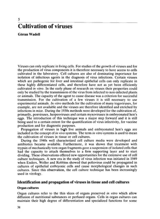
- 1. Cultivation of viruses Goran Wadell Viruses can only replicate in living cells. For studies of the growth of viruses and for the production of virus components it is therefore necessary to have access to cells cultivated in the laboratory. Cell cultures are also of dominating importance for isolation of infectious agents in the diagnosis of virus infections. Certain viruses which are pathogenic for liver and intestinal epithelial cells can only replicate in these highly differentiated cells, and therefore have not as yet been efficiently cultivated in vitro. In the early phase of research on viruses their properties could only be studied by the transmission of the virus from infected to non-infected plants or animals. The capacity of the agent to cause disease was a criterion for successful transmission. For the cultivation of a few viruses it is still necessary to use experimental animals. In vitro methods for the cultivation of many togaviruses, for example, are not available and the viruses are therefore identified and enriched by infections in mice. During the 1930s methods were developed for the cultivation of, primarily, poxviruses, herpesviruses and certain myxoviruses in embryonated hen's eggs. The introduction of this technique was a major step forward and it is still being used to a certain extent for the quantification of virus infectivity, for vaccine production and for diagnostic purposes. Propagation of viruses in bqjh live animals and embryonated hen's eggs are included in the concept of in vivo systems. The term in vitro systems is used to mean the cultivation of viruses in tissue or cell cultures. During the 1940s well characterized cell culture media were developed and antibiotics became available. Furthermore, it was shown that treatment with trypsin of mechanically torn organ fragments gave a suspension of isolated cells that had the capacity to attach themselves to a firm supporting layer and to start dividing. These observations offered new opportunities for the extensive use of cell culture techniques. A new era in the study of virus infection was initiated in 1949 when Enders, Weller and Robbins showed that pohovirus could be propagated in cultures of epithelial embryonic cells and cause morphological changes in such cultures. Since this observation, the cell culture technique has been increasingly used in virology. Identification and propagation of viruses in tissue and cell cultures Organ cultures Organ cultures refer to the thin slices of organs preserved in vitro which allow diffusion of nutritional substances or perfused organs. Cells in organ cultures can maintain their high degree of differentiation and specialized functions for some 38
- 2. Identification and propagation of viruses in tissue and cell cultures 39 weeks. The restricted length of life and in particular the difficulties involved in identifying viral changes have led to this type of culture being used only in selected cases. Epithelium from the respiratory and the intestinal tract and fragments from nervous tissue, ovaries, thyroidea, are examples of differentiated tissues that can be maintained in organ cultures. Organ cultures with ciliated respiratory epithelium are used for the cultivation of respiratory viruses (e.g. coronaviruses and certain rhinoviruses), which do not grow in cell cultures (cf. Chapter 20). Cell cultures Cell cultures are estabhshed by the propagation of dispersed cells. In some cases such cells may grow in suspension, but most often they grow as a monolayer on the supporting material. There are three different kinds of monolayer cultures: primary cell cultures, diploid cell lines and heteroploid, estabhshed cell lines {Figure 5.1). S ' (a) Figure 5.1. Cells growing in tissue cultures. Photograph {a) shows a primary cell culture from a trypsinized monkey kidney: (b) human diploid fibroblast culture; and (c) a culture of a heteroploid cell line, HeLa cells. The cultures contain a confluent monolayer on the bottom of the vessels used for cultivation. The cells are shown as they appear in a live state by light microscopy
- 3. 40 Cultivation of viruses Primary cell cultures Cultures established by cultivation of cells which have been released from organ fragments are called primary cell cultures. A tissue can be homogenized mechani cally (explant culture) or by aid of proteolytic enzymes. In the former case groups of cells are obtained, whereas enzyme treatment with, for example, trypsin, provides a monodisperse cell suspension. The cells are suspended in a nutritional solution and transferred to a cuhivation vessel. Most live cells attach themselves spontaneously to the bottom of the vessel. Different kinds of cells in a primary culture show a varying capacity to survive. Macrophages, neurons and muscular cells can remain viable for weeks to months, however, without cell division. Epithelial cells can pass through about ten cell divisions in vitro, whereas human fibroblasts can divide 50-60 times before the cell line is extinguished. Only cells that are attached to the bottom of the cultivation vessel can divide. This division of cells covers the bottom of the vessel with a cell mosaic and since the division of cells is anchorage-dependent, multiple layers of cells are not formed, the end result being therefore, a monolayer. The density of the monolayer varies with different kinds of cells. The phenomenon which leads a cell which is surrounded by other cells to stop dividing is called contact inhibition, A monolayer in a primary culture of kidney tissue, for example, displays a patchy distribution of areas, where similar cells, epithelial cells or fibroblasts, are grouped. Because of the many different cells, a primary culture can be susceptible to infection with a relatively broad spectrum of different viruses. Kidneys from embryos or amnion membranes are frequently used for establishment of human primary cell cultures. The cells from a primary culture can be increased in number by 'passaging', which means that the cells are transferred from one cultivation vessel to another, where they again are allowed to form a monolayer. The cells are suspended mechanically or by the use of proteolytic enzymes and/or chelate-forming agents, for example EDTA. The latter compound can bind Ca++ which is needed for the anchorage of cells to the supporting layer. After distribution of the suspension of cells into three or four new cultivation vessels, cells again attach themselves to the bottom of the vessels and divide until the supporting surface is covered with a layer of cells. Passage of primary cell cultures can be made within one to four weeks after their establishment. With repeated passaging a certain kind of cell eventually becomes dominant. Diploid cell lines Repeated passage of cells from fetal lung tissue leads to the dominance of fibroblasts in the culture. The cells retain their diploid character, hence the name 'diploid cell line'. A cell line of this kind is susceptible to a broad spectrum of different kinds of viruses. Diploid cells are used for the cultivation of viruses from patients and also for the production of some live virus vaccines. In order to allow an effective usage of diploid cell lines, the majority of the cells which are obtained with the early passages are stored in a frozen state in liquid nitrogen (-196°C) after the addition of, for example, dimethylsulphoxide (DMSO) to prevent cell damage caused by the freezing. Frozen cells are viable for decades and are used for the establishment of new cultures as the need arises.
- 4. Identification and propagation of viruses in embryonated hen's eggs 41 Heteroploid (established) cell lines These cell lines usually derive from tumour cells in a carcinoma or a sarcoma. In spite of the nature of tumour cells it is often difficult to adapt such cells to growth in vitro. If cells are successfully estabhshed in laboratory vessels they can continue to divide in an unlimited fashion. Cultures can be transferred after a few days and up to a week. Changes in the cell population may occur with the passage of cells. Mutants of cells, which can combine rapid division with the optimal usage of the milieu, become dominant. An established cell hne therefore is composed of a limited number of cell clones. The term clone is used to describe a population of cells which derives from a single cell and which therefore is genetically homogenous originally. Established cell hues are heteroploid, usually hypotetraploid, and have a reduced capacity to give and respond to signals, which causes contact inhibition. For this reason they can grow to a cell density which is ten times higher than in a cuhure of diploid cells. Certain established cell lines are not anchorage-dependent for cell division and they can therefore grow in suspension cultures. Established cell lines have a varying susceptibility to infection from different viruses. In each individual case it is therefore necessary to define the cell line that is most suited to the propagation of a certain virus. In some situations the susceptibihty of cells to a virus can be increased by treatment of the virus inoculum and/or the host cell with proteolytic enzymes. Established cell lines offer the advantage over other cell lines in that they grow rapidly and that an unlimited number of cells can be obtained. Among the most commonly used human established cell lines are HeLa cells claimed to be derived from a female cervix carcinoma, HEp-2 cells from a larynx carcinoma, KB cells from a nasopharynx carcinoma and A-549 cells from an 'oat cell' carcinoma. Somewhat unexpectedly it has been found that HEp-2 and KB cell lines do not derive from the assumed original tumours but are clones of HeLa cells. This is because HeLa cells not uncommonly are unintentionally transferred and contaminate other cell cultures, whereafter they can compete with the original cells. BSC-1 and Vero cells which are cell lines derived from green monkey kidney tissue are also used for cultivation of some human viruses. Vero cells have a reduced capacity to synthesize interferon and the cells therefore are suitable for cultivation of viruses which show a high sensitivity to interferon. Cell lines of human haematopoietic cells Blood leucocytes and to a certain extent cells from lymph glands in man are relatively easy to obtain. They are propagated in suspension, but can normally survive only for some days in a cell cuhure. However, cell lines can be established after an infection with Epstein-Barr (EB) virus or by using cells from tumours in the lymphatic system. EB virus infects and can immortalize normal B-cells (see Chapters 11 and 31). Haematopoietic cells in culture are divided into lymphoblas- toid, lymphoma, myeloma and leukaemia cell lines. Lymphoblastoid cell lines occasionally can be established to growth in vitro from, for example, tonsillar tissue. Cells that divide contain EB virus-DNA, are polyclonal and with few exceptions diploid. Thus the cells mainly have retained their normal properties. Lymphoma cell lines generally derive from Burkitt's lymphoma cells (BL; see Chapters 18 and 31) and to a lesser extent from lymphosarcomas. BL cell lines are readily established to growth in vitro and therefore have been the object of
- 5. 42 Cultivation of viruses extensive studies. They contain EB virus-DNA, are monoclonal and heteroploid. Furthermore they have the capacity to form colonies in agar and to grow in mice that have a deficient cell bound immunity, so-called 'naked mice' - properties that characterize tumour cells. BL cell lines are used for production of interferon (see Chapter 24). Both myeloma and leukaemia cell lines are difficult to establish and as a rule the cultures are overgrown by lymphoblastoid cells. Microbial contamination in tissue and cell cultures Cell cultures are cultivated in a nutrition-rich environment and therefore provide excellent conditions for the growth also of microorganisms other than viruses, e.g. bacteria, mycoplasmas and fungi. In order to prevent bacterial infections antibio tics are routinely added in the form of penicillin and streptomycin or, alternatively, gentamicin. Some fungus infections can be prevented by treatment with, for example, amphotericin. Mycoplasma contamination is a major problem in cell cultivation. Mycoplasmas are prokaryotes that grow at or below the cytoplasmic membrane, and they are resistant to many antibiotics. Primary cultures normally are free from mycoplasmas, whereas cell lines commonly are contaminated. This indicates that the mycoplasmas may be transferred from persons handling the cells, or that some ingredient in the medium is a source of contamination. Kanamycin and auromycin can suppress but seldom eliminate a mycoplasma infection. An efficient aseptic technique and avoidance of mouth pipetting are important measures to reduce the occurrence of mycoplasma infection in cultures. Recently it has been reported that it is possible to eliminate a mycoplasma infection by passaging cells in a medium containing specific antiserum against the contaminating mycoplasma. Thus, in this way, valuable cell lines may be rescued and preserved. This might be important, for example, in the production of monoclonal antibodies as the production of hybridoma cells, in particular, is vulnerable to contamination with mycoplasma infections. Identification and propagation of viruses in embryonated hen's eggs A technique for the cultivation of viruses in embryonated hen's eggs was introduced in the 1930s and it was used extensively for two decades. Hereafter, different cell cultures replaced the embryonated eggs for most purposes. However, cultivation in eggs is still being used to distinguish pock-forming viruses (e.g. poxviruses and herpesviruses) and for the isolation of influenza A viruses and production of a vaccine against this virus. Different kinds of viruses are propagated in either of the embryonal membranes surrounding the amnion, chorioallantois or yolk sacs (Figure 5.2). The chorioallantoic membrane is highly susceptible to infection with poxviruses and it is therefore used for their isolation and quantifica tion (see Figure 6.1). During infection newly synthesized virus spreads from the primarily infected cells to neighbouring cells to form the localized pock-like structure. Different membranous sacs are infected by inoculation of virus directly into the fluid in the sac. The amniotic fluid is in contact with the future respiratory tract of the embryo. Influenza A virus which normally initiates infections in the respiratory tract can be demonstrated with high sensitivity after being inoculated into the amnion sac. Vaccines against influenza A virus can be prepared after the inoculation into the allantoic cavity of a virus that has been adapted to grow in this milieu.
- 6. Identification and cultivation of viruses in experimental animals 43 Inoculation into the chorioallantoic membrane Shell membrane Egg white Inoculation into _ the amniotic cavity Inoculation into the yolk sac Inoculation into the allantoic cavity Figure 5.2. Different routes for inoculation of an embryonated hen's egg. The inoculation into the yolk sac is usually made by use of 5 day-old embryo, whereas inoculation into other localities usually is performed with 10-21 day-old embryos Identification and cultivation of viruses in experimental animals There is a strong tendency towards reducing the use of experimental animals for preparation of virus materials. However, there are certain disease-related viruses that cannot be cultivated either in cell cultures or in eggs. This is because the cultures which are available do not satisfy the ecological requirements for rephcation of the virus. Attempts have been made to study non-cultivatable viruses by direct analysis of materials from patients and by an analysis of the immune response {see Chapter 20). Occasionally inoculation of experimental animals provides a more sensitive method for identification of viruses than inoculation of cell cultures. For this reason one tries to isolate, for example, rabies virus, different arboviruses and Coxsackie A viruses by intracerebral inoculation of newborn mice. In order to study the different steps in the disease process and the different defence reactions one is restricted, by necessity, to using experimental animals. Monkeys have been used to analyse the disease process in connection with infections with different hepatitis viruses (cf. Chapter 30) and with certain non-immunogenic atypical infectious agents (cf. Chapter 17). The choice of animal species which is closely related genetically to man is influenced by the fact that certain infectious agents can not rephcate in more distantly related species. Experimental animals are used also for studies of the oncogenic properties of viruses. Mice and hamsters are used primarily. One advantage with the mouse system is the availability of inbred strains. Mice belonging to an inbred strain are genetically identical. Because of this they show a uniform susceptibility to infections and, furthermore, normal and tumour cells can be transplanted from one animal to the other without the development of any rejection reaction.
- 7. 44 Cultivation of viruses Bibliography FOGH, J. (Ed.) (1975). Human Tumor Cell In Vitro. New York: Plenum Press HOORN, B. and TYRRELL, D. A. J. (1969). Organ cultures in virology. Progress in Medical Virology, 11, 408 KEAY, L. (1978). The cultivation of animal cells and production of viruses in serum-free systems. Methods in Cellbiology, 20, 169 NILSSON, K. and PONTEN, J. (1975). Classification and biological nature of established human hematopoietic cell lines. International Journal of Cancer, 15, 321 PAUL, J. (1975). Cell and Tissue Culture, 5th edn. London: Churchill Livingstone POLLACK, R. (Ed.) (1975). Readings in Mammalian Cell Culture. New York: Cold Spring Harbor Press