This document describes a study that quantified the physical demands of 21 Hatha yoga postures commonly practiced by older adults. Twenty older adult participants completed a 32-week yoga intervention, where they learned introductory postures for the first 16 weeks and intermediate postures for the second 16 weeks. Biomechanical data including joint angles, joint moments of force, and muscle activation levels were collected while participants statically held each posture. The results provide physical demand profiles for each posture that illustrate the ranges of motion, efforts required to maintain the poses, and activated muscle groups. These profiles can help instructors design safe and effective yoga routines for seniors.
![Hindawi Publishing Corporation
Evidence-Based Complementary and Alternative Medicine
Volume 2013, Article ID 165763, 29 pages
http://dx.doi.org/10.1155/2013/165763
Research Article
Physical Demand Profiles of Hatha Yoga Postures Performed by
Older Adults
George J. Salem,1
Sean S.-Y. Yu,1
Man-Ying Wang,1
Sachithra Samarawickrame,1
Rami Hashish,1
Stanley P. Azen,2
and Gail A. Greendale3
1
Division of Biokinesiology and Physical Therapy, University of Southern California (USC), 1540 E. Alcazar Street,
Los Angeles, CA 90033, USA
2
Department of Preventive Medicine, Keck School of Medicine,
University of Southern California, USA
3
Division of Geriatrics, Geffen School of Medicine at the University of California, Los Angeles (UCLA),
924 Westwood Boulevard, Suite 200, Los Angeles, CA 90024, USA
Correspondence should be addressed to George J. Salem; gsalem@usc.edu
Received 3 May 2013; Revised 30 July 2013; Accepted 4 August 2013
Academic Editor: Byung-Cheul Shin
Copyright © 2013 George J. Salem et al. This is an open access article distributed under the Creative Commons Attribution License,
which permits unrestricted use, distribution, and reproduction in any medium, provided the original work is properly cited.
Understanding the physical demands placed upon the musculoskeletal system by individual postures may allow experienced
instructors and therapists to develop safe and effective yoga programs which reduce undesirable side effects. Thus, we used
biomechanical methods to quantify the lower extremity joint angles, joint moments of force, and muscle activities of 21 Hatha
yoga postures, commonly used in senior yoga programs. Twenty older adults, 70.7 years ± 3.8 years, participated in a 32-wk yoga
class (2 d/wk) where they learned introductory and intermediate postures (asanas). They then performed the asanas in a motion
analysis laboratory. Kinematic, kinetic, and electromyographic data was collected over three seconds while the participants held
the poses statically. Profiles illustrating the postures and including the biomechanical data were then generated for each asana. Our
findings demonstrated that Hatha yoga postures engendered a range of appreciable joint angles, JMOFs, and muscle activities about
the ankle, knee, and hip, and that demands associated with some postures and posture modifications were not always intuitive. They
also demonstrated that all of the postures elicited appreciable rectus abdominis activity, which was up to 70% of that induced during
walking.
1. Introduction
Yoga has traditionally been viewed as a relatively safe form of
exercise, capable of increasing strength, flexibility, endurance,
balance, and functional capacity of people in good health and
those with musculoskeletal disorders [1–5]. Supporting these
postulates, the US Department of Health and Human Ser-
vices and the National Recreation and Park Association have
recommended yoga as a form of “total-solution” exercise for
older adults [6]. Despite these dramatic claims of improved
function across a range of physiological and psychosocial
systems, little is understood regarding the physical demands,
program efficacy, and overall safety of yoga programs for
older adults. In general, older adults have less strength,
joint flexibility, and balance, compared to younger adults.
Moreover, they have a greater prevalence of osteoarthri-
tis and neurological syndromes (e.g., sciatica and spinal-
canal stenosis)—putting them at higher risk of developing
exercise-related musculoskeletal and neurological complica-
tions. Understanding the physical demands placed upon the
musculoskeletal system by individual postures may allow
experienced instructors and therapists, whom have special-
ized in training with senior populations, to develop safe and
effective yoga programs which reduce these undesirable side
effects.
This, in essence, was the primary goal of the Yoga Empow-
ers Seniors Study (YESS)—to quantify the physical demands
associated with the performance of individual postures](https://image.slidesharecdn.com/yoga-160202173128/85/Yoga-1-320.jpg)
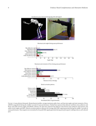
![2 Evidence-Based Complementary and Alternative Medicine
(asanas) and posture modifications frequently used in senior
yoga programs [7–9]. The purpose of the present paper
from the YESS project is to describe asana-specific lower-
extremity (LE) demands placed on the practitioners by the 9
introductory and the 12 intermediate asanas used in the YESS
project.
The physical demands of asanas are quantified biome-
chanically using 3D motion analysis, force platforms, and
electromyography (EMG). While performing an asana, grav-
itational forces tend to rotate our arms and legs and pull
our body towards the earth. In order to “hold” a posture
and prevent our limbs from rotating, we must use our
muscles and ligaments to resist these gravitational effects.
We can quantify these muscular and ligamentous “efforts”
by calculating the joint moments of force (JMOFs) produced
about the joints of the body during the performance of
an asana. Because the JMOFs are related to the torque
that a muscle must develop while holding a posture, they
provide insight into the specific muscle groups that are used
during asana performance. Knowledge of the muscles that are
working informs the beneficial adaptations (e.g., increased
strength and endurance) that we would expect to occur.
JMOFs can also provide a window to potential injury, because
excessively-high JMOFs can create detrimental loading of
articular, ligamentous, and capsular structures, essentially
overloading the musculoskeletal system. Therefore, JMOFs
can also be used to select postures that avoid usage of
injured or overtaxed muscles and tissues. We also recorded
the muscle activity of selected muscle groups using the
electromyographic (EMG) analysis. Surface recording of the
electrical activity of major muscles provides a complemen-
tary window on the physical demands of each posture.
Aggregating the biomechanical profiles (JMOF, EMG, and
maximum joint angles) of each posture will allow the design
of the asana series that are well-balanced—targeting all of
the functionally important muscle groups without repeatedly
overloading the same musculoskeletal and articular tissues.
Knowledge of the physical demands of each posture can
also be used by experienced teachers and therapists, whom
have specialized in training with senior populations, to select
optimal asanas for their students, for example, focusing
on postures that would strengthen weak muscle groups
and/or unload injured and healing structures. In addition,
especially for senior practitioners, a well-designed series
will avoid the excessive range of motion in joints that are
particularly susceptible to injury such as the knees and
hips.
2. Methods
2.1. Study Design. The design of YESS has been previously
detailed [9]. In brief, YESS was an intervention development
study designed to quantify the physical demands of selected
Hatha yoga postures and modifications in ambulatory senior
men and women. Participants attended 1-hour Yoga classes, 2
days per week, for 32 weeks. For the first 16 weeks, they were
taught an introductory series, and for the second 16 weeks
they were advanced to an intermediate series. The classes
were led by a yoga instructor (YT500 certification) with
considerable (over 10 years) experience in teaching seniors
including teaching in prior research projects conducted by
our group. A research associate assisted at the classes. The
RA had collegiate gymnastic athletic training experience
(2 years); further, she was specifically mentored in how to
assist in yoga classes by both a PI (GAG) and the yoga
instructor. Biomechanical data, including maximum joint
angles, JMOF, and muscle activation levels, were collected
after 16 (introductory postures) and 32 weeks (intermediate
postures) of yoga practice. Participant recruitment and the
yoga classes were conducted at the University of California
Los Angeles (UCLA) and TruYoga studio (Santa Monica,
CA), respectively. Biomechanical data was collected at the
Musculoskeletal Biomechanics Research Laboratory (MBRL)
at the University of Southern California (USC). Both the USC
and UCLA Institutional Review Boards approved the study
protocol, and all participants provided informed, written
consent.
2.2. Inclusion/Exclusion Criteria. Inclusion/exclusion crite-
ria were selected in order to maximize safe participation
while in the yoga classes and during the testing sessions.
Community dwelling men and women volunteers, aged 65
years or older, who were not high-level exercisers or frequent
long walkers, and were yoga novices, were eligible for the
study. High level exercisers were defined as people who
participated in active sports (e.g., aerobics, jogging, and
tennis) or higher-intensity exercises (>6 MET). Frequent long
walkers were defined as those who walked more than a mile
without resting, at least 3 times per week. The following
safety exclusions were adopted in order to decrease potential
cardiovascular, musculoskeletal, and neurological risks to
the participants: active angina; uncontrolled hypertension
(SBP greater than 160 or DBP greater than 90); high resting
pulse or respiratory rate (HR > 90 or RR > 24 after 5
minutes seated); unstable asthma or exacerbated COPD;
cervical spine instability or other significant neck injury;
rheumatoid arthritis; unstable ankle, knee, hip, shoulder,
elbow, or wrist joints; hemiparesis or paraparesis; movement
disorders (e.g., Parkinson’s disease), peripheral neuropathies,
stroke with residual deficits, and severe vision or hearing
problems; walker or wheelchair use; insufficient hearing to
permit safety in a yoga group setting; inability to attend in-
person classes; not having a checkup by regular provider
within 12 months (if not taking any prescription medications)
or in the past 6 months (if any regular medicines taken);
could not pass specific movement safety tests. The qualifying
movements were the ability to (a) get up from the floor to
standing; (b) go from standing to the floor; (c) lift both
arms to shoulder level without losing balance; (d) stand with
feet side-by-side for 30 seconds; and (e) stand with feet hip-
width apart for 60 seconds. These were assessed by the study
PI and/or an experienced research associate. The following
feasibility/adherence exclusions were also utilized: (1) the
inability to understand their commitment to the project
(laboratory visits and regular program participation) and
(2) the cognitive limitations significant enough to preclude](https://image.slidesharecdn.com/yoga-160202173128/85/Yoga-2-320.jpg)
![Evidence-Based Complementary and Alternative Medicine 3
informed consent or to raise concerns about participation
safety.
2.3. Sample Size and Recruitment. A target sample size of
20 was determined a-priori using a power-analysis of pilot
data comparing JMOFs across asanas in a sample of 3
older adults. Recruitment was initiated on January 7, 2009
and ended on March 5, 2010. Potential participants (𝑛 =
114) were contacted and initially screened over the phone.
Screening was conducted by the Project Director (PD) (with
10 years of experience) and the Research Associate (RA)
(2 years of experience). The latter was closely supervised
by the former. 79 participants passed the phone screening
exam and 26 were elected not to participate. Of these 79,
46 passed the in-person screening and were allocated to
Group 1 (𝑛 = 15), Group 2 (𝑛 = 15), or waitlisted.
Participants were again screened at the baseline to insure no
conditions had arisen that would exclude the participants,
and that no previously undetected conditions were present.
In Group 1, 12 participants passed the baseline exam, and
in Group 2, 15 participants passed the baseline exam. Thus,
27 participants were enrolled and had baseline measures
taken.
2.4. Retention. Within Group 1, 4 participants left the study
for the following reasons: (1) time commitment was deemed
too great (𝑛 = 2); (2) one subject failed to attend 3 of the 4
initial yoga classes due to travel; (3) one subject informed the
instructor during the second class that a previous spine sur-
geon had instructed her to “not rotate her neck.” She had not
disclosed this information previously and follow-up contact
with the physician resulted in her being removed from the
study. In Group 2, 3 participants left the study: (1) 1 subject
injured her knee the week prior to the beginning of the study
and could not attend the initial yoga classes; (2) 1 subject had a
return of previously diagnosed bilateral, posterior thigh pain
following the baseline testing and then found the initial yoga
classes “difficult.” After receiving epidural injection without
much improvement, physician and PI concluded yoga would
not be advisable at the present time; (3) 1 subject had low back
pain that did not resolve with rest; thus, PI decided pain could
become worse with yoga. Thus, a total of 20 participants, 8 in
Group 1 and 12 in Group 2, were able to complete the yoga
program and had biomechanics measures taken at 16 and
32 weeks. The average age of the 14 women and 6 men was
70.7 years ± 3.8 yr.
2.5. Yoga Program. The study employed Hatha yoga, which
incorporates asanas and pranayama (breathing). The pro-
gram incorporated a standard set of opening and closing
sequences and 2 ordered progressive middle sequences,
termed series I (first 16 weeks) and series II (second 16 weeks).
Predicated on our own experiences, as well as through
reviewing videos, books, and websites aimed at seniors [10–
12], each series included postures and pose modifications
that (1) were commonly used in senior yoga programs;
(2) we believed it could be performed safely by seniors
in a group environment; and (3) provided a balanced and
comprehensive fitness program that targeted muscle groups
thought to be integral to conducting activities of daily living.
The postures that were investigated are listed below. The
introductory postures were advanced by removing or modi-
fying the use of props, or moving from a bilaterally-supported
posture to a unilaterally-supported posture. For example,
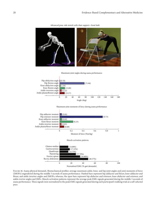
during performance of the introductory side stretch asana,
the participants supported their stance by placing their hands
approximately chest high against a wall. For the intermediate
side stretch posture, the participants lowered their support
height by placing their hands on the backrest of a chair. The
tree posture was advanced by having the participants stand on
a single limb without use of a wall in the intermediate version.
During the introductory tree posture, the participants lightly
touched a wall and had their nonsupporting limb touching
the floor. During the introductory warrior II asana, the
participants supported themselves by lightly touching a chair.
Contrastingly, the intermediate warrior II posture was per-
formed without the use of a chair. The introductory postures
are
Chair (Utkatasana) with wall (Figure 2)
Tree (Vrksasana) bilateral and wall (Figure 3)
Downward dog (Adho Mukha Svanasana) with wall
(Figure 4)
Warrior I (Virabhadrasana I) with chair (front)
(Figure 5)
Warrior I (Virabhadrasana I) with chair (back)
(Figure 6)
Warrior II (Virabhadrasana II) with chair (front)
(Figure 7)
Warrior II (Virabhadrasana II) with chair (back)
(Figure 8)
Side stretch (Parsvottanasana) with wall (front)
(Figure 9)
Side stretch (Parsvottanasana) with wall (back)
(Figure 10)
Intermediate Postures
Chair (Utkatasana) (Figure 11)
Tree (Vrksasana) unilateral and wall (Figure 12)
Tree (Vrksasana) unilateral (Figure 13)
Warrior II (Virabhadrasana II) (front) (Figure 14)
Warrior II (Virabhadrasana II) (back) (Figure 15)
Side stretch (Parsvottanasana) with chair (front)
(Figure 16)
Side stretch (Parsvottanasana) with chair (back)
(Figure 17)](https://image.slidesharecdn.com/yoga-160202173128/85/Yoga-3-320.jpg)
![4 Evidence-Based Complementary and Alternative Medicine
Figure 1: YESS participant performing the intermediate chair asana while instrumented for biomechanical analysis.
One-leg balance (Utthita Hasta Padangusthasana)
with blocks (Figure 18)
One-leg balance (Utthita Hasta Padangusthasana)
with chair (Figure 19)
One-leg balance (Utthita Hasta Padangusthasana)
unilateral (Figure 20)
Crescent (Ashta Chandrasana) (front) (Figure 21)
Crescent (Ashta Chandrasana) (back) (Figure 22)
2.6. Biomechanics. The biomechanical outcome variables
examined included (1) average maximum joint angles, (2)
average peak net JMOFs, and (3) average peak EMG activ-
ity engendered during the performance of the individual
yoga postures. Biomechanical analysis was performed at
the USC Musculoskeletal Biomechanics Research Laboratory
using standard techniques [8, 13]. Whole body kinematic
data were collected using an eleven-camera motion capture
system at 60 Hz (Qualisys Tracking System with Oqus 5
cameras; Qualisys, Gothenburg, Sweden). Reflective mark-
ers were placed on a head band and over the following
anatomical landmarks of the lower and upper extremi-
ties bilaterally: first and fifth metatarsal heads, malleoli,
femoral epicondyles, greater trochanters, acromions, greater
tubercles, humeral epicondyles, radial and ulnar styloid
processes, and third metacarpal heads. Markers were also
attached to the spinous process of the 7th cervical vertebra
(C7), jugular notch, L5/S1, bilateral iliac crests, and bilat-
eral posterior superior iliac spines, in order to define the
trunk and pelvis. Based on these markers, a total of 15
body segments were modeled, including the upper arms,
forearms, hands, head, trunk, pelvis, thighs, shanks, and
feet.
Once instrumented, the subjects performed the pose
sequences, while guided by their instructor. The sequence
of the poses was the same as when it was carried out in
the regular yoga classes. A firm but portable clear Plexiglas
wall, which permitted the capture of the markers, was
positioned for wall support in the lab visits. For each pose,
the participant was instructed to begin in a starting position,
move smoothly into the pose, hold the pose while taking one
full breath, and then return back to the original position.
Simultaneously, the instructor also performed each pose in
order to provide visual cueing. Once the participant had
moved into the pose position, the instructor provided a verbal
cue to the research associate to initiate the 3-second data
collection. Two successful trials of each pose version were
collected, and all 3 seconds of each pose were used for the
analyses.
GRFs were measured from a force platform at 1560 Hz
(AMTI, Watertown, MA). Qualisys Track Manager Software
(Qualisys, Gothenburg, Sweden) and Visual 3D (C-motion,
Rockville, MD) were used to process the raw coordinate data
and compute segmental kinematics and kinetics. Trajectory
data was filtered with a fourth-order zero lag Butterworth
12 Hz low-pass filter. In Visual 3D, the head was modeled as
a sphere, the torso and pelvis as cylinders, and the upper and
lower extremity segments as frusta of cones. The local coordi-
nate systems of body segments were derived from the stand-
ing calibration trial. Joint kinematics were computed based
upon Euler angles with the following order of rotations: flex-
ion/extension, abduction/adduction, and internal/external
rotation. The principle moments of inertia were determined
from the subject’s total body weight, segment geometry, and
anthropometric data. Using standard inverse dynamics tech-
niques, along with the International Society of Biomechanics
recommended coordinate systems, net JMOFs in the sagittal
and frontal planes, for the ankle, knee and hip, were calcu-
lated from the inertial properties, segmental kinematics, and
GRFs. JMOFs were normalized to each subject’s body weight
in kg.
Surface electromyographic (EMG) signals of the lower
gluteus medius, hamstrings, vastus lateralis, gastrocnemius,
rectus abdominis, and erector spinae muscles were collected
on the subjects’ dominant limb at 1560 Hz using active surface
electrodes (Motion Lab Systems, Baton Rouge, LA). The
electrodes were placed in the center of the muscle bellies
with the electrodes aligned in the direction of the muscle
fibers. The obtained EMG signals were amplified (×1000),
notch filtered at 60 Hz, and band-pass filtered at 20–500 Hz.
A root mean square smoothing algorithm with a 75 ms
constant window was used to smooth the EMG data over
the 3-second data collection period corresponding to the
epoch of kinematic and kinetic data. EMG processing and](https://image.slidesharecdn.com/yoga-160202173128/85/Yoga-4-320.jpg)


![Evidence-Based Complementary and Alternative Medicine 27
4. Discussion
This is the first study to use biomechanical investigation
to quantify the physical demands of introductory- and
intermediate-level Hatha asanas performed by seniors. Bio-
mechanical profiles generated from this investigation can be
used to inform experienced instructors in their design of yoga
programs for older adults. Using this information, instructors
and therapists, whom have specialized in training with senior
populations, can select appropriate postures in order to create
comprehensive programs which affect a variety of joints and
muscle groups. They can also use this information to create
balanced programs that prevent excessive repetitive loading
of the same tissues and joint structures. Lastly, the profiles
can be used to make evidenced-based decisions in order to
specifically target weak muscle groups and/or avoid the load-
ing of pathological articular and myotendinous tissues. For
example, a yoga program that comprehensively addresses the
functionally important muscle groups of the lower extremity
needs to include postures that induce appreciable JMOFs
across the hip, knee, and ankle, in multiple directions. Just
as important, the program should not put participants at
risk by repetitively loading the same muscular, tendinous,
or articular tissues without providing appropriate recovery
intervals.
Although our goal was to develop a “balanced” and com-
prehensive yoga program that targeted all of the function-
ally important muscle groups of the LE, the introductory
program, in particular, was deficient in several areas. Most
strikingly, none of the asanas developed an ankle dorsiflexor
JMOF. The ankle dorsiflexors are important muscles which
“lift the front of the foot” during the swing phase of gait in
order to clear the toes and prevent tripping accidents. Not sur-
prisingly, poor ankle dorsiflexor strength is associated with
increased fall risk in community-dwelling older adults [14].
Our findings suggest that additional unstudied postures need
to be biomechanically examined in order to identify those
which develop ankle dorsiflexor JMOFs. Once identified,
these should then be incorporated into senior programs. Else,
additional nonyoga activities/exercises should be integrated
into yoga programs in order to address dorsiflexor targeting.
Similarly, the introductory poses generated only mod-
est hip abductor JMOFs. The hip abductors are important
stabilizers of the pelvis, and their muscular performance
is correlated with balance and fall risk in seniors [15–17].
Thus, we believe it would be prudent to identify additional
“introductory level” postures, which target the hip abductors.
In contrast to the introductory program, several intermediate
asanas, including the tree with unilateral and wall support,
tree without support, and all three one-leg balance pos-
tures, generated appreciable hip abductor JMOFs and gluteus
medius activity. Notably, these were all single limb, standing
postures. Few postures also targeted the hip flexors (warrior
I, warrior II, and crescent, back limbs). The hip flexors are
important in “pulling the limb forward” during the swing
phase of gait and their performance is related to walking
speed and fall recovery in older adults [18, 19].
Some of our findings were intuitive; for example, we
demonstrated a progressive increase in the hip abductor
JMOFs and gluteus medius EMG activity across the three
one-leg balance postures. In other words, the JMOFs and
gluteus medius EMG activity associated with the one-leg
balance postures were the least with the block support, greater
with the chair support, and greatest when the posture was
done without additional support. On the other hand, some
findings were counterintuitive; for example, we expected to
see a similar progression in demand among the three tree
postures. We found, however, that although there was a large
difference in hip abductor JMOF and gluteus medius EMG
activity between the introductory posture (tree with bilateral
and wall support) and the intermediate posture (tree with
unilateral and wall support), there was no difference between
the tree with unilateral and wall support and the unsupported
tree. This finding has important clinical implications; it
suggests that the beginning participants can target their hip
abductor muscles by lifting their contralateral foot off the
floor and assisting their balance with use of a wall, even if
they do not have the balance capabilities to hold the tree
posture without the use of a wall. It also suggests that having
participants let go of the wall, while potentially increasing
their balance capabilities, is not likely to increase gluteus
medius recruitment or performance.
Another interesting and unexpected finding was that all
of the postures elicited appreciable rectus abdominis activity,
which was up to 70% of that induced during walking—an
activity requiring continuous dynamic control of the trunk.
Contrastingly, erector spinae activity was more variable
across the postures. Core stability is important because it
influences trunk orientation which in turn affects hip, knee,
and ankles position during yoga practice and joint kinematics
during ambulation. Abdominal and erector strengthening
exercises improve spinal mobility, balance, and functional
mobility in seniors [20].
The profiles can also be used to identify those asanas,
which might put seniors at risk for injury or exacerbate exist-
ing arthritic conditions. For example, frontal-plane JMOFs at
the knee joint will increase the compressional forces across
the tibia and femur and may exacerbate knee OA. The warrior
postures, in particular, generated relatively large adductor
JMOFs which are likely to increase loading on the lateral
meniscus and tibiofemoral condyles, as well as the medial
collateral ligament. Similarly, the tree postures also generated
appreciable frontal-plane JMOFs at the knee; however, these
were abductor JMOF’s and thus likely to increase the loading
across the medial meniscus and tibiofemoral condyles, and
lateral collateral ligament.
Finally, the posture profiles may be used to generate
appropriate sequences, so that the same musculoskeletal
tissues are not loaded continuously without proper rest. For
example, the chair and warrior front postures, both generated
relatively large knee extensor JMOFs and quadriceps EMG
activity; thus, it would be prudent not to sequence these
postures successively but rather to sequence another posture
in between these two, the side stretch posture, for example,
which generated a knee flexor JMOF, would be a good
candidate.](https://image.slidesharecdn.com/yoga-160202173128/85/Yoga-27-320.jpg)
![28 Evidence-Based Complementary and Alternative Medicine
Among the limitations of this study is the limited
number of asanas and modifications that we examined.
The Hatha yoga type, in particular, lends itself well to
the use of modified postures and participant-specific pro-
gram development because there is not a standard series
associated with Hatha. Thus, there are virtually hundreds
of postures and modifications that can be used in senior
programs, and future investigations should examine addi-
tional postures and modifications which are commonly
used.
We also recognize that yoga is much more than just a
series of postures but also incorporates breathing, medita-
tion, chanting, and/or spiritual components. Although we
attempted to remain as “true to the discipline” as possible, and
therefore included opening and closing sequences, controlled
breathing during the asanas, and the use of an instructor in
the laboratory, we were only able to quantify the physical
demands of the asanas and did not attempt to characterize
these other important attributes.
Lastly, we focused our study on the physical demands
associated with the postures during the 3-second interval the
posture was held. We did not examine the physical demands
associated with the transitions between the postures. In a
recent pilot study conducted in our laboratory (unpublished
data) we found that the JMOFs can be much greater during
the transition from one posture to another, than during
performance of the actual posture itself. Therefore, future
studies should also examine asana transitions in order to
provide additional sequencing information.
In conclusion, our findings demonstrate that introduc-
tory and advanced Hatha yoga postures engender a range
of appreciable joint angles, JMOFs, and muscle activities
about the ankle, knee, and hip. Further, we demonstrated
that although our goal was to develop a “balanced” and
comprehensive yoga program, the program was deficient in
several areas. For example, none of the asanas developed
an ankle dorsiflexor JMOF, and few developed hip flexor
JMOFs. We also demonstrated that some findings were
counterintuitive; for example, there was not a difference
between the JMOF engendered during the tree with unilateral
and wall support and the unsupported tree. Lastly, we found
that all of the poses elicited appreciable rectus abdominis
activity.
Profiles generated from this information may be used
by experienced instructors and therapists, whom have spe-
cialized in training with senior populations, to appropriately
design yoga programs for seniors that are comprehensive,
safe, target specific muscle groups, unload articular structures
at risk, and prevent repetitive overloading of musculoskeletal
tissues. Additional randomized and controlled trial stud-
ies are needed to determine if evidenced-based programs,
designed using biomechanical data from profiles like these,
reduce adverse events and improve participant health out-
comes. Similar biomechanical studies should also be designed
to examine additional yoga postures, other yoga types (e.g.,
Raja), additional styles (e.g., Bikram), and the transitions
between postures.
Acknowledgment
This study was supported by the National Institutes of
Health/National Center for Complementary and Alternative
Medicine Grant no. R01-AT004869-01.
References
[1] M.-Y. Wang, G. Greendale, and G. Salem, “Yoga improves upper
extremity function and scapular posturing in persons with
hyperkyphosis,” Journal of Yoga & Physical Therapy, vol. 2, no.
3, pp. 1–6, 2012.
[2] G. A. Greendale, M.-H. Huang, A. S. Karlamangla, L. Seeger,
and S. Crawford, “Yoga decreases kyphosis in senior women and
men with adult-onset hyperkyphosis: results of a randomized
controlled trial,” Journal of the American Geriatrics Society, vol.
57, no. 9, pp. 1569–1579, 2009.
[3] S. L. Kolasinski, M. Garfinkel, A. G. Tsai, W. Matz, A. van
Dyke, and H. R. Schumacher Jr., “Iyengar yoga for treating
symptoms of osteoarthritis of the knees: a pilot study,” Journal
of Alternative and Complementary Medicine, vol. 11, no. 4, pp.
689–693, 2005.
[4] J. A. Raub, “Psychophysiologic effects of hatha yoga on muscu-
loskeletal and cardiopulmonary function: a literature review,”
Journal of Alternative and Complementary Medicine, vol. 8, no.
6, pp. 797–812, 2002.
[5] M. D. Tran, R. G. Holly, J. Lashbrook, and E. A. Amsterdam,
“Effects of hatha yoga practice on the health-related aspects of
physical fitness,” Preventive Cardiology, vol. 4, no. 4, pp. 165–170,
2001.
[6] N. Tummers and F. Hendrick, “Older adults say yes to yoga,”
National Recreation and Park Association, vol. 39, no. 3, pp. 54–
60, 2004.
[7] M.-Y. Wang, S.-Y. Yu, R. Hashish et al., “Physical demands of
standing yoga poses in seniors: Yoga Empowers Seniors Study
(YESS),” BMC Complementary & Alternative Medicine, vol. 13,
article 8, 2013.
[8] S.-Y. Yu, M.-Y. Wang, S. Samarawickrame et al., “The physical
demands of the tree (vriksasana) and one leg balance (utthita
hasta padangusthasana) poses performed by seniors: a biome-
chanical examination,” Evidenced-Based Complementary and
Alternative Medicine, vol. 2012, Article ID 971896, 11 pages, 2012.
[9] G. Greendale, L. Kazadi, S. Mazdyasni et al., “The Yoga Empow-
ers Seniors Study (YESS): design and asana series,” Journal of
Yoga & Physical Therapy, vol. 2, no. 1, pp. 1–8, 2012.
[10] A. Christensen, Ed., The American Yoga Association’s Easy Does
It Yoga: The Safe and Gentle Way to Health and Well-Being,
FIRESIDE, New York, NY, USA, 1st edition, 1999.
[11] S. Francina, Ed., The New Yoga for People over 50: A Comprehen-
sive Guide for Midlife & Older Beginners, Health Communica-
tions, Deerfield Beach, Fla, USA, 1997.
[12] R. Rosen, Ed., Yoga for 50+: Modified Poses & Techniques for
a Safe Practice, Ulysses Press, Berkeley, Calif, USA, 1st edition,
2004.
[13] J. Song, S. Sigward, B. Fisher, and G. J. Salem, “Altered dynamic
postural control during step turning in persons with early-stage
Parkinson’s disease,” Parkinson’s Disease, vol. 2012, Article ID
386962, 8 pages, 2012.
[14] K. Takazawa, K. Arisawa, S. Honda, Y. Shibata, and H. Saito,
“Lower-extremity muscle forces measured by a hand-held
dynamometer and the risk of falls among day-care users in](https://image.slidesharecdn.com/yoga-160202173128/85/Yoga-28-320.jpg)
![Evidence-Based Complementary and Alternative Medicine 29
Japan: using multinomial logistic regression analysis,” Disability
and Rehabilitation, vol. 25, no. 8, pp. 399–404, 2003.
[15] M. W. Rogers and M.-L. Mille, “Lateral stability and falls in older
people,” Exercise and Sport Sciences Reviews, vol. 31, no. 4, pp.
182–187, 2003.
[16] P. G. Macrae, M. Lacourse, and R. Moldavon, “Physical per-
formance measures that predict faller status in community-
dwelling older adults,” Journal of Orthopaedic and Sports Physi-
cal Therapy, vol. 16, no. 3, pp. 123–128, 1992.
[17] B. D. Iverson, M. R. Gossman, S. A. Shaddeau, and M. E. Turner
Jr., “Balance performance, force production, and activity levels
in noninstitutionalized men 60 to 90 years of age,” Physical
Therapy, vol. 70, no. 6, pp. 348–355, 1990.
[18] C. P. Carty, R. S. Barrett, N. J. Cronin, G. A. Lichtwark, and
P. M. Mills, “Lower limb muscle weakness predicts use of a
multiple- versus single-step strategy to recover from forward
loss of balance in older adults,” Journal of Gerontology A, vol.
67, no. 11, pp. 1246–1252, 2012.
[19] L. E. Cofr´e, N. Lythgo, D. Morgan, and M. P. Galea, “Aging
modifies joint power and work when gait speeds are matched,”
Gait and Posture, vol. 33, no. 3, pp. 484–489, 2011.
[20] U. Granacher, A. Lacroix, T. Muehlbauer, K. Roettger, and
A. Gollhofer, “Effects of core instability strength training on
trunk muscle strength, spinal mobility, dynamic balance and
functional mobility in older adults,” Gerontology, vol. 59, no. 2,
pp. 105–113, 2012.](https://image.slidesharecdn.com/yoga-160202173128/85/Yoga-29-320.jpg)
![Marivo_SIF_2016[1094]](https://cdn.slidesharecdn.com/ss_thumbnails/30cca194-da59-4dbf-8bb3-e17b007a9b34-161113230150-thumbnail.jpg?width=640&height=640&fit=bounds)