Slope walking causes short-term changes in soleus H-reflex excitability. Downslope walking reduces the Hmax/Mmax ratio more than level walking, indicating decreased spinal excitability. Upslope walking increases the Hslp/Mslp ratio and decreases rate-dependent depression, suggesting increased spinal excitability. These changes recover within 45 minutes. The effects of slope walking on spinal excitability are determined by the specific slope and likely related to differences in muscle activity and afferent feedback required for different slopes.
The hamstring muscle group is the most frequently injured, representing
approximately 12 to 24% of all athletic injuries.1,2 These injuries may be due to
disproportionate training performed for the quadriceps,3 with hamstring strains
occurring more frequently in those who demonstrated hamstring weakness, and
lower hamstring-to-quadriceps strength ratios.2 Thus, hamstring strength is impor-
tant for athletic performance and injury prevention in a variety of sports.
Acute effect of different combined stretching methodsFernando Farias
The purpose of this study was to investigate the acute effect of different stretching methods, during a warm-up,
on the acceleration and speed of soccer players. The acceleration performance of 20 collegiate soccer players (body height:
177.25 ± 5.31 cm; body mass: 65.10 ± 5.62 kg; age: 16.85 ± 0.87 years; BMI: 20.70 ± 5.54; experience: 8.46 ± 1.49
years) was evaluated after different warm-up procedures, using 10 and 20 m tests. Subjects performed five types of a
warm-up: static, dynamic, combined static + dynamic, combined dynamic + static, and no-stretching. Subjects were
divided into five groups. Each group performed five different warm-up protocols in five non-consecutive days. The
warm-up protocol used for each group was randomly assigned. The protocols consisted of 4 min jogging, a 1 min
stretching program (except for the no-stretching protocol), and 2 min rest periods, followed by the 10 and 20 m sprint
test, on the same day. The current findings showed significant differences in the 10 and 20 m tests after dynamic
stretching compared with static, combined, and no-stretching protocols. There were also significant differences between
the combined stretching compared with static and no-stretching protocols. We concluded that soccer players performed
better with respect to acceleration and speed, after dynamic and combined stretching, as they were able to produce more
force for a faster execution.
Postexercise Cold Water Immersion Benefits Are Not Greater than the Placebo E...Fernando Farias
A CWI placebo is also as effective as
CWI itself in the recovery of muscle strength over 48 h.
This can likely be attributed to improved subjective ratings
of pain and readiness for exercise, suggesting that the hy-
pothesized physiological benefits surrounding CWI may
be at least partly placebo related.
Effects of seated and standing cold water immersion on recovery from repeated...Fernando Farias
There were
no significant group differences between control and either of the cold water immersion interventions. Seated cold water
immersion was associated with lower DOMS than standing cold water immersion (effect size = 1.86; P = 0.001). These
data suggest that increasing hydrostatic pressure by standing in cold water does not provide an additional recovery benefit
over seated cold water immersion, and that both seated and standing immersions have no benefit in promoting recovery
EFFECTS OF STRENGTH TRAINING ON SQUAT AND SPRINT PERFORMANCE IN SOCCER PLAYERSFernando Farias
We have demonstrated that a simple in-season strength training program resulted in an improvement in maximal back squat performance, which was reflected in improve- ments in short sprint performance, as identified by a decrease in sprint time over 5, 10, and 20 m, in professional soccer players, in line with the hypotheses. Furthermore, the changes in relative 1RM squat strength demonstrate strong associations with the changes in 5 (r = 0.62), 10 (r = 0.78), and 20-m (r = 0.60) sprint performances.
Epidemiological studies have consistently shown hamstring
strain injuries (HSIs) to have a high prevalence rate in many
sports, such as sprinting (11%; Lysholm & Wiklander, 1987),
Australian Rules Football (16–23%; Orchard, 2001; Orchard,
Marsden, Lord, & Garlick, 1997) and football (12–14%:
Ekstrand, Hagglund, & Walden, 2011; Hawkins, Hulse,
Wilkinson, Hodson, & Gibson, 2001). The epidemiology and
aetiology of HSI in football has received extensive attention in
the scientific literature (Ekstrand et al., 2011; Woods et al., 2004),
given the economic burden associated with professional
players missing training and competitive fixtures (Woods,
Hawkins, Hulse, & Hodson, 2002). b
The effects of self-myofascial release using a foam roll or roller massager on joint range of motion, muscle recovery, and performance: a systematic review
Cold water immersion alters muscle recruitment and balanceFernando Farias
The purpose of this study was to evaluate the effects of cold-water immersion on the electromyographic (EMG) response of
the lower limb and balance during unipodal jump landing. The evaluation comprised 40 individuals (20 basketball players
and 20 non-athletes). The EMG response in the lateral gastrocnemius, tibialis anterior, fibular longus, rectus femoris,
hamstring and gluteus medius; amplitude and mean speed of the centre of pressure, flight time and ground reaction force
(GRF) were analysed. All volunteers remained for 20 min with their ankle immersed in cold-water, and were re-evaluated
immediately post and after 10, 20 and 30 min of reheating
Eccentric training using external loads greater than the
relative concentric training intensity is a potent stimulus for
enhancements in muscle mechanical function, and MTU
morphological and architectural adaptations. The inclusion
of eccentric loads above maximal concentric strength is
therefore an avenue to induce novel training stimuli and
effect change in key determinants, and functional metrics,
of strength, power and speed performance. Strength
improvements are largely mode-specific and arise from a
combination of neural, morphological and architectural
adaptations [8]. Increased agonist volitional drive is posited
as the primary contributing factor to the marked increases
in eccentric strength observed following training [54].
Eccentric training improves concentric muscle power and
SSC performance to a greater extent than concentric or
traditional modalities
Impact of the Nordic hamstring and hip extension exercises on hamstring archi...Fernando Farias
The architectural and morphological adaptations of the hamstrings in response to training
33 with different exercises have not been explored. PURPOSE: To evaluate changes in biceps
34 femoris long head (BFLH) fascicle length and hamstring muscle size following 10-weeks of
35 Nordic hamstring exercise (NHE) or hip extension (HE) training. METHODS: Thirty
36 recreationally active male athletes (age, 22.0 ± 3.6 years, height, 180.4 ± 7 cm, weight, 80.8 ±
37 11.1 kg) were allocated to one of three groups: 1) HE training (n=10), NHE training (n=10),
38 or no training (CON) (n=10). BFLH fascicle length was assessed before, during (Week 5) and
39 after the intervention with 2D-ultrasound. Hamstring muscle size was determined before and
40 after training via magnetic resonance imaging.
The effect of instability training on knee joint proprioception and core stre...Fernando Farias
A general 10-week IT program utilizing Swiss balls and body mass as a resistance proved effective for improving knee proprioception as well as trunk flexion and extension strength in previously inactive individuals. The present study demonstrates that the use of body weight as a resis- tance under unstable conditions can provide significant improvements in knee proprioception (for as long as 9 months after training) and trunk strength for the untrained population that should contribute to general health and functionality.
IOSR Journal of Humanities and Social Science is an International Journal edited by International Organization of Scientific Research (IOSR).The Journal provides a common forum where all aspects of humanities and social sciences are presented. IOSR-JHSS publishes original papers, review papers, conceptual framework, analytical and simulation models, case studies, empirical research, technical notes etc.
NPSN Center, as NPSN affiliate and eOffice premium partner, is honor to serve businesses such glorious services as office spaces whether physical or virtual, as well as consultations.
A multifunction displays is a small screen in by multiple soft keys that can be used to display information to the user in numerous configurable ways. It initiated in aviation, first time it adopted by the military air craft and later were approved by commercial aircraft for general aviation and automotive use.
Bergabung Bersama Kampus Paling Beda dan Paling Inspiratif di Indonesia.Kampus Dbi Bali
Kampus DBI will be regarded as the quality, and the clear choice of Diploma Program for the best. We are responsible for creating exceptional, profitable results with the investments entrusted to us by efficiently satisfying students.
The goal of Kampus DBI has always been to consistently improve the quality of the program, quality of outputs to create as many as successful students both in career and entrepreneurship.
Miguel JuncoReferences PathophysiologyBohannon, R. W. (1983). .docxendawalling
Miguel Junco
References Pathophysiology
Bohannon, R. W. (1983). Results of Resistance Exercise on a Patient with Amyotrophic Lateral Sclerosis. Phys. Ther, 63(6), 965–968. doi: 10.1093/ptj/63.6.965
Goodman, C. C., & Marshall, C. (2017). Pathology for the physical therapist assistant. Philadelphia: Saunders.
Jette, D. U., Slavin, M. D., Andres, P. L., & Munsat, T. L. (1999). The Relationship of Lower-Limb Muscle Force to Walking Ability in Patients with Amyotrophic Lateral Sclerosis. Phys Ther, 79(7), 672–681. doi: 10.1093/ptj/79.7.672
Martin, S., & Kessler, M. (2016). Neurologic interventions for physical therapy. St. Louis: Elsevier Saunders.
Bello-Haas, V. D., Kloos, A. D., & Mitsumoto, H. (1998). Physical Therapy for a Patient Through Six Stages of Amyotrophic Lateral Sclerosis. Phys. Ther, 78(12), 1312–1324. doi: 10.1093/ptj/78.12.1312
The Relationship of Lower-Limb
Muscle Force to Walking Ability in
Patients With Amyotrophic
Lateral Sclerosis
Background and Purpose. The purpose of this study was to determine
the level of muscle force associated with ability to walk in the
community without assistance, in the community with assistance, or at
home only in individuals with amyotrophic lateral sclerosis (ALS).
Subjects and Methods. Percentage of predicted maximal muscle force
(%PMF) of lower-extremity muscles was determined, and walking
ability was categorized in 118 patients with ALS during periodic visits to
the Neuromuscular Research Unit. Data were derived from consecu-
tive visits in which subjects demonstrated declines in walking ability.
Means for %PMF of each muscle group and a limb average were
calculated at each consecutive visit. Results. The mean lower-extremity
average %PMF was: (1) 54.01% (SD512.76%) for subjects who walked
independently in the community and 50.19% (SD514.38%) during
the next visit when these same subjects required assistance in the
community (difference53.82%, 95% confidence interval [CI]5
2.4525.19);(2) 37.52% (SD515.17%) during the last visit that subjects
walked with assistance in the community and 32.18% (SD513.83%)
during the next visit when they walked only at home (differ-
ence55.33%, 95% CI53.61–7.06); and (3) 19.12% (SD59.08%) dur-
ing the visit when subjects were last able to ambulate at home versus
13.70% (SD57.36%) when they became unable to walk (differ-
ence55.42%, 95% CI52.9727.96). Conclusion and Discussion. The
findings suggest there are required levels of lower-extremity muscle
force for various categories of walking ability. Variations in forces
within and between categories of walking ability, however, indicate the
complexity of this relationship. [Jette DU, Slavin MD, Andres PL,
Munsat TL. The relationship of lower-limb muscle force to walking
ability in patients with amyotrophic lateral sclerosis. Phys Ther.
1999;79:672– 681.]
Key Words: Gait; Muscle performance, lower extremity; Neuromuscular disorders, general.
672 Physical Therapy . Volume 79 . Number 7 . July 1999
Re
se
ar.
Miguel JuncoReferences PathophysiologyBohannon, R. W. (1983). .docxaltheaboyer
Miguel Junco
References Pathophysiology
Bohannon, R. W. (1983). Results of Resistance Exercise on a Patient with Amyotrophic Lateral Sclerosis. Phys. Ther, 63(6), 965–968. doi: 10.1093/ptj/63.6.965
Goodman, C. C., & Marshall, C. (2017). Pathology for the physical therapist assistant. Philadelphia: Saunders.
Jette, D. U., Slavin, M. D., Andres, P. L., & Munsat, T. L. (1999). The Relationship of Lower-Limb Muscle Force to Walking Ability in Patients with Amyotrophic Lateral Sclerosis. Phys Ther, 79(7), 672–681. doi: 10.1093/ptj/79.7.672
Martin, S., & Kessler, M. (2016). Neurologic interventions for physical therapy. St. Louis: Elsevier Saunders.
Bello-Haas, V. D., Kloos, A. D., & Mitsumoto, H. (1998). Physical Therapy for a Patient Through Six Stages of Amyotrophic Lateral Sclerosis. Phys. Ther, 78(12), 1312–1324. doi: 10.1093/ptj/78.12.1312
The Relationship of Lower-Limb
Muscle Force to Walking Ability in
Patients With Amyotrophic
Lateral Sclerosis
Background and Purpose. The purpose of this study was to determine
the level of muscle force associated with ability to walk in the
community without assistance, in the community with assistance, or at
home only in individuals with amyotrophic lateral sclerosis (ALS).
Subjects and Methods. Percentage of predicted maximal muscle force
(%PMF) of lower-extremity muscles was determined, and walking
ability was categorized in 118 patients with ALS during periodic visits to
the Neuromuscular Research Unit. Data were derived from consecu-
tive visits in which subjects demonstrated declines in walking ability.
Means for %PMF of each muscle group and a limb average were
calculated at each consecutive visit. Results. The mean lower-extremity
average %PMF was: (1) 54.01% (SD512.76%) for subjects who walked
independently in the community and 50.19% (SD514.38%) during
the next visit when these same subjects required assistance in the
community (difference53.82%, 95% confidence interval [CI]5
2.4525.19);(2) 37.52% (SD515.17%) during the last visit that subjects
walked with assistance in the community and 32.18% (SD513.83%)
during the next visit when they walked only at home (differ-
ence55.33%, 95% CI53.61–7.06); and (3) 19.12% (SD59.08%) dur-
ing the visit when subjects were last able to ambulate at home versus
13.70% (SD57.36%) when they became unable to walk (differ-
ence55.42%, 95% CI52.9727.96). Conclusion and Discussion. The
findings suggest there are required levels of lower-extremity muscle
force for various categories of walking ability. Variations in forces
within and between categories of walking ability, however, indicate the
complexity of this relationship. [Jette DU, Slavin MD, Andres PL,
Munsat TL. The relationship of lower-limb muscle force to walking
ability in patients with amyotrophic lateral sclerosis. Phys Ther.
1999;79:672– 681.]
Key Words: Gait; Muscle performance, lower extremity; Neuromuscular disorders, general.
672 Physical Therapy . Volume 79 . Number 7 . July 1999
Re
se
ar ...
Effect of aerobic exercise on walking capacity in subjects with parkinsonism-...Sports Journal
Background Parkinson disease is a neurological syndrome usually resulting from deficiency of
neurotransmitter dopamine as the consequence of degenerative, vascular or inflammatory changes in the
basal ganglia. Aerobic exercise is physical exercise of low to high intensity that depends primarily on the
aerobic energy-generating process. Objective of the study was to review the effect of aerobic exercise on
walking capacity in subject with Parkinson disease. Methods. Total of 7 articles reviewed, which
supports aerobic exercise and walking capacity and conclusion made on the basis of results reviewed.
Conclusion. Thus based on the various results achieved in the previous studies, this literature review
concluded that the aerobic exercises can be a great way to improve walking capacity in patients with
Parkinsonism.
Nikos Malliaropoulos - Rehabilitation of hamstring injuries MuscleTech Network
Nikos Malliaropoulos
Director of the Athletics National Sports Medicine Centre Thessaloniki Greece. Consultant SEM Physician Barts and The London Clinical Senior Lecturer QMUL CSEM.
-
The rehabilitation of Hamstring injuries - Can we be more injury specific?
(6th MuscleTech Network Workshop)
14th October, Barcelona
To Compare The Effect Of Proprioceptive Neuromuscular Facilitation Program Ve...IOSR Journals
Abstract: Low back pain has been a matter of concern, affecting up to 90% of population at some point in
their lifetime, up to 50% have more than one episode. People of all age group can be affected by this menace
irrespective to their gender and quality of life. It has become one of the leading causes for the visit to physician
thus also puts a heavy burden on the currency of the country. Physiotherapy is the most widely used form of
treatment adopted for gaining relief from low back pain. The exercises include stretching, strengthening, range
of motion exercises, McKenzie therapy and core stability exercises other techniques like Proprioceptive
neuromuscular facilitation program etc. It has been concluded in various studies core stability exercises and
Proprioceptive neuromuscular facilitation are beneficial in low back pain patients but comparison of their effect
needs to be established to provide early and better relief from the disability. Therefore objective of the study was
to compare the effect of Proprioceptive neuromuscular facilitation program and Core stabilization exercises on
low back pain patients. 40 subjects aged 30 – 50 years with low back pain for more than 4 weeks were made
part of the study based on inclusion and exclusion criteria and were then divided into two groups named A, B.
Group A received Proprioceptive neuromuscular facilitation and group B received Core stabilization exercises
and hot pack given initially for 10-15 minutes to the lower back. The exercise program was given for 4 weeks
with a total of 24 sessions and progression of the activity was made within the tolerance of the patient. Pre and
post treatment readings were taken of pain, Oswestry Disability Questionnaire and Functional Reach Test.
Results were analyzed using paired, unpaired t- test. Results showed that there is significant effect on pain,
Oswestry Disability Questionnaire and Functional Reach Test in the two groups but group A was clinically
more significant than groups B. The study concluded that patients with low back pain are benefitted more by
Proprioceptive neuromuscular facilitation program. So, Proprioceptive neuromuscular facilitation program
should be practiced more.
Keywords: Low Back Pain, Core Stabilization Exercises, Proprioceptive Neuromuscular Facilitation.
Acupuncture for the Treatment of Foot Drop- A case Report
phy212308
1. ORIGINAL RESEARCH
Slope walking causes short-term changes in soleus H-reflex
excitability
Manning J. Sabatier, Wesley Wedewer, Ben Barton, Eric Henderson, John T. Murphy & Kar Ou
Department of Rehabilitation Medicine, Emory University School of Medicine, Atlanta, Georgia
Keywords
Homosynaptic depression, locomotion, rate-
dependent depression, spinal excitability,
treadmill.
Correspondence
Manning J. Sabatier, Division of Physical
Therapy, Department of Rehabilitation
Medicine, Emory University School of
Medicine, 1441 Clifton Rd, NE, Rm 209,
Atlanta, GA 30322.
Tel: 404 712 5675
Fax: 404 712 4130
E-mail: manning.sabatier@emory.edu
Funding Information
National Multiple Sclerosis Society grant
PP2321.
Received: 12 November 2014; Revised: 22
January 2015; Accepted: 27 January 2015
doi: 10.14814/phy2.12308
Physiol Rep, 3(3), 2015, e12308,
doi: 10.14814/phy2.12308
Abstract
The purpose of this study was to test the hypothesis that downslope treadmill
walking decreases spinal excitability. Soleus H-reflexes were measured in six-
teen adults on 3 days. Measurements were taken before and twice after
20 min of treadmill walking at 2.5 mph (starting at 10 and 45 min post). Par-
ticipants walked on a different slope each day [level (Lv), upslope (Us) or
downslope (Ds)]. The tibial nerve was electrically stimulated with a range of
intensities to construct the M-response and H-reflex curves. Maximum evoked
responses (Hmax and Mmax) and slopes of the ascending limbs (Hslp and
Mslp) of the curves were evaluated. Rate-dependent depression (RDD) was
measured as the % depression of the H-reflex when measured at a rate of
1.0 Hz versus 0.1 Hz. Heart rate (HR), blood pressure (BP), and ratings of
perceived exertion (RPE) were measured during walking. Ds and Lv walking
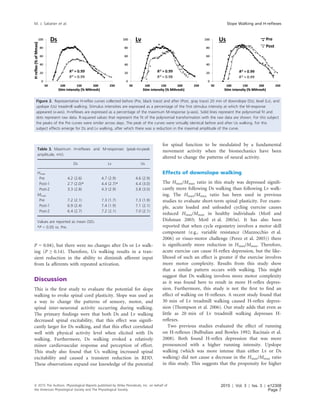
reduced the Hmax/Mmax ratio (P = 0.001 & P = 0.02), although the reduction
was larger for Ds walking (29.3 Æ 6.2% vs. 6.8 Æ 5.2%, P = 0.02). The reduc-
tion associated with Ds walking was correlated with physical activity level as
measured via questionnaire (r = À0.52, P = 0.04). Us walking caused an
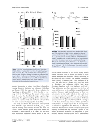
increase in the Hslp/Mslp ratio (P = 0.03) and a decrease in RDD (P = 0.04).
These changes recovered by 45 min. Exercise HR and BP were highest during
Us walking. RPE was greater during Ds and Us walking compared to Lv walk-
ing, but did not exceed “Fairly light” for Ds walking. In conclusion, in healthy
adults treadmill walking has a short-term effect on soleus H-reflex excitability
that is determined by the slope of the treadmill surface.
Introduction
The spinal cord is a major locus of activity-dependent
neural plasticity associated with motor learning and
skilled performance improvements (Windhorst 1996;
Wolpaw and Tennissen 2001). Activity-dependent spinal
plasticity can be evoked in the short-term and the long-
term, and spinal excitability reflects the regular patterns
of motor activity in which people engage (Zehr 2002).
For example, soleus H-reflexes are smaller in explosively
trained athletes (Casabona et al. 1990) and skilled ballet
dancers (Nielsen et al. 1993), and larger in endurance
trained athletes, compared to other athletes and to
untrained controls (Casabona et al. 1990; Maffiuletti et al.
2001). Walking is a ubiquitous and fundamental rhythmic
activity that supports independence and quality of life
(Yildiz 2012) and also has been found to evoke spinal
plasticity. For example, a recent study reported that
30 min of level treadmill walking causes short-term
H-reflex depression in healthy adults (Thompson et al.
2006). Another study found that 20 min of over-ground
walking caused an increase in rate-dependent depression
of H-reflexes, a form of presynaptic inhibition (Phadke
et al. 2009). However, it is not known if the response of
the H-reflex pathway could be augmented by changing
the parameters of the walking task, for example, surface
slope.
During downslope (Ds), upslope (Us), and level (Lv)
walking lower extremity muscles express unique patterns
of electromyographic (EMG) activity that reflect differences
ª 2015 The Authors. Physiological Reports published by Wiley Periodicals, Inc. on behalf of
the American Physiological Society and The Physiological Society.
This is an open access article under the terms of the Creative Commons Attribution License,
which permits use, distribution and reproduction in any medium, provided the original work is properly cited.
2015 | Vol. 3 | Iss. 3 | e12308
Page 1
Physiological Reports ISSN 2051-817X
2. in motor output and afferent feedback with different
slopes (Akima et al. 2005; Gregor et al. 2006; Lay et al.
2007). For example, an increase in extensor muscle EMG
activity during Us walking, and a decrease during Ds
walking, has been reported for both quadrupeds (Gregor
et al. 2006; Sabatier et al. 2011) and humans (Lay et al.
2007; Franz and Kram 2012). Moreover, during Ds walk-
ing there is increased muscle length-dependent afferent
feedback compared to Lv or Us walking due to increased
reliance on eccentric muscle contractions (Kuster et al.
1995; Abelew et al. 2000; Gregor et al. 2001; McIntosh
et al. 2006). Downslope walking might also require more
cortical activity than either Lv or Us walking due to this
reliance on eccentric muscle contractions (Fang et al.
2001, 2004). This might cause an increase in cortical
mediated spinal inhibition that would manifest as a
reduction in spinal excitability (Wolpaw 2007). Another
way Ds walking could affect reduced spinal excitability is
through activation of the Ia spinal reflex arc. For exam-
ple, activation of the Ia spinal reflex arc through stimula-
tion of muscle spindles by vibration results in decreased
spinal excitability (Shinohara 2005). Thus, Ds walking,
through a more natural form of persistent activation of
the spinal Ia reflex arc, may have a similar effect. Finally,
Ds walking is less metabolically demanding and evokes a
smaller cardiovascular response than level or Us walking
(Knuttgen et al. 1971; Navalta et al. 2004). Therefore, as
a potential adjunct for exercise prescription, Ds walking
could be more accommodating for people with reduced
exercise capacity.
It is also possible that Us walking might have a unique
ability to evoke spinal plasticity. During Us walking the
body’s center of mass is moved vertically, against gravity,
and there is increased motor unit recruitment in lower
extremity muscles compared to Lv and Ds walking (Lay
et al. 2007; Franz and Kram 2012). Increased effort
required for Us walking would be expected to promote
increased serotonergic and noradrenergic signaling
(Aston-Jones et al. 2000; Jacobs et al. 2002), potentially
leading to a change in spinal excitability. An increase in
spinal excitability would facilitate muscle stiffness and
force transmission in the lower extremity extensor mus-
cles to serve the Us walking movement pattern. At this
time, whether or not walking slope modulates the effect
of walking on spinal synaptic transmission remains
unknown.
The purpose of this study was to test the hypothesis
that Ds walking evokes a decrease in soleus H-reflex excit-
ability. This study also hypothesized an increase in soleus
H-reflex excitability as a result of Us walking. Soleus
H-reflexes were measured, whereas participants were rest-
ing in the recumbent seated position both before and
after treadmill slope walking.
Methods
Participants
Sixteen participants (nine men and seven women)
between the ages of 23 and 44, who did not participate in
competitive sports, and with no neurologic disease or
injury were tested on 3 days within a period of 1 week.
Participant characteristics were as follows (mean Æ SD):
age: 27.3 Æ 6.0 years; height: 177.8 Æ 10.5 cm; mass:
76.3 Æ 15.6 kg; BMI: 23.9 Æ 3.4 kg/m2
. All participants
were tested before and after treadmill walking. However,
four of these participants were tested for only the pre and
the first postwalking time point. Each visit started within
the same 1-h window of time to avoid the effects of
diurnal variation in reflex size (Wolpaw and Seegal 1982;
Lagerquist et al. 2006; Thompson et al. 2013). All partici-
pants were familiar with treadmill walking. The right leg
was tested in all but one participant, who asked for the
left leg to be tested. Written informed consent was
obtained from all participants, and the study was
approved by the Institutional Review Board of Emory
University.
Procedures
Physical activity (PA) during the week prior to testing
was assessed with the 7-day PA recall questionnaire (Blair
et al. 1985; Motl et al. 2003b; Cureton et al. 2009). There
was a range of values for PA (40–425 kJ/day), but the
group average (212 Æ 113 kJ/day) was similar to healthy
college-aged nonathletes as reported in other studies
(Motl et al. 2003b; Cureton et al. 2009). Participants
walked at a slow speed, 4.0 km/h (2.5 mph), on a Sole
Fitness F85 Folding Treadmill (Niagra Falls, ON) down-
slope (À8.5°, À15%), level (0%), and upslope (2.9À8.5°,
i.e., 5–15%), for 20 min. Duration was selected based on
previous reports of altered H-reflex responses with other
activities (treadmill running (Bulbulian and Darabos
1986), cycle ergometry (Motl et al. 2006)). For Us walk-
ing the treadmill slope changed every 5 min. The order of
slopes was 2.9°, 8.5°, 5.7°, and 2.9°. This pattern was used
to control for the much larger increase in effort that is
associated with Us walking, and to ensure that all partici-
pants could maintain a full 20 min of uninterrupted Us
walking (Navalta et al. 2004).
A different walking condition was tested for each test
session. Upslope and Lv walking were randomized to days
1 and 2. Downslope walking was always reserved for day
3 because of the potential for delayed onset muscle sore-
ness (Whitehead et al. 2001; Farr et al. 2002; Nottle and
Nosaka 2005) that could have then affected walking and/
or H-reflexes on the other 2 days of testing (Vangsgaard
2015 | Vol. 3 | Iss. 3 | e12308
Page 2
ª 2015 The Authors. Physiological Reports published by Wiley Periodicals, Inc. on behalf of
the American Physiological Society and The Physiological Society.
Slope Walking and H-reflexes M. J. Sabatier et al.
3. et al. 2013). None of our participants had ever engaged
in uninterrupted Ds treadmill walking as used in this
study. Therefore, to acclimate participants to the Ds walk-
ing task, each subject walked Ds for 3 min at the end of
the day-2 session. We have previously found that this
duration of Ds walking does not cause exercise-induced
muscle injury or delayed onset muscle soreness (Sabatier
and Black 2012). Prior to walking a wireless tri-axial
accelerometer was strapped securely to the right ankle
(BN-ACCL3; Biopac Systems Inc., Goleta, CA). Stride fre-
quency was computed online for each step using the time
period separating consecutive accelerations resulting from
foot contact with the treadmill.
During the 5th
, 10th
, 15th
, and 20th
min of walking
heart rate was measured using a Polar Ft1 Heart Rate
Monitor (Polar Electro Inc.; Lake Success, NY). Arterial
pressure was measured at the brachial artery by manual
sphygmomanometry using an appropriate-sized cuff
(Perloff et al. 1993). Participants were also asked to rate
their perceived exertion using the Borg scale, with 6 being
“very, very light” to 19 being “very, very hard” (Borg
1978). After walking participants were immediately seated
and prepared for post H-reflex testing. Heart rate and
blood pressure were measured once more 5 min after
walking stopped. H-reflex collection began again at
10 min (n = 16), and then again at 45 min (n = 12).
Soleus H-reflexes
The H-reflex was measured at the soleus while the partici-
pant was seated in a semireclined position with the hip at
120° and the knee at 30°. The soleus was chosen because
it is strongly modulated during slope walking (Lay et al.
2007). A band was secured around the legs at the distal
thigh to prevent the legs from falling into external rota-
tion or abduction. Electromyographic (EMG) activity was
recorded from the soleus using bipolar electrodes (EL503;
Biopac Systems Inc.) placed 2 cm apart along the poster-
ior lateral aspect of the muscle, 2 cm inferior to the lower
border of the lateral gastrocnemius (Basmajian and Blu-
menstein 1989). Identical day-to-day EMG and stimula-
tion electrode placement was ensured by outlining
electrode locations on the skin with a permanent marker.
EMG signals were band-pass filtered (5–1000 Hz) and
amplified by 2000 (BN-EMG2; Biopac Systems Inc.). Low
impedance (<10 kO) was verified for all stimulating and
recording electrodes using an electrode impedance meter
(UFI MkIII Checktrode, Model 1089).
Reflexes were evoked by stimulating the tibial nerve in
the popliteal fossa through a monopolar electrode (round,
2.5 cm) with the anode (square, 5 cm) placed above the
patella. Both were self-adhering carbon rubber TENS/
NMES electrodes (Medical Products Online, Danbury,
CT). Cathode placement was determined prior to testing
using a pen electrode (Model G.MPPE, Digitimer, Hert-
fordshire, AL7 3BE, England) to find the location yielding
an H-reflex without an M-response and plantar flexion
without eversion or inversion. Single, 1-msec rectangular
pulses were delivered at pseudo-random intervals (5–8 sec
in duration) using a constant-current electrical stimulator
(STMISOLA; BIOPAC Systems Inc.) controlled with cus-
tom-written scripts in AcqKnowledge software (Biopac
Systems Inc.).
The amplitude of the H-reflex depends on motoneuron
pool excitability (as measured via background EMG activ-
ity) when the H-reflex is elicited. Therefore, when evalu-
ating H-reflexes it is important to standardize for the
level of motoneuron pool excitability (Schieppati 1987;
Burke et al. 1989; Zehr 2002). In this study soleus back-
ground EMG activity was maintained at a constant low
level as measured from participants during quiet standing
(Thompson et al. 2009). The ankle was maintained at 10o
of plantar flexion using a foot brace that the subject con-
tracted against to adjust soleus EMG activity to the
required level. Prior to H-reflex testing participants stood
quietly for 30 seconds while soleus and tibialis anterior
EMG activity were measured. The average rectified soleus
EMG activity was recorded. During collection of H-reflex
recruitment curves participants were provided with visual
feedback to maintain average rectified soleus EMG activ-
ity at standing level.
Spinal excitability
The H-reflex recruitment curve was acquired by progres-
sively increasing the intensity of electrical stimulation in
0.5–1.0 mA increments to find the largest obtainable
H-reflexes and M-responses, measured as the peak-to-peak
amplitude of the raw EMG signal. The Hmax was taken as
the average of the largest three H-reflexes, and the Mmax as
the average of the largest three M-responses.
The M-response increases with increasing stimulus
intensity, eventually reaching a plateau (referred to as
Mmax subsequently). Mmax is an estimate of the motoneu-
ron pool. Longitudinal studies generally express Hmax as a
percentage of the Mmax to account for potential changes
in ability to deliver current to the peripheral nerve across
time and for differences in muscle geometry across sub-
jects (Crone et al. 1999; Zehr 2002; Palmieri et al. 2004).
This is also done to control for intersubject differences in
the efficacy of nerve stimulation and total number of
MUs accessible by nerve stimulation (Palmieri et al.
2004). Therefore, one expression of spinal excitability
used in this study is the Hmax/Mmax ratio.
A drawback to characterizing spinal excitability in the
manner described above is that stimulating above motor
ª 2015 The Authors. Physiological Reports published by Wiley Periodicals, Inc. on behalf of
the American Physiological Society and The Physiological Society.
2015 | Vol. 3 | Iss. 3 | e12308
Page 3
M. J. Sabatier et al. Slope Walking and H-reflexes
4. threshold results in collision between orthodromic and
antidromic impulse transmission. This may contribute to
inter and intrasubject variability in Hmax, and to the
decreased sensitivity of the H-reflex facilitation and inhi-
bition at Hmax (Crone et al. 1990). The rising slope of the
H recruitment curve (Hslp) has been suggested as a good
alternative because it is free of this collision effect (Funase
et al. 1994). Also, in previous studies, an increase in
spinal excitability was detected using the Hslp/Mslp
approach, but not with the Hmax/Mmax ratio (Kalmar and
Cafarelli 1999; Walton et al. 2003). Therefore, in order to
optimize this study’s potential to detect an increase in
spinal excitability the slope of the H-reflex recruitment
curve was also evaluated. Recruitment curves were fitted
with 8th
order polynomial transformations starting at the
first response of the ascending limb using custom-written
scripts in Microsoft Excel. R-squared values for raw data
versus computed curves were high (mean ÆSE,
0.96 Æ 0.01). The ratio of the Hslp to the M recruitment
curve slope (Mslp) was computed to standardize for
motoneuron excitability. This renders the Hslp/Mslp ratio
a metric of spinal excitability. Each slope was derived
from the linear regression line that included values from
the transformed curve between 25% and 75% of the Hmax
and Mmax, for the Hslp and Mslp, respectively.
Rate-dependent depression (RDD)
Rate-dependent depression (also known as postactivation
depression) was measured as a segmental presynaptic
mechanism of activity-dependent synaptic efficacy in the
H-reflex pathway (Kohn et al. 1997; Hultborn and Niel-
sen 1998; Aymard et al. 2000). Ten H-reflexes were elic-
ited at a low stimulus frequency of 0.1 Hz, and at the
stimulus intensity that elicited an H-reflex that was
between 20% of Mmax and 50% of Hmax (Sosnoff and
Motl 2010). The H-reflex was elicited 11 times at the
same stimulus intensity, but with a stimulus frequency of
1 Hz (i.e., high-frequency stimulation), and the last 10
H-reflexes of this series were averaged. The average
amplitude of these H-reflexes is expressed as a percentage
of the average H-reflex amplitude when evoked at a fre-
quency of 0.1 Hz (Sosnoff and Motl 2010). Voluntary
muscle contraction decreases RDD (Hultborn and Nielsen
1998). Therefore, participants were instructed to keep the
leg at complete rest throughout RDD testing (Meunier
et al. 2007; Lamy et al. 2009).
Muscle soreness
Downslope walking is associated with more eccentric
muscle contractile activity than Lv or Us walking (Abelew
et al. 2000; Gregor et al. 2001; Akima et al. 2005).
Because eccentric muscle contractions are associated with
increased risk for delayed onset muscle soreness (DOMS)
(Proske and Morgan 2001), Ds walking might cause
DOMS. In fact, previous studies have used a variety of Ds
locomotion patterns as a way to induce muscle soreness
(Whitehead et al. 2001; Farr et al. 2002; Nottle and
Nosaka 2005). Although no previous study to our knowl-
edge has used the Ds walking pattern described in this
study, we anticipated muscle soreness might result in
lower extremity muscles after Ds walking. Therefore, mus-
cle pain intensity in the lower extremities (anterior and
posterior leg, anterior thigh) was assessed using a visual
analogue scale (VAS). The VAS consists of a 10-cm line
ranging from 0 (no pain) to 10 (worst pain imaginable).
Participants rated pain intensity felt during daily life
activity during the 4 days after the last session.
Statistical analysis
This study used a repeated measures design with out-
comes collected on all 3 days. The repeated measures
design was modeled using a general linear model with
slope as a fixed effect and random effects for subject
nested within time point using R version 3.1.0 (The R
Foundation for Statistical Computing) (Bates et al. 2014).
If the interaction term for the model was significant, each
post time point was compared to the pre time point
using a model adjusted t-test where the standard errors
and degrees of freedom were estimated from the model
with restricted maximum likelihood methodology. Intra-
class correlation coefficients were computed using Statisti-
ca data analysis software system (version 10, StatSoft,
Inc., 2013) to evaluate day-to-day reliability of dependent
measures. Linear correlation analysis was carried out to
determine the relationship between variables of interest.
The T-distribution was used to determine the statistical
significance of correlations. The significance level was set
at P ≤ 0.05 for all statistical tests.
Results
Study participants were healthy adults who did not par-
ticipate in competitive sports. Nevertheless, we anticipated
regular PA levels might vary significantly and contribute
to variability in starting values of our dependent mea-
sures, or the way these values responded to slope walking.
Therefore, this study also measured PA via questionnaire
to determine if there are relationships between regular PA
and our dependent measures. There was a significant neg-
ative correlation between the change in Hmax/Mmax with
Ds walking and PA (r = À0.52, P = 0.04, Fig. 1). There-
fore, participants who reported more PA had a larger
reduction in Hmax/Mmax after Ds walking. There was no
2015 | Vol. 3 | Iss. 3 | e12308
Page 4
ª 2015 The Authors. Physiological Reports published by Wiley Periodicals, Inc. on behalf of
the American Physiological Society and The Physiological Society.
Slope Walking and H-reflexes M. J. Sabatier et al.
5. correlation between PA and Hmax/Mmax (r = 0.39,
P = 0.20), Hslp/Mslp (r = 0.22, P = 0.49), or RDD
(r = 0.06, P = 0.85). There was no correlation between
PA and the change in Hmax/Mmax for Lv (r = À0.14,
P = 0.60) and Us walking (r = 0.11, P = 0.69). There
were also no significant correlations between PA and the
change in Hslp/Mslp (r = À0.26, P = 0.33; r = 0.01,
P = 0.98; r = À0.07, P = 0.79) or RDD (r = 0.27,
P = 0.31; r = À0.20, P = 0.45; r = 0.23, P = 0.39) with
Ds, Lv or Us walking, respectively.
During treadmill walking there were several slope-
related changes in measures associated with effort. Effort
during slope walking was monitored by measuring blood
pressure (BP), heart rate (HR) and ratings of perceived
exertion (RPEs) (Table 1). Heart rate was highest during
Us walking, and not significantly different between Lv
and Ds walking. An exception was after treadmill walking.
Five minutes after cessation of both Ds and Us walking
HR remained elevated compared to after Lv walking.
Heart rate increased during the Ds walking bout and was
highest during the 3rd
and 4th
epochs. Like HR, systolic
blood pressure (SBP) was also highest during Us walking.
However, SBP was similar between Lv and Ds walking.
Diastolic blood pressure (DBP) was higher during Ds
walking than during both Lv and Us walking. However,
DBP fell to a lower level at 5 min after cessation of Ds
walking compared to either Lv or Us walking. Ratings of
perceived exertion were higher for both Ds and Us walk-
ing compared to Lv walking. For Ds walking RPE did not
exceed 10 (9 was anchored as “Very light,” and 11 was
“Fairly light”). Thus, although there was increased per-
ceived effort associated with walking on either slope, it
remaind light for Ds walking.
Figure 1. The depressive effect of Ds walking (ordinate) as a
function of physical activity level (7-d PAR, abscissa) is illustrated in
this scatterplot. The post-pre Hmax/Mmax difference is expressed as a
percentage of pre on the ordinate. A horizontal dashed line is
aligned at zero on the ordinate, or an absence of change in Hmax/
Mmax. Values below zero reflect a reduction in Hmax/Mmax after Ds
walking. There was a significant negative correlation between the
percent change in Hmax/Mmax with Ds walking and physical activity
(r = À0.52, P = 0.04).
Table 1. Cardiovascular function and perceived exertion during each 5-min epoch of slope walking.
Pre
Treadmill walking
Post1–5 min 6–10 min 11–15 min 16–20 min
HR
Us 61 (10) 94 (14) 117 (23) 114 (21) 105 (17) 67 (13)
Lv 63 (11) 83 (13)* 87 (13)* 88 (14)* 87 (15)* 61 (10)
Ds 63 (9) 81 (12)* 84 (11)* 88 (10)* 87 (13)* 66 (11)*
SBP
Us 117 (15) 123 (19) 133 (18) 125 (17) 119 (14) 119 (15)
Lv 113 (13)* 116 (16)* 112 (15)* 113 (16)* 114 (15)* 115 (13)
Ds 113 (10) 117 (13)* 117 (16)* 114 (15)* 116 (15) 114 (9)*
DBP
Us 76 (9) 68 (11) 64 (13) 61 (10) 61 (11) 75 (11)
Lv 71 (9)* 66 (9) 65 (10) 64 (11) 63 (9) 74 (9)
Ds 71 (11)* 70 (9)#
68 (9) 69 (9)*#
69 (9)*#
71 (8)*#
RPE
Us – 7.8 (1.0) 12.0 (2.0) 10.7 (1.1) 9.3 (1.7) –
Lv – 7.3 (1.1) 7.6 (1.2)* 7.6 (1.2)* 7.6 (1.1)* –
Ds – 8.8 (1.6)*#
9.6 (2.0)*#
9.9 (2.0)* 10.0 (2.2)* –
Values are reported as mean (SD).
SBP, systolic blood pressure; DBP, diastolic blood pressure.
*P < 0.05 vs. Us.
#
P < 0.05 vs. Lv.
ª 2015 The Authors. Physiological Reports published by Wiley Periodicals, Inc. on behalf of
the American Physiological Society and The Physiological Society.
2015 | Vol. 3 | Iss. 3 | e12308
Page 5
M. J. Sabatier et al. Slope Walking and H-reflexes
6. Step cycle timing
Stride frequency was measured to determine if basic step
cycle timing for walking at different slopes in this study is
consistent with previous reports on the biomechanics of
slope walking. Stride frequency was significantly greater
for Ds walking (1.04 Æ 0.07 Hz) compared to Lv
(0.85 Æ 0.02 Hz) and Us walking (0.83 Æ 0.07 Hz),
P ≤ 0.05 for all comparisons. These results are consistent
with previous investigations of the biomechanics of walk-
ing on different slopes (Hunter et al. 2010; Franz and
Kram 2012, 2013). Thus, although comprehensive biome-
chanical measures of lower body movements were not
made here, the results of step cycle timing, effort and
DOMS support the idea that this study compared well to
the studies referenced above.
Lower extremity muscle soreness
There were no reports of soreness or other discomfort
after either Lv or Us walking. Although five participants
reported no soreness, all other participants reported
soreness ranging from 0.5 cm to 8.4 cm on the VAS
scale after Ds walking. Soreness only occurred after Ds
walking, and the majority of soreness was experienced in
the tibialis anterior and the triceps surae (Table 2). This
effect subsided during the 4 days after Ds walking, sup-
porting the notion that the Ds walking pattern used in
this study involved more eccentric muscle contraction
than either Lv or Us walking. Incidently, one subject
also reported hip flexor soreness near the inguinal liga-
ment. Another subject reported no soreness in the quad-
riceps, but rather a generalized feeling of fatigue in the
legs.
H-reflexes and M-responses
Changes in H-reflex amplitude occurred as a result of Ds
and Lv walking. In Fig. 2 H-reflex curves from a repre-
sentative subject are expressed as a percentage of the max-
imum M-response for pre and post Ds, Lv, and Us
walking. The average Mmax and Hmax values across partic-
ipants for Ds, Lv, and Us walking are shown in Table 3.
Pre Mmax and Hmax values did not change across days
and were very reliable (Mmax, ICC = 0.97; Hmax,
ICC = 0.94; Hmax/Mmax = 0.95). For Hmax the model
interaction term was statistically significant (P = 0.02),
and there was a statistically significant effect (Post-1 vs.
Pre) for Ds and Lv walking only (P < 0.001 for Ds walk-
ing, and P = 0.02 for Lv walking). The change in Hmax
after Ds walking was larger than the change after Lv walk-
ing (36.5.2 Æ 20.0% vs. 8.4 Æ 17.0%, mean Æ SEM,
P = 0.03). The interaction term was not statistically sig-
nificant for Mmax.
Hmax/Mmax ratios are illustrated in Fig. 3A. The model
interaction term was statistically significant for Hmax/
Mmax (P = 0.01). There was a reduction in Hmax/Mmax
for Post-1 versus Pre for both Ds walking (P < 0.001),
and Lv walking (P = 0.02). The change after Ds walking
was larger than the change after Lv walking (29.3 Æ 6.2%
vs. 6.8 Æ 5.2%, mean Æ SEM, P = 0.02). EMG biofeed-
back was used to standardize background EMG activity
during the collection of H-reflex recruitment curves. As a
result, background EMG activity did not change across
bouts of testing (Fig. 3C). Therefore, changes in back-
ground motor neuron recruitment can be ruled out as a
cause of changes in H-reflex size. Results for Hslp/Mslp
ratios are illustrated in Fig. 3B. The model interaction
term was statistically significant for Hslp/Mslp (P = 0.05).
There was an increase in Hslp/Mslp for Us walking only
(Post-1 vs. Pre, P = 0.04). Therefore, when spinal excit-
ability was characterized using the Hslp/Mslp method, Us
walking had an effect that was not detected using the
Hmax/Mmax approach.
Rate-dependent depression
The final objective of this study was to evaluate the effect
of slope walking on RDD (Fig. 4). There was no detect-
able background EMG activity during the 100 ms preced-
ing electrical stimulation (data not shown). This is
consistent with the study protocol as subjects were asked
to keep the leg at complete rest. RDD is expressed as a
percentage (i.e., % depression when H-reflexes were elic-
ited at 1 Hz vs. when they were elicited at 0.1 Hz) in
Fig. 4B. The model interaction term was statistically sig-
nificant for RDD (P = 0.01). There was a significant
reduction in RDD after Us walking (Pre vs. Post-1,
Table 2. Muscle soreness after downslope walking.
Four days following Ds treadmill walking
24 h 48 h 72 h 96 h
Tibialis Anterior
R 1.4 (2.3) 0.9 (1.6) 0.2 (0.4)* 0.0 (0.1)*
L 1.1 (1.8) 0.9 (1.9) 0.3 (0.6)* 0.1 (0.2)*
Triceps Surae
R 1.4 (2.2) 0.9 (1.7)* 0.3 (0.7)* 0.1 (0.2)*
L 1.4 (2.3) 0.9 (1.9)* 0.3 (0.6)* 0.1 (0.2)*
Quadriceps
R 0.3 (0.8) 0.2 (0.5) 0.1 (0.2)* 0.1 (0.2)*
L 0.3 (0.6) 0.3 (0.7) 0.1 (0.2)* 0.1 (0.2)*
Values are reported as mean (SD).
*P < 0.05 vs. 24 h.
2015 | Vol. 3 | Iss. 3 | e12308
Page 6
ª 2015 The Authors. Physiological Reports published by Wiley Periodicals, Inc. on behalf of
the American Physiological Society and The Physiological Society.
Slope Walking and H-reflexes M. J. Sabatier et al.
7. P = 0.04), but there were no changes after Ds or Lv walk-
ing (P ≥ 0.14). Therefore, Us walking results in a tran-
sient reduction in the ability to diminish afferent input
from Ia afferents with repeated activation.
Discussion
This is the first study to evaluate the potential for slope
walking to evoke spinal cord plasticity. Slope was used as
a way to change the patterns of sensory, motor, and
spinal inter-neuronal activity occurring during walking.
The primary findings were that both Ds and Lv walking
decreased spinal excitability, that this effect was signifi-
cantly larger for Ds walking, and that this effect correlated
well with physical activity level when elicited with Ds
walking. Furthermore, Ds walking evoked a relatively
minor cardiovascular response and perception of effort.
This study also found that Us walking increased spinal
excitability and caused a transient reduction in RDD.
These observations expand our knowledge of the potential
for spinal function to be modulated by a fundamental
movement activity when the biomechanics have been
altered to change the patterns of neural activity.
Effects of downslope walking
The Hmax/Mmax ratio in this study was depressed signifi-
cantly more following Ds walking than following Lv walk-
ing. The Hmax/Mmax ratio has been used in previous
studies to evaluate short-term spinal plasticity. For exam-
ple, acute loaded and unloaded cycling exercise causes
reduced Hmax/Mmax in healthy individuals (Motl and
Dishman 2003; Motl et al. 2003a). It has also been
reported that when cycle ergometry involves a motor skill
component (e.g., variable resistance (Mazzocchio et al.
2006) or visuo-motor challenge (Perez et al. 2005)) there
is significantly more reduction in Hmax/Mmax. Therefore,
acute exercise can cause H-reflex depression, but the like-
lihood of such an effect is greater if the exercise involves
more motor complexity. Results from this study show
that a similar pattern occurs with walking. This might
suggest that Ds walking involves more motor complexity
as it was found here to result in more H-reflex depres-
sion. Furthermore, this study is not the first to find an
effect of walking on H-reflexes. A recent study found that
30 min of Lv treadmill walking caused H-reflex depres-
sion (Thompson et al. 2006). Our study adds that even as
little as 20 min of Lv treadmill walking depresses H-
reflexes.
Two previous studies evaluated the effect of running
on H-reflexes (Bulbulian and Bowles 1992; Racinais et al.
2008). Both found H-reflex depression that was more
pronounced with a higher running intensity. Upslope
walking (which was more intense than either Lv or Ds
walking) did not cause a decrease in the Hmax/Mmax ratio
in this study. This suggests that the propensity for higher
Table 3. Maximum H-reflexes and M-responses (peak-to-peak
amplitude, mV).
Ds Lv Us
Hmax
Pre 4.2 (2.6) 4.7 (2.9) 4.6 (2.9)
Post-1 2.7 (2.0)* 4.4 (2.7)* 4.4 (3.0)
Post-2 3.3 (2.8) 4.3 (2.9) 3.8 (3.0)
Mmax
Pre 7.2 (2.1) 7.3 (1.7) 7.3 (1.9)
Post-1 6.9 (2.4) 7.4 (1.9) 7.1 (2.1)
Post-2 6.4 (2.7) 7.2 (2.1) 7.0 (2.1)
Values are reported as mean (SD).
*P < 0.05 vs. Pre.
Figure 2. Representative H-reflex curves collected before (Pre, black trace) and after (Post, gray trace) 20 min of downslope (Ds), level (Lv), and
upslope (Us) treadmill walking. Stimulus intensities are expressed as a percentage of the first stimulus intensity at which the M-response
appeared (x-axis). H-reflexes are expressed as a percentage of the maximum M-response (y-axis). Solid lines represent the polynomial fit and
dots represent raw data. R-squared values that represent the fit of the polynomial transformation with the raw data are shown. For this subject
the peaks of the Pre curves were similar across days. The peak of the curves were virtually identical before and after Us walking. For this
subject effects emerge for Ds and Lv walking, after which there was a reduction in the maximal amplitude of the curve.
ª 2015 The Authors. Physiological Reports published by Wiley Periodicals, Inc. on behalf of
the American Physiological Society and The Physiological Society.
2015 | Vol. 3 | Iss. 3 | e12308
Page 7
M. J. Sabatier et al. Slope Walking and H-reflexes
8. intensity locomotion to reduce Hmax/Mmax is limited to
running. However, Bulbulian and colleagues (Bulbulian
and Bowles 1992) also reported a larger reduction in
Hmax/Mmax for 20 min of Ds (À10%) than for Lv run-
ning, both at 50% VO2max. The results of this study
make it is clear that the effect of using a negative slope
for running also applies to walking, despite numerous dif-
ferences in neural control and mechanics between these
two forms of locomotion (Cappellini et al. 2006).
This study also found that participants who reported
more PA had a larger reduction in the Hmax/Mmax ratio
after Ds walking. This supports the idea that there are
chronic central nervous system adaptations related to
increased PA levels (Adkins et al. 2006), and also that
such adaptations predispose healthy adults to the Ds
walking effect discovered in this study. Highly trained
cohorts have been found to present with smaller or larger
resting H-reflexes than untrained cohorts, depending on
the nature of their training. For instance, competitive
power athletes (Casabona et al. 1990; Maffiuletti et al.
2001) and ballet dancers (Nielsen et al. 1993) have smal-
ler H-reflexes, and endurance athletes have larger
H-reflexes (Casabona et al. 1990; Maffiuletti et al. 2001).
These differences have been attributed to the level of
motor skill involved in these athletes’ competitive physical
activities. However, highly trained cohorts as these were
not evaluated in this study. Indeed, in this study there
were no correlations between PA and any baseline mea-
sures of the H-reflex pathway. As this study is the first to
report a decrease in spinal excitability with Ds walking, it
is not clear if these very unique athletic populations
would respond in unique ways to the Ds walking stimu-
lus. We would hypothesize that there may be subtle dif-
ferences in the amounts of skilled activity our participants
regularly undertake that may have been captured in self-
reports of more PA. However, it is not possible to quan-
tify such potential differences with the PA questionnaire
A
B
C
Figure 3. H-reflex results before and after 20 min of downslope
(Ds), level (Lv), and upslope (Us) treadmill walking. (A) Hmax/Mmax
ratio, mean + SE, *P < 0.05 versus pre. There was a significant
reduction after Ds walking and after Lv walking. (B) Hslp/Mslp ratio,
mean + SE, *P < 0.05versus pre. There was a significant increase
after Us walking. Prestimulus background EMG activity (100 msec
prior to electrical stimulation) (C). There was no change in
prestimulus background EMG activity across time.
A
B
Figure 4. Rate-dependent depression (RDD) of the H-reflex before
and after downslope (Ds), level (Lv), and upslope (Us) treadmill
walking. (A) The average of 10 sweeps at 0.1 Hz (dotted line) and
at 1.0 Hz (solid line), pre and post walking for one representative
subject. Average H-reflex size was similar for 0.1 Hz, but depression
with 1.0 Hz stimulation frequency was less at Post. (B) Average
RDD results expressed as percent depression (i.e., % depression
when H-reflexes were elicited at 1 Hz vs. when they were elicited
at 0.1 Hz), mean + SE, *P < 0.05versus pre. There was a reduction
in RDD after Us walking.
2015 | Vol. 3 | Iss. 3 | e12308
Page 8
ª 2015 The Authors. Physiological Reports published by Wiley Periodicals, Inc. on behalf of
the American Physiological Society and The Physiological Society.
Slope Walking and H-reflexes M. J. Sabatier et al.
9. used in this study. Future studies could evaluate the effect
of slope walking in distinct athletic populations to deter-
mine if high volumes of PA involving more or less motor
control impact the slope-walking response. Such investi-
gations could provide insight into the physiological basis
of the slope-walking effects.
Effects of upslope walking
This study found that Us walking increased the Hslp/
Mslp ratio and reduced RDD. Increased effort may have
prompted these effects as systolic blood pressure, heart
rate, and RPE were elevated during Us walking. Increased
MU recruitment and effort associated with Us walking
may have resulted in increased serotonergic signaling
from neurons of the raphe neuclei of the brain stem
(Mazzardo-Martins et al. 2010) which have excitatory
projections on alpha motoneurons (Barasi and Roberts
1974; White and Neuman 1980). Activity in the descend-
ing serotonergic system has been shown to increase in
proportion to motor output (Jacobs et al. 2002). Further-
more, Cardona and Rudomin (Cardona and Rudomin
1983) reported a decrease in RDD in response to activa-
tion of brainstem serotonergic pathways in the isolated
frog preparation. Norepinephrine system activity may
have a similar effect as it also projects monosynaptically
to motoneurons throughout the spinal cord (Holstege
and Kuypers 1987) and increases with increased arousal
(Aston-Jones et al. 2000). Both serotonin and norepi-
nephrine enhance the effects of excitatory inputs to spinal
motoneurons, producing long-lasting changes in moto-
neuron excitability (White and Neuman 1980). Therefore,
in this study the motoneuron pool may have been less
dependent on Ia synaptic transmission to reach firing
threshold as a result of Us walking. This would facilitate a
more sustained response to repetitive afferent inputs.
Decreased RDD resulting from Us walking as found in
this study suggests the overall pattern of MU recruitment
and incoming sensory information during Us walking
results in a change in gaiting of afferent feedback that del-
egates more control of movement to peripheral reflex
pathways. The increase in Hslp/Mslp found after Us walk-
ing would also contribute to this neural strategy. This is
consistent with the idea that Us walking in healthy adults
involves little skill development or motor learning.
Rather, Us walking is unique in that it is associated with
more MU recruitment (Gregor et al. 2006; Franz and
Kram 2012) and force related feedback compared to Lv
or Ds walking (Gregor et al. 2006). As such, a decrease in
RDD could support an overall strategy to optimize mus-
cle stiffness during Us walking and improve force trans-
mission across skeletal muscle.
In conclusion, this study provides the first evidence
that in healthy human participants walking for 20 min at
a slow speed, treadmill slope determines the nature of
resulting changes in the H-reflex pathway. Although spec-
ulative at this time, it is possible that these effects consti-
tute the initial stage of activity-dependent spinal
plasticity. Future studies should determine if repeated
exposures of either Ds or Us walking convert the out-
comes reported here into more permanent adaptations, as
happens with motor or exercise training.
Acknowledgments
We wish to thank Jennifer F. Sabatier, MApStat, for her
assistance with statistical analysis; Dr Trisha Kesar and Dr
Michael Borich for their comments on this manuscript;
Ken Graap and Mickey Rowe of Biopac Systems Inc for
their assistance with scripts for data acquisition; and Katie
Moise, Philip Ikoku, and Sultan Budhwani for assistance
with data collection and analysis. We would also like to
thank all the participants who volunteered to be part of
this study for their patience, time, and efforts. The
authors thank the financial support from the Emory
University Dept of Rehabilitation Medicine, Division of
Physical Therapy.
Conflict of Interest
The authors have no conflict of interest to declare.
References
Abelew, T. A., M. D. Miller, T. C. Cope, and T. R. Nichols.
2000. Local loss of proprioception results in disruption of
interjoint coordination during locomotion in the cat. J.
Neurophysiol. 84:2709–2714.
Adkins, D. L., J. Boychuk, M. S. Remple, and J. A. Kleim.
2006. Motor training induces experience-specific patterns of
plasticity across motor cortex and spinal cord. J. Appl.
Physiol. 101:1776–1782.
Akima, H., R. Kinugasa, and S. Kuno. 2005. Recruitment of
the thigh muscles during sprint cycling by muscle functional
magnetic resonance imaging. Int. J. Sports Med. 26:245–252.
Aston-Jones, G., J. Rajkowski, and J. Cohen. 2000. Locus
coeruleus and regulation of behavioral flexibility and
attention. Prog. Brain Res. 126:165–182.
Aymard, C., R. Katz, C. Lafitte, E. Lo, A. Penicaud, P. Pradat-
Diehl, et al. 2000. Presynaptic inhibition and homosynaptic
depression: a comparison between lower and upper limbs in
normal human subjects and patients with hemiplegia. Brain
123(Pt 8):1688–1702.
Barasi, S., and M. H. Roberts. 1974. The modification of
lumbar motoneurone excitability by stimulation of a
ª 2015 The Authors. Physiological Reports published by Wiley Periodicals, Inc. on behalf of
the American Physiological Society and The Physiological Society.
2015 | Vol. 3 | Iss. 3 | e12308
Page 9
M. J. Sabatier et al. Slope Walking and H-reflexes
10. putative 5-hydroxytryptamine pathway. Br. J. Pharmacol.
52:339–348.
Basmajian, J. V., and R. Blumenstein. 1989. Electrode
placement in electromyographic biofeedback. Pp. 369–382 in
J. V. Basmajian, ed. Biofeedback, principles and practice for
clinicians. Williams & Wilkins, Baltimore, MD.
Bates, D., M. Maechler, B. Bolker, and S. Walker. 2014. lme4:
Linear mixed-effects models using Eigen: S4. R package
version 1.1-5.
Blair, S. N., W. L. Haskell, P. Ho, R. S. Jr Paffenbarger,
K. M. Vranizan, J. W. Farquhar, et al. 1985. Assessment of
habitual physical activity by a seven-day recall in a
community survey and controlled experiments. Am. J.
Epidemiol. 122:794–804.
Borg, G. 1978. Subjective effort and physical abilities. Scand. J.
Rehabil. Med. Suppl. 6:105–113.
Bulbulian, R., and D. K. Bowles. 1992. Effect of downhill
running on motoneuron pool excitability. J. Appl. Physiol.
73:968–973.
Bulbulian, R., and B. L. Darabos. 1986. Motor neuron
excitability: the Hoffmann reflex following exercise of low
and high intensity. Med. Sci. Sports Exerc. 18:697–702.
Burke, D., R. W. Adams, and N. F. Skuse. 1989. The effects of
voluntary contraction on the H reflex of human limb
muscles. Brain 112(Pt 2):417–433.
Cappellini, G., Y. P. Ivanenko, R. E. Poppele, and
F. Lacquaniti. 2006. Motor patterns in human walking and
running. J. Neurophysiol. 95:3426–3437.
Cardona, A., and P. Rudomin. 1983. Activation of brainstem
serotoninergic pathways decreases homosynaptic depression
of monosynaptic responses of frog spinal motoneurons.
Brain Res. 280:373–378.
Casabona, A., M. C. Polizzi, and V. Perciavalle. 1990.
Differences in H-reflex between athletes trained for
explosive contractions and non-trained subjects. Eur.
J. Appl. Physiol. 61:26–32.
Crone, C., H. Hultborn, L. Mazieres, C. Morin, J. Nielsen, and
E. Pierrot-Deseilligny. 1990. Sensitivity of monosynaptic test
reflexes to facilitation and inhibition as a function of the
test reflex size: a study in man and the cat. Exp. Brain Res.
81:35–45.
Crone, C., L. L. Johnsen, H. Hultborn, and G. B. Orsnes.
1999. Amplitude of the maximum motor response (Mmax)
in human muscles typically decreases during the course of
an experiment. Exp. Brain Res. 124:265–270.
Cureton, K. J., P. D. Tomporowski, A. Singhal, J. D. Pasley,
K. A. Bigelman, K. Lambourne, et al. 2009. Dietary
quercetin supplementation is not ergogenic in untrained
men. J. Appl. Physiol. 107:1095–1104.
Fang, Y., V. Siemionow, V. Sahgal, F. Xiong, and G. H. Yue.
2001. Greater movement-related cortical potential during
human eccentric versus concentric muscle contractions.
J. Neurophysiol. 86:1764–1772.
Fang, Y., V. Siemionow, V. Sahgal, F. Xiong, and G. H. Yue.
2004. Distinct brain activation patterns for human maximal
voluntary eccentric and concentric muscle actions. Brain
Res. 1023:200–212.
Farr, T., C. Nottle, K. Nosaka, and P. Sacco. 2002. The effects
of therapeutic massage on delayed onset muscle soreness
and muscle function following downhill walking. J Sci Med
Sport 5:297–306.
Franz, J. R., and R. Kram. 2012. The effects of grade and speed
on leg muscle activations during walking. Gait Posture.
35:143–147.
Franz, J. R., and R. Kram. 2013. How does age affect leg
muscle activity/coactivity during uphill and downhill
walking? Gait Posture. 37:378–384.
Funase, K., K. Imanaka, and Y. Nishihira. 1994. Excitability of
the soleus motoneuron pool revealed by the developmental
slope of the H-reflex as reflex gain. Electromyogr. Clin.
Neurophysiol. 34:477–489.
Gregor, R. J., J. L. Smith, D. W. Smith, A. Oliver, and B. I.
Prilutsky. 2001. Hindlimb kinetics and neural control during
slope walking in the cat: unexpected findings. J. Appl.
Biomech. 17:277–286.
Gregor, R. J., D. W. Smith, and B. I. Prilutsky. 2006.
Mechanics of slope walking in the cat: quantification of
muscle load, length change, and ankle extensor EMG
patterns. J. Neurophysiol. 95:1397–1409.
Holstege, J. C., and H. G. Kuypers. 1987. Brainstem
projections to spinal motoneurons: an update. Neuroscience
23:809–821.
Hultborn, H., and H. B. Nielsen. 1998. Modulation of
transmitter release from Ia afferents by their preceding
activity - a ‘postactivation depression’. Pp. 178–191 in
P. Rudomin and L. Mendell, eds. Presynaptic inhibition &
neural control. Oxford University Press, New York.
Hunter, L. C., E. C. Hendrix, and J. C. Dean. 2010. The cost
of walking downhill: is the preferred gait energetically
optimal? J. Biomech. 43:1910–1915.
Jacobs, B. L., F. J. Martin-Cora, and C. A. Fornal. 2002.
Activity of medullary serotonergic neurons in freely moving
animals. Brain Res. Brain Res. Rev. 40:45–52.
Kalmar, J. M., and E. Cafarelli. 1999. Effects of caffeine on
neuromuscular function. J. Appl. Physiol. 87:801–808.
Knuttgen, H. G., F. B. Petersen, and K. Klausen. 1971. Oxygen
uptake and heart rate responses to exercise performed with
concentric and eccentric muscle contractions. Med. Sci.
Sports 3:1–5.
Kohn, A. F., M. K. Floeter, and M. Hallett. 1997. Presynaptic
inhibition compared with homosynaptic depression as an
explanation for soleus H-reflex depression in humans. Exp.
Brain Res. 116:375–380.
Kuster, M., S. Sakurai, and G. A. Wood. 1995. Kinematic and
kinetic comparison of downhill and level walking. Clin.
Biomech. (Bristol, Avon) 10:79–84.
2015 | Vol. 3 | Iss. 3 | e12308
Page 10
ª 2015 The Authors. Physiological Reports published by Wiley Periodicals, Inc. on behalf of
the American Physiological Society and The Physiological Society.
Slope Walking and H-reflexes M. J. Sabatier et al.
11. Lagerquist, O., E. P. Zehr, E. R. Baldwin, P. M. Klakowicz,
and D. F. Collins. 2006. Diurnal changes in the amplitude
of the Hoffmann reflex in the human soleus but not in the
flexor carpi radialis muscle. Exp. Brain Res. 170:1–6.
Lamy, J. C., I. Wargon, D. Mazevet, Z. Ghanim, P. Pradat-
Diehl, and R. Katz. 2009. Impaired efficacy of spinal
presynaptic mechanisms in spastic stroke patients. Brain
132:734–748.
Lay, A. N., C. J. Hass, T. Richard Nichols, and R. J. Gregor.
2007. The effects of sloped surfaces on locomotion: an
electromyographic analysis. J. Biomech. 40:1276–1285.
Maffiuletti, N. A., A. Martin, N. Babault, M. Pensini, B. Lucas,
and M. Schieppati. 2001. Electrical and mechanical H(max)-
to-M(max) ratio in power- and endurance-trained athletes.
J. Appl. Physiol. 90:3–9.
Mazzardo-Martins, L., D. F. Martins, R. Marcon, U. D. Dos
Santos, B. Speckhann, V. M. Gadotti, et al. 2010. High-
intensity extended swimming exercise reduces pain-related
behavior in mice: involvement of endogenous opioids and
the serotonergic system. J. Pain 11:1384–1393.
Mazzocchio, R., T. Kitago, G. Liuzzi, J. R. Wolpaw, and
L. G. Cohen. 2006. Plastic changes in the human H-reflex
pathway at rest following skillful cycling training. Clin.
Neurophysiol. 117:1682–1691.
McIntosh, A. S., K. T. Beatty, L. N. Dwan, and D. R. Vickers.
2006. Gait dynamics on an inclined walkway. J. Biomech.
39:2491–2502.
Meunier, S., J. Kwon, H. Russmann, S. Ravindran,
R. Mazzocchio, and L. Cohen. 2007. Spinal use-dependent
plasticity of synaptic transmission in humans after a single
cycling session. J. Physiol. 579:375–388.
Motl, R. W., and R. K. Dishman. 2003. Acute leg-cycling
exercise attenuates the H-reflex recorded in soleus but not
flexor carpi radialis. Muscle Nerve 28:609–614.
Motl, R. W., B. D. Knowles, and R. K. Dishman. 2003a. Acute
bouts of active and passive leg cycling attenuate the
amplitude of the soleus H-reflex in humans. Neurosci. Lett.
347:69–72.
Motl, R. W., P. J. O’Connor, and R. K. Dishman. 2003b.
Effect of caffeine on perceptions of leg muscle pain during
moderate intensity cycling exercise. J. Pain 4:316–321.
Motl, R. W., E. M. Snook, M. L. Hinkle, and E. McAuley.
2006. Effect of acute leg cycling on the soleus H-reflex and
modified Ashworth scale scores in individuals with multiple
sclerosis. Neurosci. Lett. 406:289–292.
Navalta, J. W., D. A. Sedlock, and K.-S. Park. 2004.
Physiological responses to downhill walking in older and
younger individuals. J. Exerc. Physiol. Online 7:45–51.
Nielsen, J., C. Crone, and H. Hultborn. 1993. H-reflexes are
smaller in dancers from The Royal Danish Ballet than in
well-trained athletes. Eur. J. Appl. Physiol. 66:116–121.
Nottle, C., and K. Nosaka. 2005. The magnitude of muscle
damage induced by downhill backward walking. J. Sci. Med.
Sport 8:264–273.
Palmieri, R. M., C. D. Ingersoll, and M. A. Hoffman.
2004. The Hoffmann Reflex: methodologic Consider-
ations and Applications for Use in Sports Medicine
and Athletic Training Research. J. Athl. Train. 39:268–
277.
Perez, M. A., B. K. Lungholt, and J. B. Nielsen. 2005.
Presynaptic control of group Ia afferents in relation to
acquisition of a visuo-motor skill in healthy humans.
J. Physiol. 568:343–354.
Perloff, D., C. Grim, J. Flack, E. D. Frohlich, M. Hill,
M. McDonald, et al. 1993. Human blood pressure
determination by sphygmomanometry. Circulation 88:2460–
2470.
Phadke, C. P., S. M. Flynn, F. J. Thompson, A. L. Behrman,
M. H. Trimble, and C. G. Kukulka. 2009. Comparison of
single bout effects of bicycle training versus locomotor
training on paired reflex depression of the soleus H-reflex
after motor incomplete spinal cord injury. Arch. Phys. Med.
Rehabil. 90:1218–1228.
Proske, U., and D. L. Morgan. 2001. Muscle damage from
eccentric exercise: mechanism, mechanical signs, adaptation
and clinical applications. J. Physiol. 537:333–345.
Racinais, S., A. Bringard, K. Puchaux, T. D. Noakes, and
S. Perrey. 2008. Modulation in voluntary neural drive in
relation to muscle soreness. Eur. J. Appl. Physiol. 102:439–
446.
Sabatier, M. J., and C. Black. 2012. Exercise induced
quadriceps injury disrupts lower body EMG activity during
downslope walking. In: Society for Neuroscience 2012
Annual Meeting. New Orleans, Louisiana.
Sabatier, M. J., B. N. To, J. Nicolini, and A. W. English. 2011.
Effect of axon misdirection on recovery of EMG activity and
kinematics after peripheral nerve injury. Cells Tissues
Organs 193:298–309.
Schieppati, M. 1987. The Hoffmann reflex: a means of
assessing spinal reflex excitability and its descending control
in man. Prog. Neurobiol. 28:345–376.
Shinohara, M. 2005. Effects of prolonged vibration on motor
unit activity and motor performance. Med. Sci. Sports
Exerc. 37:2120–2125.
Sosnoff, J. J., and R. W. Motl. 2010. Effect of acute
unloaded arm versus leg cycling exercise on the soleus H-
reflex in adults with multiple sclerosis. Neurosci. Lett.
479:307–311.
StatSoft, Inc. 2013. STATISTICA (data analysis software
system), version 12. Available at: www.statsoft.com.
Thompson, A. K., B. Doran, and R. B. Stein. 2006. Short-term
effects of functional electrical stimulation on spinal
excitatory and inhibitory reflexes in ankle extensor and
flexor muscles. Exp. Brain Res. 170:216–226.
Thompson, A. K., X. Y. Chen, and J. R. Wolpaw. 2009.
Acquisition of a simple motor skill: task-dependent
adaptation plus long-term change in the human soleus
H-reflex. J. Neurosci. 29:5784–5792.
ª 2015 The Authors. Physiological Reports published by Wiley Periodicals, Inc. on behalf of
the American Physiological Society and The Physiological Society.
2015 | Vol. 3 | Iss. 3 | e12308
Page 11
M. J. Sabatier et al. Slope Walking and H-reflexes
12. Thompson, A. K., F. R. Pomerantz, and J. R. Wolpaw. 2013.
Operant conditioning of a spinal reflex can improve
locomotion after spinal cord injury in humans. J. Neurosci.
33:2365–2375.
Vangsgaard, S., L. T. Norgaard, B. K. Flaskager, K. Sogaard,
J. L. Taylor, and P. Madeleine. 2013. Eccentric exercise
inhibits the H reflex in the middle part of the trapezius
muscle. Eur. J. Appl. Physiol. 113:77–87.
Walton, C., J. Kalmar, and E. Cafarelli. 2003. Caffeine increases
spinal excitability in humans. Muscle Nerve 28:359–364.
White, S. R., and R. S. Neuman. 1980. Facilitation of spinal
motoneurone excitability by 5-hydroxytryptamine and
noradrenaline. Brain Res. 188:119–127.
Whitehead, N. P., N. S. Weerakkody, J. E. Gregory,
D. L. Morgan, and U. Proske. 2001. Changes in passive
tension of muscle in humans and animals after eccentric
exercise. J. Physiol. 533:593–604.
Windhorst, U.. 1996. The spinal cord and its brain:
representations and models. To what extent do forebrain
mechanisms appear at brainstem spinal cord levels?. Prog.
Neurobiol. 49:381–414.
Wolpaw, J. R. 2007. Spinal cord plasticity in acquisition
and maintenance of motor skills. Acta Physiol. (Oxf)
189:155–169.
Wolpaw, J. R., and R. F. Seegal. 1982. Diurnal rhythm in the
spinal stretch reflex. Brain Res. 244:365–369.
Wolpaw, J. R., and A. M. Tennissen. 2001. Activity-dependent
spinal cord plasticity in health and disease. Annu. Rev.
Neurosci. 24:807–843.
Yildiz, M. 2012. The impact of slower walking speed on
activities of daily living in patients with multiple sclerosis.
Int. J. Clin. Pract. 66:1088–1094.
Zehr, P. E. 2002. Considerations for use of the Hoffmann
reflex in exercise studies. Eur. J. Appl. Physiol. 86:455–468.
2015 | Vol. 3 | Iss. 3 | e12308
Page 12
ª 2015 The Authors. Physiological Reports published by Wiley Periodicals, Inc. on behalf of
the American Physiological Society and The Physiological Society.
Slope Walking and H-reflexes M. J. Sabatier et al.