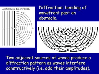
Diffraction: bending of
wavefrontpast an
obstacle.
Two adjacent sources of waves produce a
diffraction pattern as waves interfere
constructively (i.e. add their amplitudes).
3.
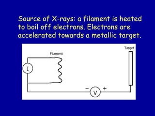
Source of X-rays:a filament is heated
to boil off electrons. Electrons are
accelerated towards a metallic target.
4.
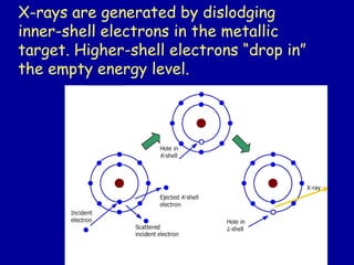
X-rays are generatedby dislodging
inner-shell electrons in the metallic
target. Higher-shell electrons “drop in”
the empty energy level.
5.
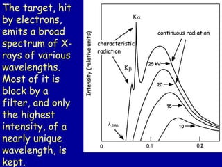
The target, hit
byelectrons,
emits a broad
spectrum of X-
rays of various
wavelengths.
Most of it is
block by a
filter, and only
the highest
intensity, of a
nearly unique
wavelength, is
kept.
6.
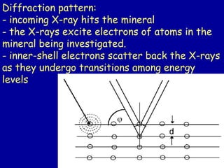
Diffraction pattern:
- incomingX-ray hits the mineral
- the X-rays excite electrons of atoms in the
mineral being investigated.
- inner-shell electrons scatter back the X-rays
as they undergo transitions among energy
levels
7.
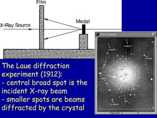
The Laue diffraction
experiment(1912):
- central broad spot is the
incident X-ray beam
- smaller spots are beams
diffracted by the crystal
8.
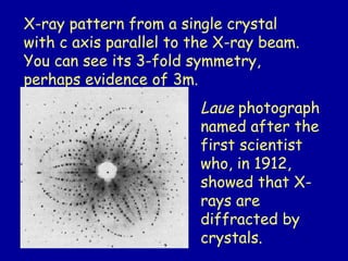
X-ray pattern froma single crystal
with c axis parallel to the X-ray beam.
You can see its 3-fold symmetry,
perhaps evidence of 3m.
Laue photograph
named after the
first scientist
who, in 1912,
showed that X-
rays are
diffracted by
crystals.
9.
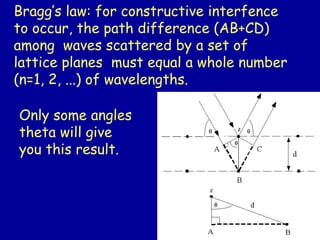
Bragg’s law: forconstructive interfence
to occur, the path difference (AB+CD)
among waves scattered by a set of
lattice planes must equal a whole number
(n=1, 2, ...) of wavelengths.
Only some angles
theta will give
you this result.
10.
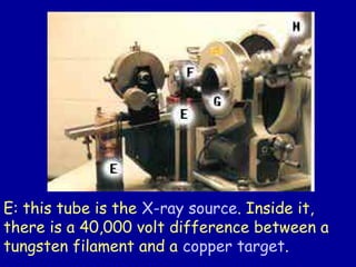
E: this tubeis the X-ray source. Inside it,
there is a 40,000 volt difference between a
tungsten filament and a copper target.
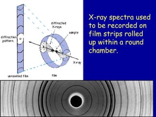
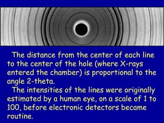
The distance fromthe center of each line
to the center of the hole (where X-rays
entered the chamber) is proportional to the
angle 2-theta.
The intensities of the lines were originally
estimated by a human eye, on a scale of 1 to
100, before electronic detectors became
routine.
14.
The first informationwe get from XRD
is whether or not the solid being
investigated is crystalline or amorphous.
Silica glass, for example, has SiO4
tetrahedra like quartz.
Synthetic (i.e. human-made) quartz is
indistinguishable from natural quartz by
XRD if their structure (space group)
and composition are the same.
15.
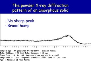
The powder X-raydiffraction
pattern of an amorphous solid
- No sharp peak
- Broad hump
17.
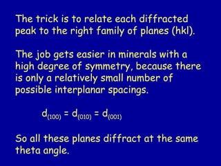
The trick isto relate each diffracted
peak to the right family of planes (hkl).
The job gets easier in minerals with a
high degree of symmetry, because there
is only a relatively small number of
possible interplanar spacings.
d(100) = d(010) = d(001)
So all these planes diffract at the same
theta angle.
18.
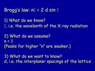
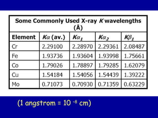
Bragg’s law: n= 2 d sin
1) What do we know?
, i.e. the wavelenth of the X-ray radiation
2) What do we assume?
n = 1
(Peaks for higher “n” are weaker.)
3) What do we want to know?
d, i.e. the interplanar spacings of the lattice
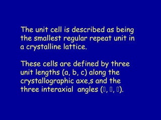
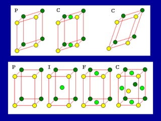
The unit cellis described as being
the smallest regular repeat unit in
a crystalline lattice.
These cells are defined by three
unit lengths (a, b, c) along the
crystallographic axe,s and the
three interaxial angles (, , ).
21.
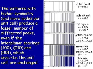
The patterns with
highersymmetry
(and more nodes per
unit cell) produce a
lesser number of
diffracted peaks,
even if the
interplanar spacings
(100), (010) and
(001), which
describe the unit
cell, are unchanged.
22.
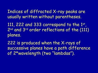
Indices of diffractedX-ray peaks are
usually written without parentheses.
111, 222 and 333 correspond to the 1st
,
2nd
and 3rd
order reflections of the (111)
planes.
222 is produced when the X-rays of
successive planes have a path difference
of 2*wavelength (two “lambdas”).
23.
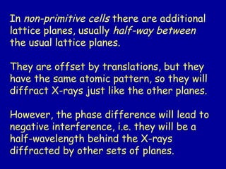
In non-primitive cellsthere are additional
lattice planes, usually half-way between
the usual lattice planes.
They are offset by translations, but they
have the same atomic pattern, so they will
diffract X-rays just like the other planes.
However, the phase difference will lead to
negative interference, i.e. they will be a
half-wavelength behind the X-rays
diffracted by other sets of planes.
25.
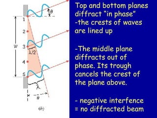
Top and bottomplanes
diffract “in phase”
-the crests of waves
are lined up
-The middle plane
diffracts out of
phase. Its trough
cancels the crest of
the plane above.
- negative interfence
= no diffracted beam
26.
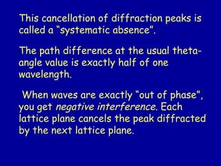
This cancellation ofdiffraction peaks is
called a “systematic absence”.
The path difference at the usual theta-
angle value is exactly half of one
wavelength.
When waves are exactly “out of phase”,
you get negative interference. Each
lattice plane cancels the peak diffracted
by the next lattice plane.
27.
The patterns with
highersymmetry
(and more nodes per
unit cell) produce a
lesser number of
diffracted peaks,
even if the
interplanar spacings
(100), (010) and
(001), which
describe the unit
cell, are unchanged.
28.
Powder X-ray diffractionis a routine
technique to measure the amount of
crystalline SiO2 (quartz) present in
mineral dust or soil.
A chemical analysis will not distinguish
the SiO2 of quartz from the silicate
portion present in the structure of clays
and many other minerals.
29.
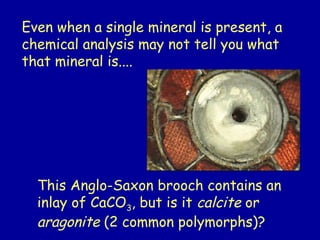
Even when asingle mineral is present, a
chemical analysis may not tell you what
that mineral is....
This Anglo-Saxon brooch contains an
inlay of CaCO3, but is it calcite or
aragonite (2 common polymorphs)?
30.
Bragg’s law predictsat which angles
the peaks will be diffracted, but not
their intensities.
Diffraction intensities are influenced
by the atomic number (Z) of the atoms
in the structure, by the shape and size
of the specimen, and by other factors
related to the machine.
We use the peak intensities to
determine where the atoms are in the
unit cell.
31.
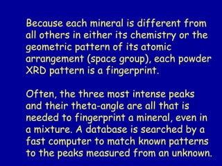
Because each mineralis different from
all others in either its chemistry or the
geometric pattern of its atomic
arrangement (space group), each powder
XRD pattern is a fingerprint.
Often, the three most intense peaks
and their theta-angle are all that is
needed to fingerprint a mineral, even in
a mixture. A database is searched by a
fast computer to match known patterns
to the peaks measured from an unknown.
32.
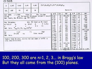
100, 200, 300are n=1, 2, 3... in Bragg’s law
But they all come from the (100) planes.
33.
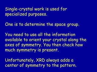
Single-crystal work isused for
specialized purposes.
One is to determine the space group.
You need to use all the information
available to orient your crystal along the
axes of symmetry. You then check how
much symmetry is present.
Unfortunately, XRD always adds a
center of symmetry to the pattern.
34.
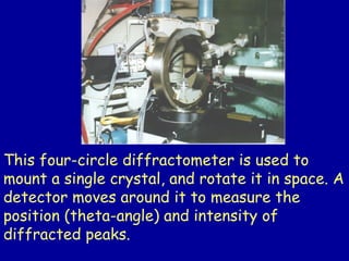
This four-circle diffractometeris used to
mount a single crystal, and rotate it in space. A
detector moves around it to measure the
position (theta-angle) and intensity of
diffracted peaks.
35.
How to solvecrystal structures?
The electron density ( ) at a point X, Y, Z in a
unit cell of volume “V” is;
(X,Y,Z) = 1/V Fhkl cos [ 2 (h X + k Y + l Z) - ]
Therefore if we know Fhkl and (for each h, k, l)
we can compute for all values of X, Y, and Z
and plot the values obtained to give a three-
dimensional electron density map. Then,
assuming atoms to be at the centres of the
electron density peaks, we would have the entire
structure.
36.
The presence orabsence of a center
of inversion is usually determined from
properties such as :
-presence of polar forms (e.g.
pyramids, monohedra) which indicate
that the “+” end of a crystallographic
axis is different from the “-” end.
- piezoelectricity which can only exist
in crystalline structures having at
least one polar axis.
37.
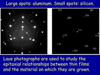
Laue photographs areused to study the
epitaxial relationships between thin films
and the material on which they are grown.
Large spots: aluminum. Small spots: silicon.
38.
When detecting twinningmatters !
Piezoelectric crystals may not display that
property if they are twinned.
Twinning can show up in
- external forms
- re-entrant angles (non-convex morphology)
39.
Ion order-disorder canbe detected by
X-ray diffraction.
This is very different from the lack of
order found in an amorphous solid.
40.
A cathode filamentis heated so that it
boils off electrons. A large voltage (20-
100kV) is maintained between the filament
and the target (a metal such as Mo, Cu, Co,
Fe or Cr).
The electrons are accelerated and hit the
target metal.
41.
Structures with lighterelements can
be studied using neutron diffraction.
Neutrons are scattered by the nucleus,
and their scattering varies less from
element to element.
whereas
X-rays are scattered by the electron
cloud, and light elements barely re-
emit them.


![How to solve crystal structures?
The electron density ( ) at a point X, Y, Z in a
unit cell of volume “V” is;
(X,Y,Z) = 1/V Fhkl cos [ 2 (h X + k Y + l Z) - ]
Therefore if we know Fhkl and (for each h, k, l)
we can compute for all values of X, Y, and Z
and plot the values obtained to give a three-
dimensional electron density map. Then,
assuming atoms to be at the centres of the
electron density peaks, we would have the entire
structure.](https://image.slidesharecdn.com/xrd-part2-250903083500-a13f113a/85/XRD-basic-operation-and-methods-for-analysis-35-320.jpg)