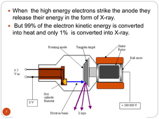
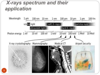
MD SHARIQUE AHMAD
FAC NO :- 17EEIM041
DEPT OF
ELECTRICAL ENGG
Z.H.C.ET AMU
X-Rays
1
Contents:
 Introduction
 History of X-ray
 Generation of X-ray
 Application
 Working
 Challenges
2
INTRODUCTION:
 X-rays are electromagnetic waves but they are more
energetic so they can penetrate many materials.
 When the X-rays hit the film, they expose it just as
light would.
 Since bone, fat, muscle, tumours and other masses
all absorb X-rays at different levels, the image on the
film lets you see different (distinct) structures inside
the body because of the different levels of exposure
on the film.
3
History of X-ray:
 X-ray technology was invented completely by
accident. In
1895, a German physicist “Wilhelm Roentgen”
made the discovery while experimenting with
electron beams in a gas discharge tube.
 He noticed that a fluorescent screen in his lab
started to glow when the electron beam was
turned on.
 fluorescent material normally glows in reaction to
electromagnetic radiation, but Roentgen's tube
was surrounded by heavy black cardboard.4
 Roentgen placed various objects between the
tube and the screen, and the screen still glowed.
 Finally, he put his hand in front of the tube, and
saw the outline of his bones projected onto the
fluorescent screen.
 The new rays he temporarily termed "X-rays",
using the mathematical designation ("X") for
something unknown.
 Immediately after discovering X-rays themselves,
he had discovered their most beneficial
application.
5
Generation of X-ray:
 The system is designed to provide a large number
of electron at cathode with high kinetic energy
focused to a small target at anode.
 The cathode is heated to release a large number of
electons by a process known as thermionic
emission.
 Electrons emitted from the cathode are accelerated
by high voltage towards anode.
 The whole process needs to be conducted in a
high vaccum.
6
 When the high energy electrons strike the anode they
release their energy in the form of X-ray.
 But 99% of the electron kinetic energy is converted
into heat and only 1% is converted into X-ray.
7
Application of X-rays:
 X-rays are used in medicine like identifying bone
structures ,dental radiography, mammography,
Radiotherapy etc.
 X-ray machines are used in airports and various
stations to check luggage etc.
 In Science x-rays are used to analyze the arrangement
of atoms in many kinds of substances particularly
crystals.
 Archaeologists used X-rays to examine ancient objects
covered by a crust of dirt.
 Used in research involving quantum mechanics theory
crystallography and cosmology.
8
X-rays spectrum and their
application
9
Working of X-rays:
 X-rays are able to penetrate through materials of light
atoms like flesh. The heavier atoms like metal absorb
them.
 A filter near the x-ray source blocks the low energy
rays so only the high energy rays pass through a
patient toward a sheet of film.
 Calcium in bones is considered a type of metal so
they absorbs X-ray photons. when photographic film
is placed on the body, this allows the technician to
take the picture.
10
Challenges:
11
 Diagnostic X-rays radiation can cause cell
mutations that may lead to cancer and tumours.
 Thyroid glands, which are the primary glands of
metabolism and energy, are also known victims of
x-rays.
 Multiple X-rays have been linked with multiple
myeloma – a form of bone marrow cancer.
 Old X-ray equipment gives off 20 to 30 times as
high a dose of radiation as new one.
12

X rays

  • 1.
    MD SHARIQUE AHMAD FACNO :- 17EEIM041 DEPT OF ELECTRICAL ENGG Z.H.C.ET AMU X-Rays 1
  • 2.
    Contents:  Introduction  Historyof X-ray  Generation of X-ray  Application  Working  Challenges 2
  • 3.
    INTRODUCTION:  X-rays areelectromagnetic waves but they are more energetic so they can penetrate many materials.  When the X-rays hit the film, they expose it just as light would.  Since bone, fat, muscle, tumours and other masses all absorb X-rays at different levels, the image on the film lets you see different (distinct) structures inside the body because of the different levels of exposure on the film. 3
  • 4.
    History of X-ray: X-ray technology was invented completely by accident. In 1895, a German physicist “Wilhelm Roentgen” made the discovery while experimenting with electron beams in a gas discharge tube.  He noticed that a fluorescent screen in his lab started to glow when the electron beam was turned on.  fluorescent material normally glows in reaction to electromagnetic radiation, but Roentgen's tube was surrounded by heavy black cardboard.4
  • 5.
     Roentgen placedvarious objects between the tube and the screen, and the screen still glowed.  Finally, he put his hand in front of the tube, and saw the outline of his bones projected onto the fluorescent screen.  The new rays he temporarily termed "X-rays", using the mathematical designation ("X") for something unknown.  Immediately after discovering X-rays themselves, he had discovered their most beneficial application. 5
  • 6.
    Generation of X-ray: The system is designed to provide a large number of electron at cathode with high kinetic energy focused to a small target at anode.  The cathode is heated to release a large number of electons by a process known as thermionic emission.  Electrons emitted from the cathode are accelerated by high voltage towards anode.  The whole process needs to be conducted in a high vaccum. 6
  • 7.
     When thehigh energy electrons strike the anode they release their energy in the form of X-ray.  But 99% of the electron kinetic energy is converted into heat and only 1% is converted into X-ray. 7
  • 8.
    Application of X-rays: X-rays are used in medicine like identifying bone structures ,dental radiography, mammography, Radiotherapy etc.  X-ray machines are used in airports and various stations to check luggage etc.  In Science x-rays are used to analyze the arrangement of atoms in many kinds of substances particularly crystals.  Archaeologists used X-rays to examine ancient objects covered by a crust of dirt.  Used in research involving quantum mechanics theory crystallography and cosmology. 8
  • 9.
    X-rays spectrum andtheir application 9
  • 10.
    Working of X-rays: X-rays are able to penetrate through materials of light atoms like flesh. The heavier atoms like metal absorb them.  A filter near the x-ray source blocks the low energy rays so only the high energy rays pass through a patient toward a sheet of film.  Calcium in bones is considered a type of metal so they absorbs X-ray photons. when photographic film is placed on the body, this allows the technician to take the picture. 10
  • 11.
    Challenges: 11  Diagnostic X-raysradiation can cause cell mutations that may lead to cancer and tumours.  Thyroid glands, which are the primary glands of metabolism and energy, are also known victims of x-rays.  Multiple X-rays have been linked with multiple myeloma – a form of bone marrow cancer.  Old X-ray equipment gives off 20 to 30 times as high a dose of radiation as new one.
  • 12.