Download to read offline
![Principles and mechanisms of hair follicle neogenesis X. Wang et al.
such as in the process of dedifferentiation−redifferentiation.
The former mechanism is observed upon mouse digit tip re-
generation, another well-known example of embryonic-like
regeneration in mammals (Lehoczky et al. 2011; Rinkevich
et al. 2011; Takeo et al. 2013; Leung et al. 2014). Similarly,
during limb regeneration in the axolotl (Ambystoma mexi-
canum), skeletal muscle regenerates from preexisting Pax7+
muscle-fated stem cells (Kragl et al. 2009; Sandoval-Guzman
et al. 2014). An example of embryonic-like regeneration via
cellular reprogramming is the regeneration of an amputated
eye lens in newts and frogs, which occurs via reprogramming
of the iris pigmented epithelial cells (Freeman 1963; Eguchi
et al. 1974; Henry & Elkins 2001; Tsonis & Del Rio-Tsonis
2004). Additionally, radial neuroglia cells can reprogram into
muscle and cartilage during tail regeneration (Echeverri &
Tanaka 2002), and dermal fibroblasts can reprogram into
chondrocytes during limb regeneration in the axolotl (Kragl
et al. 2009; Hirata et al. 2010).
The latter mechanism of regeneration is commonly asso-
ciated with the formation of a blastema. Although the term
“blastema” is deeply rooted in the context of Urodele regen-
eration biology, broadly speaking it defines a mass of prolif-
erating multipotent progenitors at the site of amputation that
serves as the cellular source for de novo regeneration (Hay
& Fischman 1961; O’Steen & Walker 1961; Gardiner et al.
1986; Muneoka et al. 1986; Roensch et al. 2013). In Urodeles,
the formation of the blastema requires the induction of a spe-
cialized epidermis known as the apical epithelial cap (Singer
& Inoue 1964), which secretes a number of signaling mor-
phogens including fibroblast growth factors (FGFs) (Chris-
tensen et al. 2002; Satoh et al. 2011), bone morphogenic
proteins (BMPs) (Makanae et al. 2014), and Wingless-Int
(WNTs) (Ghosh et al. 2008; Shimokawa et al. 2013). Cellu-
lar reprogramming is thought to be one of the mechanisms
by which blastema cells acquire multipotency (Satoh et al.
2008a; Satoh et al. 2008b; McCusker & Gardiner 2013). To
date, reprogramming is yet to be confirmed for embryonic-
like regeneration in mammals; however, blastema-like his-
tological features have been noted during ear regeneration
in the African spiny mouse (Acomys) (Seifert et al. 2012;
Tanaka 2012). Below, we argue that the phenomenon of HF
neogenesis in large skin wounds, in combination with the
plethora of genetic tools available in Mus musculus, presents
itself as a highly promising and tractable experimental model
to further probe for blastema-like regeneration in mammals.
Basic features of the WIHN model
Although neither was recognized at the time as WIHN, de
novo regeneration of HFs was first observed in adult rats
by Dann et al. (1941) following excisional wounding and
by Taylor (1949) following full-thickness skin cryo-injury.
Several years later, the WIHN phenomenon was reported
and explicitly recognized in rabbits by Breedis (1954) and
Billingham & Russell (1956). Breedis (1954) wrote that fol-
lowing excisional wounding “functioning hair follicles and
sebaceous glands appeared in the scars, sometimes in great
profusion.” Similarly, Billingham & Russell (1956) reported
that “with the production of these [neogenic] hairs the origi-
nally smooth scars may be said to have become transformed
into a sort of ad hoc skin.” Additional studies confirmed
WIHN in sheep (Brook et al. 1960), rarely in humans (Klig-
man & Strauss 1956; Kligman 1959), and once again in
rats (Mikhail 1963) and rabbits (Stenb¨ack et al. 1967). Im-
portantly, around the time of its discovery, the WIHN phe-
nomenon was not universally accepted. After repeating full-
thickness wounding experiments in rabbits, Straile (1959)
concluded that “uninjured follicles moved from the periph-
ery into the wounds and repopulated them without evidence
of a neoformation.” However, Billingham (1958) argued that
“there can be little doubt that an interaction of epidermis and
dermis is involved in initiating the development of hairs, and
the process of hair neogenesis, as seen in wounds in adult
rabbits, probably does not differ significantly from that which
occurs normally in neonatal life.”
Surprisingly, these early accounts of the WIHN phe-
nomenon went largely forgotten, and during the next four
decades the prevailing dogma was that HFs form only once
in ontogenesis—during embryonic development—and that
skin wounds in adults inevitably heal into hairless scars. Only
recently, following the landmark study by Ito et al. (2007),
did the re-discovery of the WIHN phenomenon in adult mice
occur. Through careful observations and with the help of
an array of genetic mouse tools, Ito et al. (2007) unequiv-
ocally confirmed that HFs in the wound’s center regenerate
de novo via a process that recapitulates normal embryonic
hair morphogenesis. A series of recent studies describing the
cellular and signaling aspects of hair neogenesis have dramat-
ically raised awareness of the WIHN phenomenon, carrying
it into the broader scope of stem cell biology and regenera-
tive medicine (Fan et al. 2011; Sun et al. 2011; Seifert et al.
2012; Driskell et al. 2013; Fuchs et al. 2013; Gay et al. 2013;
Myung et al. 2013; Nelson et al. 2013; Takeo et al. 2015). A
study by Seifert et al. (2012) on hair neogenesis following
autotomy-like skin shedding in Acomys is of particular inter-
est (also reviewed in Tanaka 2012; Seifert & Maden 2014).
It demonstrates that WIHN can be an indispensable part of a
natural adaptation against predation—spiny mice have very
fragile skin that breaks easily, leaving large full-thickness
wounds that efficiently regenerate numerous HFs.
In Mus musculus, WIHN is typically observed follow-
ing large wounding on the lower back, when a circular re-
gion of skin, at least 1 cm in diameter, is excised (Ito et
al. 2007). Wound size appears to be the key factor deter-
mining WIHN activation, with excisional wounds smaller
than 1 cm generally failing to regenerate de novo HFs. In
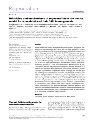
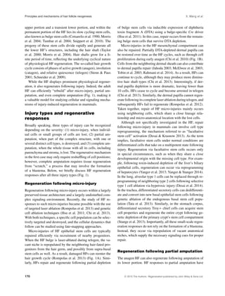
172 C 2015 The Authors. Regeneration published by John Wiley & Sons Ltd.](https://image.slidesharecdn.com/773fbdc6-06d5-4fa5-bee4-3589e5baf800-160111182842/85/Wang_et_al-2015-Regeneration-4-320.jpg)
![X. Wang et al. Principles and mechanisms of hair follicle neogenesis
adult mice older than 2 months, wounds 1.5 cm in diame-
ter are recommended for more efficient WIHN. Early post-
wounding events in the WIHN model are typical of all ex-
cisional wounds and include reepithelialization over newly
formed granulation tissue. These processes culminate in full
reepithelialization and scab detachment on post-wounding
day 13−14 (PWD13−14) (Fig. 2). This time point, also re-
ferred to as scab detachment day 0 (SD0) (Fan et al. 2011),
coincides with the onset of HF neogenesis. Placodes of the
first de novo follicles appear on day SD1 and continue to
emerge asynchronously over the course of the following
week, until the process plateaus at around PWD21. Indeed,
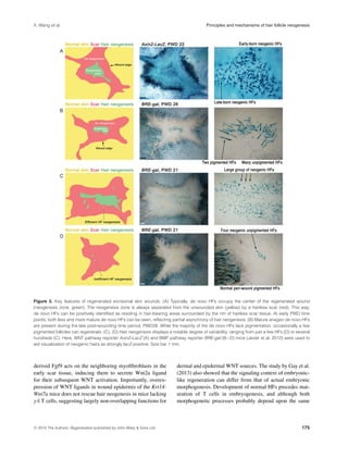
as exemplified in Figure 3A, neogenic hairs at various stages
of morphogenesis can be seen within the wound’s center at
PWD22. In vivo temporal dynamics of HF neogenesis in
the WIHN model are comprehensively covered by Fan et al.
(2011).
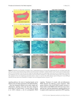
Typically, de novo follicles form in the very center of the
wound (Fig. 3A, B); however, more peripheral locations are
not uncommon (Figs 3D, 4B). Importantly, in all instances,
de novo follicles are separated from the preexisting folli-
cles at the wound’s edge by a circular, hairless scar (Fig. 3).
All neogenic hairs have zigzag morphology (note that four
distinct hair morphologies exist in normal mouse pelage:
guard, awl, auchene, and zigzag [Sundberg & Hogan 1994]),
and typically lack pigmentation in otherwise pigmented mice
(Ito et al. 2007). In rare instances, a few pigmented neogenic
hairs can also form (Fig. 3B). Commonly, de novo HFs form
one large cluster with (Figs 3C, 4A) or without a few small
satellite clusters (Fig. 3A, B). Rarely, multiple small clusters
scattered throughout the wound can be observed (Fig. 4B).
Importantly, even in age, gender, and strain matched litter-
mates, the efficiency of hair neogenesis varies, ranging from
just a few follicles (Fig. 3D) to several hundred (Fig. 3C), sug-
gesting a stochastic component to the WIHN phenomenon.
Classic accounts of WIHN in rabbits indicate that neoge-
nesis can be very efficient with as many as 3500 de novo
follicles forming per injured area (Billingham 1958) (note
that neogenesis-inducing wounds in rabbits are larger than in
mice, usually 2.5 cm in diameter).
Importantly, de novo HFs contain functional bulge stem
cells and undergo repetitive hair growth cycles, similar to
normal HFs (Ito et al. 2007). Typically, de novo follicles
enter first telogen at around PWD35 (albeit asynchronously
due to their asynchronous morphogenesis), and then enter
second anagen at around PWD45 (Fig. 2). Orientation is
another important aspect of neogenic hairs. While normal
hairs in the mouse dorsum follow the same cranial−caudal
orientation (Guo et al. 2004), the axial position of neogenic
hairs varies significantly. Although sometimes they appear
to lack any specific orientation (Figs 3B and 4D, purple sub-
domain), they often share a common orientation within one
cluster (Fig. 4B) or a sub-cluster (Fig. 4A, C and D). The
latter observation indicates that a rudimental hair patterning
mechanism functions in the WIHN model.
Cellular basis of de novo hair
neogenesis—the possibility of a
blastema-like mechanism
Neogenic HFs in the wound’s center regenerate all key ep-
ithelial and mesenchymal cell types characteristic of normal
body hair. Critically, each neogenic follicle forms a new
bulge populated by functional epithelial stem cells and a new
mesenchymal dermal papilla (reviewed in Chuong 2007; Ito
et al. 2007). Although the definitive origin of these de novo
follicular cell populations remains to be elucidated, a few
clues are beginning to emerge.
In their original study, Ito et al. (2007) elegantly showed
that preexisting Krt15+ bulge stem cells in HFs located on
the wound edge do not give rise to neogenic hairs. Although
Krt15+ stem cells are efficiently recruited from peri-wound
HFs to the newly forming wound epidermis (Ito et al. 2005;
reviewed in Plikus et al. 2012), their progeny are short-
lived and distinctly fail to contribute toward hair neogenesis.
Therefore, de novo follicles do not regenerate from hair-fated
bulge epithelial progenitors. One possibility is that neogenic
hairs regenerate from the progeny of Lrig1+ (Jensen et al.
2009), Lgr6+ (Snippert et al. 2010), and/or Gli1+ stem cells
(Brownell et al. 2011) residing in the upper, supra-bulge com-
partments of the peri-wound follicles. Indeed, all of these
stem/progenitor cell types were previously shown to give
rise to long-lasting cell clones in the wound epidermis (re-
viewed in Plikus et al. 2012). Another possibility is that
progeny of bona fide interfollicular epidermal cells expand
their lineage plasticity and acquire competence to regenerate
hair lineages de novo—a level of cellular plasticity not nor-
mally observed in unwounded skin. The latter possibility im-
plies some degree of epigenetic reprogramming in the wound
epidermis and a blastema-like mechanism for HF neogene-
sis. Although Shaw and Martin (2009) observed prominent
changes in the epigenetic makeup of new wound epidermis,
the overall mechanisms for epigenetic reprogramming during
mouse wound healing remain largely unexplored (reviewed
in Plikus et al. 2014).
Similarly, the lineage origin of neogenic dermal papil-
lae in the WIHN model remains to be established. A re-
cent study by Driskell et al. (2013) identified two principal
dermal fibroblast lineages in the normal skin termed upper
papillary and lower reticular populations. Lineage studies fol-
lowing wounding indicate that progeny of both reticular and
papillary fibroblasts contribute to the wound’s granulation
tissue in successive waves. Although normal dermal papil-
lae derive from the papillary lineage during embryonic hair
morphogenesis, and papillary, but not reticular, fibroblasts
display hair-inducing properties in the so-called chamber hair
C 2015 The Authors. Regeneration published by John Wiley & Sons Ltd. 173](https://image.slidesharecdn.com/773fbdc6-06d5-4fa5-bee4-3589e5baf800-160111182842/85/Wang_et_al-2015-Regeneration-5-320.jpg)


This document reviews the principles and mechanisms of regeneration in the mouse model for wound-induced hair follicle neogenesis. It discusses how wounding in mice leads to the de novo formation of hair follicles through a process that duplicates many aspects of embryonic hair development. This regeneration requires activation of WNT signaling, similar to hair development, but depends on FGF9 signaling from immune cells unlike development. The cellular mechanisms are not fully understood but could involve epigenetic reprogramming of wound cells into an embryonic-like state. Future studies of this model may provide insights into mammalian regeneration.


