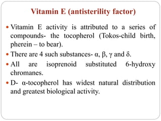
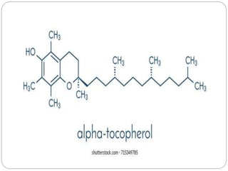
Ms. Funde A.P
M. Pharm (QAT)
Vitamin D, E and K
Vitamin D
 Vitamin D is a group of steroids (D1, D2, D3,
D4 and D5) chiefly occuring in animals but also
in plants and yeast.
 The physiologically active forms are D2 and D3.
 Vitamin D posses cyclopentanophenanthrene
ring in the structure.
 Provitamins D are ergosterol (plant) and 7
dehydrocholesterol (in skin). These on UV-
irradiation or sunlight exposure give rise
ergocalciferol (D2) and cholecalciferol (D3)
respectively.
Sources
 Richest sources are liver and viscera of fish.
 Other sources are egg and cheese.
 Its vitamin D content can be incresed by UV
irradiation.
Daily requirements
• Infants and children- 400IU
• Adults- do not require under normal conditions.
• During pregnancy and lactation- 500-600 IU
(1 IU= activity caused by 0.025 μg of calciferol)
Physiological functions
 Vitamin D has a direct effect on calcification.
 It increases calcium and phosphorus absorption
from intestine.
 It has a role in citrate metabolism as
administration of vitamin D increases citrate
contents of blood, bones, kidney, heart and small
intestine.
 Vitamin D is involved in renal handling of
phosphorus.
Deficiency diseases
Rickets in children
• Ricket is characterised by faulty calcification of
bones due to low vitamin D content of body, a
deficiency of calcium and phosphorus in diet or
due to both.
• The features are; infant becomes restless, fretful
and pale with flabby and toneless muscles.
• Abdomen is distended and there is extension and
widening of epiphysis at growing points.
Osteomalacia in adults
• Disease is limited to female sex.
• The bones specially pelvic girdle, ribs and
femora become soft, painful and deformed.
• Softening of bones is primarily due to deficiency
of vitamin D and to a lesser extent to the
deficiency of calcium.
Vitamin E (antisterility factor)
 Vitamin E activity is attributed to a series of
compounds- the tocopherol (Tokos-child birth,
pherein – to bear).
 There are 4 such substances- α, β, γ and δ.
 All are isoprenoid substituted 6-hydroxy
chromanes.
 D- α-tocopherol has widest natural distribution
and greatest biological activity.
Sources
 Rich source of vitamin E are milk, egg, muscle
meat, fish, cereals and leafy vegetables.
 Daily requirements- 30mg
Functions
 vitamin E act as natural antioxidant.
 Vitamin E and selenium act synergistically and
reduce requirement for each other in the body.
 It prevent hepatic necrosis produced by lack of
S containing amino acids in dietary proteins.
Deficiency
• In huamn deficiency causes muscle weakness,
muscular dystrophy, fragility of erythrocytes
with mild anemia.
 In animal deficiency causes resorption of
foetus in females and atrophy of spermatogenic
tissue leading to permanant sterility in male.
Vitamin K
 Vitamin K are napthoquinone derivatives with
polyisoprenoid side chains.
 Fat soluble vitamin K1 (phylloquinone) and K2
(menaquinione) are naturally occuring.
 Water soluble K3 (menadione) is synthetic and
used clinically.
sources
 The chief source of vitamin k are liver, green
leafy vegetables, peas, cereals, egg yok and
cheese. Fruits are poor sources.
Functrions
 Vitamin K is required for the biosynthesis of
blood clotting factors II, VII, IX and X.
 All are synthesised in the liver initially as
inactive precursor proteins.
 Biologically active clotting factors involves post
translational modification of glutamate to γ-
carboxy glutamate residue mediated by a
specific vitamin K dependant carboxylase.

Vitamin D, E and K

  • 1.
    Ms. Funde A.P M.Pharm (QAT) Vitamin D, E and K
  • 2.
    Vitamin D  VitaminD is a group of steroids (D1, D2, D3, D4 and D5) chiefly occuring in animals but also in plants and yeast.  The physiologically active forms are D2 and D3.  Vitamin D posses cyclopentanophenanthrene ring in the structure.
  • 3.
     Provitamins Dare ergosterol (plant) and 7 dehydrocholesterol (in skin). These on UV- irradiation or sunlight exposure give rise ergocalciferol (D2) and cholecalciferol (D3) respectively.
  • 5.
    Sources  Richest sourcesare liver and viscera of fish.  Other sources are egg and cheese.  Its vitamin D content can be incresed by UV irradiation.
  • 6.
    Daily requirements • Infantsand children- 400IU • Adults- do not require under normal conditions. • During pregnancy and lactation- 500-600 IU (1 IU= activity caused by 0.025 μg of calciferol)
  • 7.
    Physiological functions  VitaminD has a direct effect on calcification.  It increases calcium and phosphorus absorption from intestine.  It has a role in citrate metabolism as administration of vitamin D increases citrate contents of blood, bones, kidney, heart and small intestine.  Vitamin D is involved in renal handling of phosphorus.
  • 8.
    Deficiency diseases Rickets inchildren • Ricket is characterised by faulty calcification of bones due to low vitamin D content of body, a deficiency of calcium and phosphorus in diet or due to both. • The features are; infant becomes restless, fretful and pale with flabby and toneless muscles. • Abdomen is distended and there is extension and widening of epiphysis at growing points.
  • 9.
    Osteomalacia in adults •Disease is limited to female sex. • The bones specially pelvic girdle, ribs and femora become soft, painful and deformed. • Softening of bones is primarily due to deficiency of vitamin D and to a lesser extent to the deficiency of calcium.
  • 10.
    Vitamin E (antisterilityfactor)  Vitamin E activity is attributed to a series of compounds- the tocopherol (Tokos-child birth, pherein – to bear).  There are 4 such substances- α, β, γ and δ.  All are isoprenoid substituted 6-hydroxy chromanes.  D- α-tocopherol has widest natural distribution and greatest biological activity.
  • 12.
    Sources  Rich sourceof vitamin E are milk, egg, muscle meat, fish, cereals and leafy vegetables.  Daily requirements- 30mg Functions  vitamin E act as natural antioxidant.  Vitamin E and selenium act synergistically and reduce requirement for each other in the body.  It prevent hepatic necrosis produced by lack of S containing amino acids in dietary proteins.
  • 13.
    Deficiency • In huamndeficiency causes muscle weakness, muscular dystrophy, fragility of erythrocytes with mild anemia.  In animal deficiency causes resorption of foetus in females and atrophy of spermatogenic tissue leading to permanant sterility in male.
  • 14.
    Vitamin K  VitaminK are napthoquinone derivatives with polyisoprenoid side chains.  Fat soluble vitamin K1 (phylloquinone) and K2 (menaquinione) are naturally occuring.  Water soluble K3 (menadione) is synthetic and used clinically.
  • 16.
    sources  The chiefsource of vitamin k are liver, green leafy vegetables, peas, cereals, egg yok and cheese. Fruits are poor sources. Functrions  Vitamin K is required for the biosynthesis of blood clotting factors II, VII, IX and X.  All are synthesised in the liver initially as inactive precursor proteins.
  • 17.
     Biologically activeclotting factors involves post translational modification of glutamate to γ- carboxy glutamate residue mediated by a specific vitamin K dependant carboxylase.