VIP ℂall Girls Arekere Bangalore 6378878445 WhatsApp: Me All Time Serviℂe Ava...
VIÊM RUỘT THỪA - SCANS
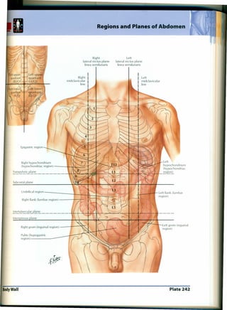
1. BodyWall
Regions and Planes of Abdomen
Right
lateral rectus plane
linea semilunaris
Left
lateral rectus plane
linea semilunaris
Right
midclavicular
line
Left
midclavicular
line
Epigastric region
Right hypochondrium
(hypochondriac region) } ~
~nspyloricp~n~ ~~ ~
Left
hypochondrium
(hypochondriac
_re~~
Subcostalplane
Umbilical region + ~'...l, 1 ..•..
L i r Left flank (lumbar
region)
Right flank (lumbar region) r'
~tertubercula'!:"p~n~ ---tL-~ ...•c
Right groin (inguinal region) • <i",
Pubis (hypogastric
region) -
~~~
Plate 242
2. Regions and Planes of Abdomen
Right
lateral rectus plane
linea semilunaris
Left
lateral rectus plane
linea semilunaris
Right
midclavicular
line
Left
midclavicular
line
Epigastric region
Right hypochondrium
(hypochondriac region) ~
Transpyloric p~n~_______ 'I ~
Left
hypochondrium
(hypochondriac
~~lJ..I- regior:!l
Subcostal plane
Umbilical region l ----tt=:::!- ~
• I ~, -
Right flank (lumbar region) ~.
~ertubercul~~~ ~~~
Right groin (inguinal region) ! '~
Left groin (inguinal
region)
Pubis (hypogastric
region) -
~~p
Body Wall Plate 242
3. Greater Omentum and Abdominal Viscera
See also Plates 267, 323, 330
~------'''k-- 11I---;--- Right lobe of liver
i'l-'-rt-.Mt--- Greater omentum overlying
transverse colon and small
intestine (jejunum and ileum)
Transverse colon (turned up) _-----;~~M~.Ji~::~G~5~~
Greater omentum (turned up)
Transverse mesocolon __ ---rT
Left colic (splenic) flexure
Right colic (hepatic) flexure
Small intestine (jejunum and ileum)
Ascending colon
Cecum
Small intestine (covering descending colon)
Sigmoid colon
Urinary bladder
Falciform ligament
Left lobe of liver
Stomach
Gallbladder
Plate 261 Peritoneal Cavity
------- - ---
4. Mesenteric Relations of Intestines (continued)
Cecum ---~~¥--~t--
Rectum
Transverse colon
(elevated)
Greater
omentum
(elevated)
Transverse mesocolon
(elevated over pancreas)
Jejunum (cut)
Free taenia
Mesentery (cut and
small intestine removed)
Omental
(epiploic)
appendices Left colic (splenic) flexure
Right colic
(hepatic) flexure --+-~~ Left paracolic gutter
Right paracolic
gutter Descending colon
Ascending colon _~·'U· .•
---.,::L-l~-:.Mr-Sigmoid mesocolon
Termi nal pactlr~t~o~f--P.~iI--=---::----:
ileum (cut) Sigmoid colon
Retrocecal
recess ----;~----4~~C'
Vermiform
appendix
Sigmoid mesocolon
Intersigmoid recess
Common iliac vessels
Plate 263 PeritonealCavi~
5. Stomach in Situ
Left lobe of liver Hepatoduodenalligament }
Lesser omentum
Hepatogastric ligament
Round
ligament
of liver---_....
(obliterated
left umbilical
vein)
Cardiac notch (incisure)
Fundus of stomach
Diaphragm
Spleen
Inferior
border
of liver _--wnTflJ
Right lobe
of liver
Gallbladder Body
of stomach
Pylorus
Duodenum
Right kidney
(retro-
peritoneal)
Greater omentum Left colic (splenic) flexure
Plate 267 Viscera (Gut)
7. Schema of muscle fibers at ileal orifice ~~p
Ileocecal Region (continued)
Ileocecal lips: labial form of ileal orifice
(as seen commonly postmortem
and occasionally in vivo)
1 I' . Free taenia, .•.....~
Orifice of vermiform appendix
Free taenia
Vermiform appendix
Circular muscle of colon
Fibers to papilla
Fibers to ileum
Fibers around ileocecal junction
Fibers to taenia t Longitudinal
Fibers to papilla ~ muscle of ileum
Circular muscle of ileum
Ileal papilla: papillary
form of ileal orifice
(found most commonly
in vivo)
Viscera (Gut) Plate 274
8. (Vermiform) Appendix
-
Mesoappendix
----..Serosa (visceral peritoneum)
~Continuous longitudinal muscle
r
I
,
McBurney's
point (on
spinoumbilical
line)
,'--"
I
/ ) I
/
, ...../ I
I
I
I
I
I
",--
/ I
I I
( t
t
' ,;
I ,---,,"
I
,
,
I
/
, I
I
Variations in position of appendix
Barium radiograph of unusually long appendix
Cecum Appendix
Fixed retrocecal
appendix
Submucosa
Circular muscle
Aggregate lymphoid
nodules
Crypts of LieberkUhn
Plate 275
9. External anal sphincter muscle ""
~~p
Mucosa and Musculature of Large Intestine
- For rectum and anal canal see Plates 371-376
Greater omentum (cut away)
Right colic
(hepatic) flexure Left colic
(splenic)
flexure
Ascending
colon (
Ileal
orifice
Rectosigmoid junction
(taeniae spread out and
unite to form longitudinal
~:Ielayer)
~ ~~."
Vermiform appendix
Sigmoid colon
Sigmoid mesocolon
Levator ani muscle i'4i:'"
Viscera (Gut) Plate 276
10. l.eft
triangular
ligament
Fissure for
ligamentum
venosum
Suprarenal gland
Right kidney
Right triangular
ligament ------y
Surfaces and Bed of Liver
-Diaphragm (pulled up)
Costal
impressions
Left triangular ligament
Fibrous appendix of liver
Round ligament (ligamentum teres) of liver
(obliterated left umbilical vein) forming free
border of falciform ligament
Anterior view
Gallbladder
(fundus)
Colic impression --+--.,;¥-..
Round ligament of liver- I--'I
Porta hepatis
Fissure for ligamentum teres
Hepatic portal vein
Proper hepatic artery
Caudate process
Caudate lobe
Fissure for
ligamentum venosum
Esophageal
impression
Gastric impression
__ - Left triangular
ligament
Gallbladder
Quadrate lobe
Duodenal impression
Renal impression
Cystic duct
Common hepatic duct
Common bile duct
Right triangular ligament
Hepatorenal portion of
coronary ligament
Suprarenal impression
Bare area
Inferior vena cava
Hepatic veins Coronary
ligament Left triangular
ligamentVisceral surface
Falciform ligament
Coronary ligament
Bare area
Duodenum
Posterior view
Plate 277 Viscera (Accessory Organs)
11. Liver in Situ: Vascular and Duct Systems
Roundligament (ligamentum teres) of liver _
(obliterated left umbilical vein) . . ~
Quadrate lobe
Gallbladder
Left lobe of liver
Caudate lobe seen through lesser
omentum (hepatogastric ligament)
Window cut in lesser omentum
(hepatoduodenalligament)
Proper hepatic artery
Omental
(epiploic)
foramen
(of Winslow)
Kidney
(retro-
peritoneal)
Lesser
omentum
(hepatogastric
ligament)
Spleen
Greater
omentum
Left colic
(splenic)
flexure
~~~
Hepatic vein
Portal
triad
Sinusoids
Portal
triad
Sinusoids
1~ Central
vein
Portal triads
(Hepatic artery,
portal vein branch,
bile ductule)
Perivascular fibrous (Glisson's)
capsule extending into porta
hepatis along vessels and bile duct
Proper hepatic artery
Hepatic portal vein
Common hepatic duct
Viscera (Accessory Organs) Plate 278