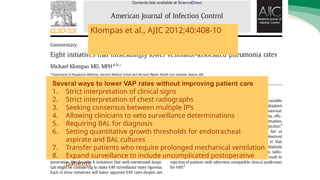
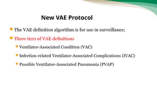
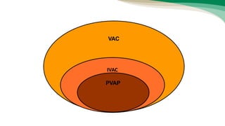
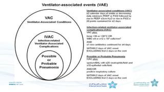
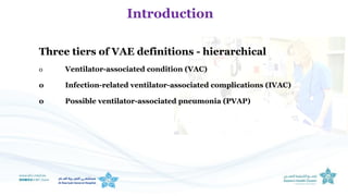
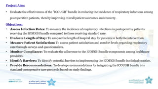
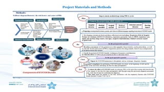
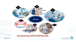
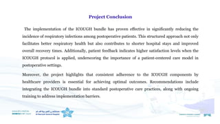
Ventilator Associated Event
Diagnosis of VAP
Limited clinically-defined VAP
• pulmonary edema, atelectasis, and/or acute respiratory distress
syndrome.
• Clinical manifestation
• X-rays
• Microbiology
• The Centers for Disease Control and Prevention (CDC) recently
transitioned from ventilator-associated pneumonia surveillance to
ventilator-associated event (VAE) surveillance in adult inpatient
settings.
• The VAE surveillance definition algorithm is based on objective criteria and
includes three tiers: Ventilator-Associated Conditions, Infection-related
Ventilator-Associated Complications, and Possible and Probable VAP.
Inter-observer agreement in VAP surveillance
Several ways to lower VAP rates without improving patient care
1. Strict interpretation of clinical signs
2. Strict interpretation of chest radiographs
3. Seeking consensus between multiple IP’s
4. Allowing clinicians to veto surveillance determinations
5. Requiring BAL for diagnosis
6. Setting quantitative growth thresholds for endotracheal
aspirate and BAL cultures
7. Transfer patients who require prolonged mechanical ventilation
8. Expand surveillance to include uncomplicated postoperative
patients
Klompas et al., AJIC 2012;40:408-10
Surveillance settings:
 VAE surveillance can be done in any adult inpatient location
where denominator data can be collected.
 Currently it is implemented in adult ICUs, step-down units, and
wards.
 Pediatric and neonatal units should conduct PedVAE
surveillance.
11
Patient Safety
Component
Others
Procedure-
Associated
Module
Device-
Associated
Module
AUR MDRO
SSI
CLABSI VAP CAUTI DE
VAE
 The VAE definition algorithm is for use in surveillance;
 Three tiers of VAE definitions
 Ventilator-Associated Condition (VAC)
 Infection-related Ventilator-Associated Complications (IVAC)
 Possible Ventilator-Associated Pneumonia (PVAP)
New VAE Protocol
VAEs are identified by using a combination of objective
criteria: deterioration in respiratory status after a period
of stability or improvement on the ventilator, evidence
of infection or inflammation, and laboratory evidence of
respiratory infection
VAE definition
VAC
IVAC
PVAP
 Ventilator: A device to assist or control respiration, inclusive of the
weaning period, through a tracheostomy or by endotracheal intubation
 Lung expansion devices such as intermittent positive- pressure breathing
(IPPB); nasal positive end- expiratory pressure (nasal PEEP); and
continuous nasal positive airway pressure (CPAP, hypoCPAP) are not
considered ventilators unless delivered via tracheostomy or endotracheal
intubation (e.g., ET- CPAP).
Ventilator
 After a period of stability or improvement on the ventilator
(defined by ≥ 2 calendar days of stable or decreasing daily
minimum FiO2 or PEEP values), the patient has at least one
of the following indicators of worsening oxygenation:
 Increase in daily minimum FiO2 of ≥ 0.20 (20 points) over the
daily minimum FiO2 in the baseline period, sustained for ≥ 2
calendar days.
 Increase in daily minimum* PEEP values of ≥ 3 cmH2O over the
daily minimum PEEP in the baseline period†, sustained for ≥ 2
calendar days.
Ventilator-Associated Condition (VAC)
Fraction of Inspired Oxygen (FiO2) is oxygen concentration (%) is typically
maintained below 0.5 even with ventilation, to avoid oxygen toxicity. Natural air
includes 20.9% oxygen, which is equivalent to FiO2 of 0.21.
Positive end-expiratory pressure (PEEP) is the pressure in the lungs above
atmospheric pressure applied by a ventilator. A small amount of applied PEEP (0 to
5 cmH2O) is used in most mechanically ventilated patients to mitigate end-
expiratory alveolar collapse
In hypoxia:
Worsening FiO2 means increase by 20%
Worsening PEEP means increase by 3 cmH20
Oxygen requirement
 On or after calendar day 3 of mechanical ventilation and within 2 calendar
days before or after the onset of worsening oxygenation, the patient meets
both of the following criteria:
 Temperature > 38 °C or < 36°C, OR white blood cell count ≥ 12,000
cells/mm3 or ≤ 4,000 cells/mm3.
AND
 A new antimicrobial agent(s) is started, and is continued for ≥ 4
calendar days.
IVAC: Infection-related Ventilator-
Associated Complication
One of three criteria for PVAP:
 Criterion 1: Positive culture meeting specific quantitative or semi-quantitative
threshold
 Criterion 2: Purulent respiratory secretions AND a positive culture NOT meeting the
quantitative or semi-quantitative thresholds
 Criterion 3: Positive pleural fluid culture, positive lung histopathology, positive diagnostic
test for Legionella species or selected respiratory viruses
Possible VAP (PVAP)
Ventilator Bundle
3.1. Elements of Ventilator Bundle
3.1.1. Elevate the head of the bed to 30-45 degrees.
3.1.1.1. Reduces potential for aspiration.
3.1.1.2. Potential to improve ventilation.
3.1.2. Provide a Daily Sedation Vacation and Assess the Readiness to Exudate.
3.1.2.1. Has been demonstrated to reduce overall patient
sedation.
3.1.2.2. Promotes early weaning.
3.1.3. Provide Peptic Ulcer Disease Prophylaxis.
3.1.3.1. Patients with Respiratory Failure have an
increased risk of “stress ulcers” and associated GI bleeding.
3.1.3.2. Acid-Suppressive Therapy (H2 blockers, sucralfate, PPI) decrease the risk of GI
Bleeding.
3.1.4. Provide Deep Vein Thrombosis Prophylaxis (unless contraindicated).
3.1.4.1. Patients with Respiratory Failure have an increased risk of Deep Vein Thrombosis
3.1.4.2. Treatment with anticoagulants (e.g., heparin) has been shown to reduce this risk
and the potential for pulmonary emboli.
3.1.5. Daily Oral Care.
3.1.5.1. The US FDA recommends 0.12% oral chlorhexidine for use as mouth rinse.
3.1.5.2. Dental plaque develops in patients that are mechanically
ventilated because of the lack of mechanical chewing and the
absence of saliva, which minimizes the development of biofilm on the teeth.
3.1.5.3. Dental plaque can be a significant reservoir for potential respiratory that cause
ventilator- associated pneumonia (VAP).
Limitation of VAP definition
Ventilator settings used in meeting the VAC definition:-
Electronic tools for surveillance
• CDC developed a web-based “VAE Calculator.”
• The Calculator, which was updated recently to reflect the 2014
protocol modifications, allows users to enter VAE data elements for a
single patient and receive a VAE determination along with an
explanation regarding why the determination was made. Patient
identifiers are not entered into the Calculator; it is primarily intended
to be an educational tool, and does not store patient data or submit
data to the NHSN.
Three tiers of VAE definitions - hierarchical
o Ventilator-associated condition (VAC)
o Infection-related ventilator-associated complications (IVAC)
o Possible ventilator-associated pneumonia (PVAP)
Introduction
Project Aim:
• Evaluate the effectiveness of the "ICOUGH" bundle in reducing the incidence of respiratory infections among
postoperative patients, thereby improving overall patient outcomes and recovery.
Objectives:
• Assess Infection Rates: To measure the incidence of respiratory infections in postoperative patients
receiving the ICOUGH bundle compared to those receiving standard care.
• Evaluate Length of Stay: To analyze the length of hospital stay for patients in both the intervention
• Measure Patient Satisfaction: To assess patient satisfaction and comfort levels regarding respiratory
care through surveys and questionnaires.
• Monitor Compliance: To evaluate the adherence to the ICOUGH bundle components among healthcare
providers.
• Identify Barriers: To identify potential barriers to implementing the ICOUGH bundle in clinical practice.
• Provide Recommendations: To develop recommendations for integrating the ICOUGH bundle into
standard postoperative care protocols based on study findings.
Project Materials and Methods
Project Results
Project Results
Percentages of PRTS pre and post implementing ICOUGH bundle
Surgery type Pre
intervention
(%)
Post-
intervention
(%)
T-test P-value
Abdominal
surgeries
25 7 5.23 0.001
Cesarean
section
13 3 4.67 0.005
Orthopedic
surgeries
5 1 3.50 0.015
Total 14.33 3.67 -- --
Project Conclusion
The implementation of the ICOUGH bundle has proven effective in significantly reducing the
incidence of respiratory infections among postoperative patients. This structured approach not only
facilitates better respiratory health but also contributes to shorter hospital stays and improved
overall recovery times. Additionally, patient feedback indicates higher satisfaction levels when the
ICOUGH protocol is applied, underscoring the importance of a patient-centered care model in
postoperative settings.
Moreover, the project highlights that consistent adherence to the ICOUGH components by
healthcare providers is essential for achieving optimal outcomes. Recommendations include
integrating the ICOUGH bundle into standard postoperative care practices, along with ongoing
training to address implementation barriers.
• Adoption of the ICOUGH Bundle: Healthcare facilities should integrate the
ICOUGH bundle into standard postoperative care protocols to enhance
respiratory health and reduce infection rates among patients.
• Training and Education: Ongoing training programs for healthcare providers
are essential to ensure proper implementation of the ICOUGH components,
fostering adherence and maximizing effectiveness.
• Monitoring and Evaluation: Regular monitoring and evaluation of the
ICOUGH bundle's implementation should be conducted to assess compliance,
identify areas for improvement, and ensure that best practices are being followed.
Recommendations
Patient Education: Develop educational materials for patients that explain the importance
of each component of the ICOUGH bundle, empowering them to actively participate in their
recovery.
Address Barriers: Identify and address potential barriers to implementation, such as staff
workload or resource limitations, to facilitate a smoother adoption of the ICOUGH bundle.
Further Research: Conduct additional studies to explore the long-term outcomes of the
ICOUGH bundle and its applicability across different surgical specialties and patient
demographics, helping to refine and expand its use.
Recommendations

ventilator associated event.pptxaaaaaddddffffgghh

  • 1.
  • 2.
    Diagnosis of VAP Limitedclinically-defined VAP • pulmonary edema, atelectasis, and/or acute respiratory distress syndrome. • Clinical manifestation • X-rays • Microbiology
  • 3.
    • The Centersfor Disease Control and Prevention (CDC) recently transitioned from ventilator-associated pneumonia surveillance to ventilator-associated event (VAE) surveillance in adult inpatient settings. • The VAE surveillance definition algorithm is based on objective criteria and includes three tiers: Ventilator-Associated Conditions, Infection-related Ventilator-Associated Complications, and Possible and Probable VAP.
  • 4.
  • 5.
    Several ways tolower VAP rates without improving patient care 1. Strict interpretation of clinical signs 2. Strict interpretation of chest radiographs 3. Seeking consensus between multiple IP’s 4. Allowing clinicians to veto surveillance determinations 5. Requiring BAL for diagnosis 6. Setting quantitative growth thresholds for endotracheal aspirate and BAL cultures 7. Transfer patients who require prolonged mechanical ventilation 8. Expand surveillance to include uncomplicated postoperative patients Klompas et al., AJIC 2012;40:408-10
  • 6.
    Surveillance settings:  VAEsurveillance can be done in any adult inpatient location where denominator data can be collected.  Currently it is implemented in adult ICUs, step-down units, and wards.  Pediatric and neonatal units should conduct PedVAE surveillance.
  • 7.
  • 8.
     The VAEdefinition algorithm is for use in surveillance;  Three tiers of VAE definitions  Ventilator-Associated Condition (VAC)  Infection-related Ventilator-Associated Complications (IVAC)  Possible Ventilator-Associated Pneumonia (PVAP) New VAE Protocol
  • 9.
    VAEs are identifiedby using a combination of objective criteria: deterioration in respiratory status after a period of stability or improvement on the ventilator, evidence of infection or inflammation, and laboratory evidence of respiratory infection VAE definition
  • 10.
  • 11.
     Ventilator: Adevice to assist or control respiration, inclusive of the weaning period, through a tracheostomy or by endotracheal intubation  Lung expansion devices such as intermittent positive- pressure breathing (IPPB); nasal positive end- expiratory pressure (nasal PEEP); and continuous nasal positive airway pressure (CPAP, hypoCPAP) are not considered ventilators unless delivered via tracheostomy or endotracheal intubation (e.g., ET- CPAP). Ventilator
  • 12.
     After aperiod of stability or improvement on the ventilator (defined by ≥ 2 calendar days of stable or decreasing daily minimum FiO2 or PEEP values), the patient has at least one of the following indicators of worsening oxygenation:  Increase in daily minimum FiO2 of ≥ 0.20 (20 points) over the daily minimum FiO2 in the baseline period, sustained for ≥ 2 calendar days.  Increase in daily minimum* PEEP values of ≥ 3 cmH2O over the daily minimum PEEP in the baseline period†, sustained for ≥ 2 calendar days. Ventilator-Associated Condition (VAC)
  • 13.
    Fraction of InspiredOxygen (FiO2) is oxygen concentration (%) is typically maintained below 0.5 even with ventilation, to avoid oxygen toxicity. Natural air includes 20.9% oxygen, which is equivalent to FiO2 of 0.21. Positive end-expiratory pressure (PEEP) is the pressure in the lungs above atmospheric pressure applied by a ventilator. A small amount of applied PEEP (0 to 5 cmH2O) is used in most mechanically ventilated patients to mitigate end- expiratory alveolar collapse In hypoxia: Worsening FiO2 means increase by 20% Worsening PEEP means increase by 3 cmH20 Oxygen requirement
  • 14.
     On orafter calendar day 3 of mechanical ventilation and within 2 calendar days before or after the onset of worsening oxygenation, the patient meets both of the following criteria:  Temperature > 38 °C or < 36°C, OR white blood cell count ≥ 12,000 cells/mm3 or ≤ 4,000 cells/mm3. AND  A new antimicrobial agent(s) is started, and is continued for ≥ 4 calendar days. IVAC: Infection-related Ventilator- Associated Complication
  • 15.
    One of threecriteria for PVAP:  Criterion 1: Positive culture meeting specific quantitative or semi-quantitative threshold  Criterion 2: Purulent respiratory secretions AND a positive culture NOT meeting the quantitative or semi-quantitative thresholds  Criterion 3: Positive pleural fluid culture, positive lung histopathology, positive diagnostic test for Legionella species or selected respiratory viruses Possible VAP (PVAP)
  • 16.
    Ventilator Bundle 3.1. Elementsof Ventilator Bundle 3.1.1. Elevate the head of the bed to 30-45 degrees. 3.1.1.1. Reduces potential for aspiration. 3.1.1.2. Potential to improve ventilation. 3.1.2. Provide a Daily Sedation Vacation and Assess the Readiness to Exudate. 3.1.2.1. Has been demonstrated to reduce overall patient sedation. 3.1.2.2. Promotes early weaning. 3.1.3. Provide Peptic Ulcer Disease Prophylaxis. 3.1.3.1. Patients with Respiratory Failure have an increased risk of “stress ulcers” and associated GI bleeding.
  • 17.
    3.1.3.2. Acid-Suppressive Therapy(H2 blockers, sucralfate, PPI) decrease the risk of GI Bleeding. 3.1.4. Provide Deep Vein Thrombosis Prophylaxis (unless contraindicated). 3.1.4.1. Patients with Respiratory Failure have an increased risk of Deep Vein Thrombosis 3.1.4.2. Treatment with anticoagulants (e.g., heparin) has been shown to reduce this risk and the potential for pulmonary emboli. 3.1.5. Daily Oral Care. 3.1.5.1. The US FDA recommends 0.12% oral chlorhexidine for use as mouth rinse. 3.1.5.2. Dental plaque develops in patients that are mechanically ventilated because of the lack of mechanical chewing and the absence of saliva, which minimizes the development of biofilm on the teeth. 3.1.5.3. Dental plaque can be a significant reservoir for potential respiratory that cause ventilator- associated pneumonia (VAP).
  • 18.
  • 20.
    Ventilator settings usedin meeting the VAC definition:-
  • 21.
    Electronic tools forsurveillance • CDC developed a web-based “VAE Calculator.” • The Calculator, which was updated recently to reflect the 2014 protocol modifications, allows users to enter VAE data elements for a single patient and receive a VAE determination along with an explanation regarding why the determination was made. Patient identifiers are not entered into the Calculator; it is primarily intended to be an educational tool, and does not store patient data or submit data to the NHSN.
  • 23.
    Three tiers ofVAE definitions - hierarchical o Ventilator-associated condition (VAC) o Infection-related ventilator-associated complications (IVAC) o Possible ventilator-associated pneumonia (PVAP) Introduction
  • 24.
    Project Aim: • Evaluatethe effectiveness of the "ICOUGH" bundle in reducing the incidence of respiratory infections among postoperative patients, thereby improving overall patient outcomes and recovery. Objectives: • Assess Infection Rates: To measure the incidence of respiratory infections in postoperative patients receiving the ICOUGH bundle compared to those receiving standard care. • Evaluate Length of Stay: To analyze the length of hospital stay for patients in both the intervention • Measure Patient Satisfaction: To assess patient satisfaction and comfort levels regarding respiratory care through surveys and questionnaires. • Monitor Compliance: To evaluate the adherence to the ICOUGH bundle components among healthcare providers. • Identify Barriers: To identify potential barriers to implementing the ICOUGH bundle in clinical practice. • Provide Recommendations: To develop recommendations for integrating the ICOUGH bundle into standard postoperative care protocols based on study findings.
  • 25.
  • 27.
  • 28.
    Project Results Percentages ofPRTS pre and post implementing ICOUGH bundle Surgery type Pre intervention (%) Post- intervention (%) T-test P-value Abdominal surgeries 25 7 5.23 0.001 Cesarean section 13 3 4.67 0.005 Orthopedic surgeries 5 1 3.50 0.015 Total 14.33 3.67 -- --
  • 29.
    Project Conclusion The implementationof the ICOUGH bundle has proven effective in significantly reducing the incidence of respiratory infections among postoperative patients. This structured approach not only facilitates better respiratory health but also contributes to shorter hospital stays and improved overall recovery times. Additionally, patient feedback indicates higher satisfaction levels when the ICOUGH protocol is applied, underscoring the importance of a patient-centered care model in postoperative settings. Moreover, the project highlights that consistent adherence to the ICOUGH components by healthcare providers is essential for achieving optimal outcomes. Recommendations include integrating the ICOUGH bundle into standard postoperative care practices, along with ongoing training to address implementation barriers.
  • 30.
    • Adoption ofthe ICOUGH Bundle: Healthcare facilities should integrate the ICOUGH bundle into standard postoperative care protocols to enhance respiratory health and reduce infection rates among patients. • Training and Education: Ongoing training programs for healthcare providers are essential to ensure proper implementation of the ICOUGH components, fostering adherence and maximizing effectiveness. • Monitoring and Evaluation: Regular monitoring and evaluation of the ICOUGH bundle's implementation should be conducted to assess compliance, identify areas for improvement, and ensure that best practices are being followed. Recommendations
  • 31.
    Patient Education: Developeducational materials for patients that explain the importance of each component of the ICOUGH bundle, empowering them to actively participate in their recovery. Address Barriers: Identify and address potential barriers to implementation, such as staff workload or resource limitations, to facilitate a smoother adoption of the ICOUGH bundle. Further Research: Conduct additional studies to explore the long-term outcomes of the ICOUGH bundle and its applicability across different surgical specialties and patient demographics, helping to refine and expand its use. Recommendations