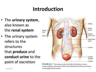
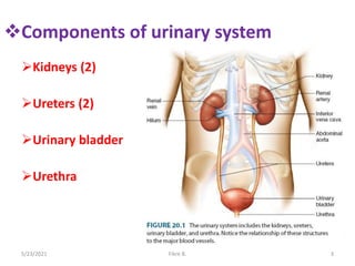
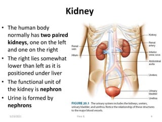
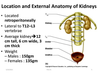
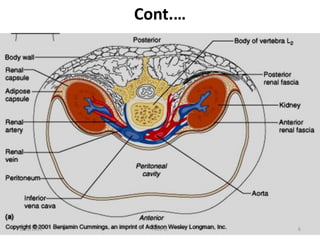
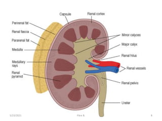
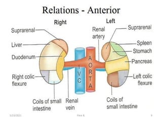
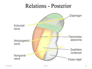
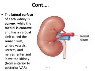
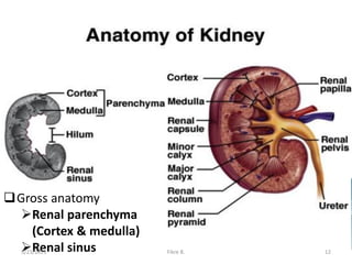
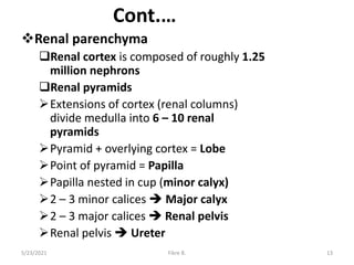
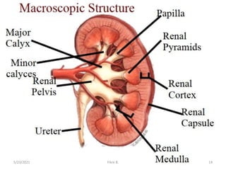
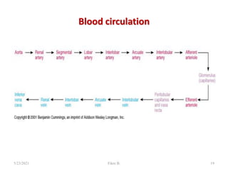
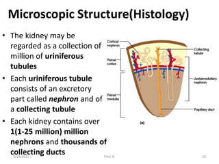
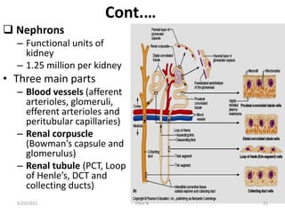
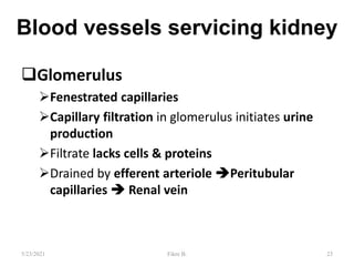
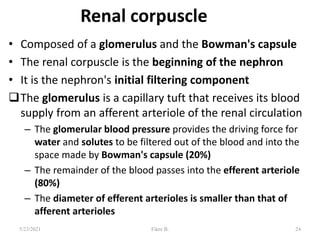
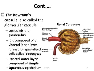
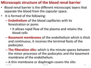
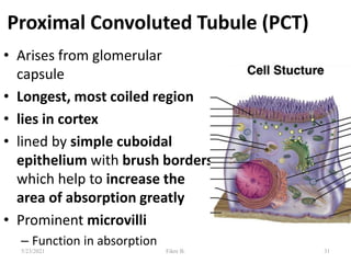
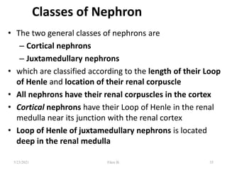
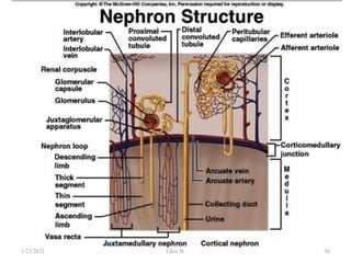
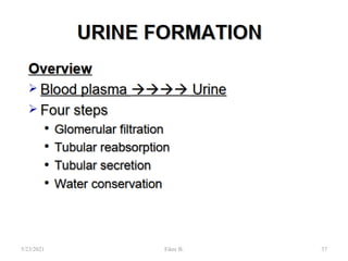
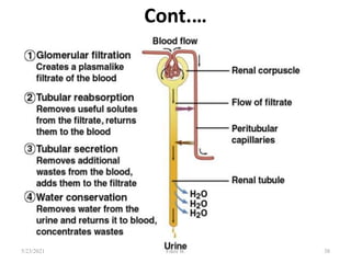
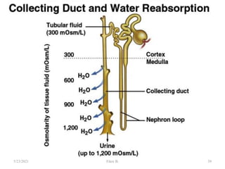
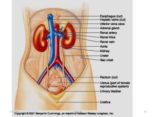
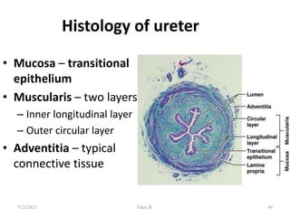
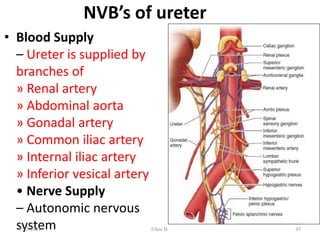
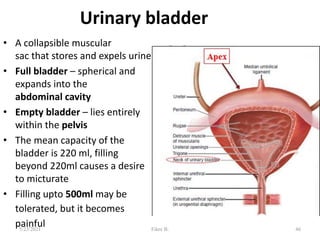
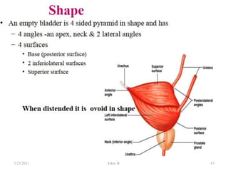
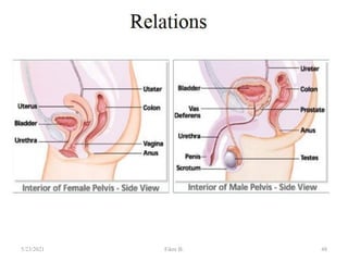
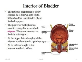
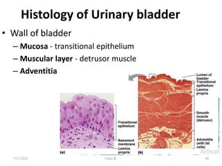
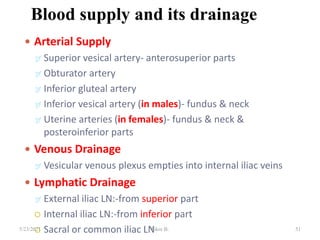
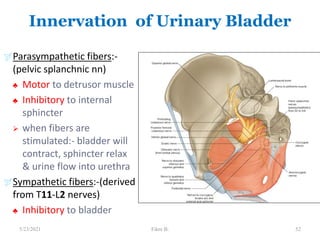
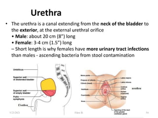
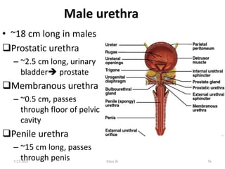
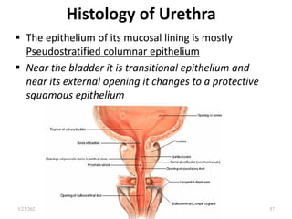
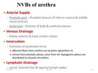
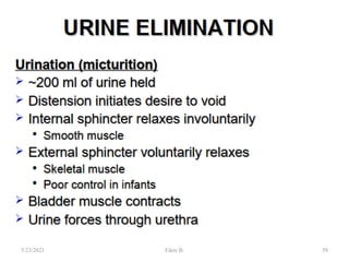
Urinary system
By: Fikre Bayu(MSc)
5/23/2021 Fikre B. 1
Introduction
• The urinary system,
also known as
the renal system
• The urinary system
refers to the
structures
that produce and
conduct urine to the
point of excretion
5/23/2021 Fikre B. 2
Components of urinary system
Kidneys (2)
Ureters (2)
Urinary bladder
Urethra
5/23/2021 Fikre B. 3
Kidney
• The human body
normally has two paired
kidneys, one on the left
and one on the right
• The right lies somewhat
lower than left as it is
positioned under liver
• The functional unit of
the kidney is nephron
• Urine is formed by
nephrons
5/23/2021 Fikre B. 4
Location and External Anatomy of Kidneys
• Located
retroperitoneally
• Lateral to T12–L3
vertebrae
• Average kidney12
cm tall, 6 cm wide, 3
cm thick
• Weight
– Males : 150gm
– Females : 135gm
5/23/2021 Fikre B. 5
Cont.…
5/23/2021 Fikre B. 6
Protective and supportive layers of kidney
• A thin, tough layer of dense connective tissue called the
fibrous capsule adheres directly to the kidney’s surface,
maintaining its shape and forming a barrier that can inhibit
the spread of infection from the surrounding regions
• Just external to the renal capsule is the perirenal fat and
external to that is an envelope of renal fascia
• The renal fascia contains an external layer of fat, the
pararenal fat
• The perirenal and pararenal fat layers cushion the kidney
against blows and help hold the kidneys in place
5/23/2021 Fikre B. 7
5/23/2021 Fikre B. 8
5/23/2021 Fikre B. 9
5/23/2021 Fikre B. 10
Cont.…
 The lateral surface
of each kidney is
convex, while the
medial is concave
and has a vertical
cleft called the
renal hilum,
where vessels,
ureters, and
nerves enter and
leave the kidney
(from anterior to
posterior VAR) 11
5/23/2021 Fikre B.
Gross anatomy
Renal parenchyma
(Cortex & medulla)
Renal sinus
5/23/2021 Fikre B. 12
Cont.…
Renal parenchyma
Renal cortex is composed of roughly 1.25
million nephrons
Renal pyramids
Extensions of cortex (renal columns)
divide medulla into 6 – 10 renal
pyramids
Pyramid + overlying cortex = Lobe
Point of pyramid = Papilla
Papilla nested in cup (minor calyx)
2 – 3 minor calices  Major calyx
2 – 3 major calices  Renal pelvis
Renal pelvis  Ureter
5/23/2021 Fikre B. 13
5/23/2021 Fikre B. 14
Cont.…
• Renal sinus
– Surrounded by renal parenchyma
– Contains blood & lymph vessels, nerves,
urine-collecting structures
• Hilus
– On concave surface
– Vessels and nerves enter and exit
5/23/2021 Fikre B. 15
Blood supply
• The kidney continuously cleanse the blood
and adjust its composition
• Kidneys possess an extensive blood supply
• Under normal resting conditions, the renal
arteries deliver approximately one-fourth of
the total systemic cardiac output (1200 ml) to
the kidneys each minute
5/23/2021 Fikre B. 16
5/23/2021 Fikre B. 17
Blood Circulation
5/23/2021 Fikre B. 18
Blood circulation
5/23/2021 Fikre B. 19
Microscopic Structure(Histology)
• The kidney may be
regarded as a collection of
million of uriniferous
tubules
• Each uriniferous tubule
consists of an excretory
part called nephron and of
a collecting tubule
• Each kidney contains over
1(1-25 million) million
nephrons and thousands of
collecting ducts
5/23/2021 Fikre B. 20
Cont.…
 Nephrons
– Functional units of
kidney
– 1.25 million per kidney
• Three main parts
– Blood vessels (afferent
arterioles, glomeruli,
efferent arterioles and
peritubular capillaries)
– Renal corpuscle
(Bowman’s capsule and
glomerulus)
– Renal tubule (PCT, Loop
of Henle’s, DCT and
collecting ducts)
5/23/2021 Fikre B. 21
22
5/23/2021 Fikre B.
Blood vessels servicing kidney
Glomerulus
Fenestrated capillaries
Capillary filtration in glomerulus initiates urine
production
Filtrate lacks cells & proteins
Drained by efferent arteriole Peritubular
capillaries  Renal vein
23
5/23/2021 Fikre B.
Renal corpuscle
• Composed of a glomerulus and the Bowman's capsule
• The renal corpuscle is the beginning of the nephron
• It is the nephron's initial filtering component
The glomerulus is a capillary tuft that receives its blood
supply from an afferent arteriole of the renal circulation
– The glomerular blood pressure provides the driving force for
water and solutes to be filtered out of the blood and into the
space made by Bowman's capsule (20%)
– The remainder of the blood passes into the efferent arteriole
(80%)
– The diameter of efferent arterioles is smaller than that of
afferent arterioles
24
5/23/2021 Fikre B.
Cont.…
 The Bowman's
capsule, also called the
glomerular capsule
– surrounds the
glomerulus
– It is composed of a
visceral inner layer
formed by specialized
cells called podocytes
– Parietal outer layer
composed of simple
squamous epithelium 25
5/23/2021 Fikre B.
Microscopic structure of the blood renal barrier
• Blood renal barrier is the different microscopic layers that
separate the blood from the capsular space.
• It is formed of the following:
– Endothelium of the blood capillaries with its
fenestration or pores
• It allows rapid flow of the plasma and retains the
blood cells
– Basement membranes of the endothelium which is thick
and continuous, it receives the terminal foots of the
podocytes
– The filteration slits: which is the minute spaces between
the minor processes of the podocytes and the basement
membrane of the endothelium.
– A thin membrane or diaphragm covers the slits
5/23/2021 Fikre B. 26
Microscopic structure of the mesangial cells
• are branched cells present between the blood
capillaries
• The cells are faintly stained and have flat nuclei
• can be easily identified by the EM and their
dense scattering iron containing protein
especially after the injection of ferritin
• They have the following functions:
–Regeneration of the basement membrane of
the glomerular capillaries
–Phagocytic function
–Supportive function
–They may be of hormonal importance
5/23/2021 Fikre B. 27
28
5/23/2021 Fikre B.
Cont.…
 The filtration membrane
is the actual filter that
lies between the blood
and the interior of the
glomerular capsule
 It is a porous membrane
that allows free passage
of water and solutes
smaller than plasma
proteins
 The capillary pores
prevent passage of blood
cells, but plasma
components are allowed
to pass 29
5/23/2021 Fikre B.
Renal tubules
• Leads from
glomerular
capsule
– Ends at tip of
medullary
pyramid
• Four major regions
– Proximal
convoluted tubule
– Nephron loop
– Distal convoluted
tubule
– Collecting duct 30
5/23/2021 Fikre B.
Proximal Convoluted Tubule (PCT)
• Arises from glomerular
capsule
• Longest, most coiled region
• lies in cortex
• lined by simple cuboidal
epithelium with brush borders
which help to increase the
area of absorption greatly
• Prominent microvilli
– Function in absorption
31
5/23/2021 Fikre B.
Nephron loop or Loop of Henle
• “U” – shaped, distal to PCT
• lies in medulla and has 2 parts
– Descending limb of loop of Henle (thin and thick limbs)
– Ascending limb of loop of Henle (thin and thick limbs)
– Thick ascending limb of loop of Henle (enters cortex and
becomes DCT-distal convoluted tubule
 Thick segments
– Thick limb is lined by simple cuboidal epithelium
– Active transport of salts
– High metabolism, many mitochondria
 Thin segments
– Thin limb is lined by simple squamous epithelium
– Permeable to water
– Low metabolism
32
5/23/2021 Fikre B.
Distal Convoluted Tubule (DCT)
• Coiled, distal to nephron loop
• Shorter than PCT
• Less coiled than PCT
• Very few microvilli
• Contacts afferent and efferent arterioles
• Contact with peritubular capillaries
33
5/23/2021 Fikre B.
Collecting ducts
• DCTs of several nephrons empty into a
collecting duct
• Passes into medulla
• Several merge into papillary duct (~30 per
papilla)
• Drain into minor calyx
34
5/23/2021 Fikre B.
Classes of Nephron
• The two general classes of nephrons are
– Cortical nephrons
– Juxtamedullary nephrons
• which are classified according to the length of their Loop
of Henle and location of their renal corpuscle
• All nephrons have their renal corpuscles in the cortex
• Cortical nephrons have their Loop of Henle in the renal
medulla near its junction with the renal cortex
• Loop of Henle of juxtamedullary nephrons is located
deep in the renal medulla
35
5/23/2021 Fikre B.
36
5/23/2021 Fikre B.
37
5/23/2021 Fikre B.
Cont.…
38
5/23/2021 Fikre B.
39
5/23/2021 Fikre B.
Functions of kidney
 Regulates blood volume and pressure
 Filters blood plasma, eliminates waste, returns
useful chemicals to blood
• Regulates PCO2 and acid base balance
• Regulates osmolarity of body fluids
• Secretes renin, activates angiotensin and
aldosterone (to controls BP, electrolyte balance)
• Secretes erythropoietin, controls RBC count
• Synthesize calcitriol (Vitamin D)
• Detoxifies free radicals and drugs
• Gluconeogenesis 40
5/23/2021 Fikre B.
41
5/23/2021 Fikre B.
Ureters
• The Ureters are a pair of narrow , thick
walled muscular tubes which convey
urine from the kidneys to urinary bladder
• Each Ureters is about 25cm (10 inch)long
• The upper half lies in the abdomen and the
lower half in the pelvis
• It measures 3mm diameter, but it slightly
constricted at three places:
– At the pelviureteric junction
– At the brim of lesser pelvis
– At its passage through the bladder wall 42
5/23/2021 Fikre B.
43
5/23/2021 Fikre B.
Histology of ureter
• Mucosa – transitional
epithelium
• Muscularis – two layers
– Inner longitudinal layer
– Outer circular layer
• Adventitia – typical
connective tissue
44
5/23/2021 Fikre B.
NVB’s of ureter
• Blood Supply
– Ureter is supplied by
branches of
» Renal artery
» Abdominal aorta
» Gonadal artery
» Common iliac artery
» Internal iliac artery
» Inferior vesical artery
• Nerve Supply
– Autonomic nervous
system 45
5/23/2021 Fikre B.
Urinary bladder
• A collapsible muscular
sac that stores and expels urine
• Full bladder – spherical and
expands into the
abdominal cavity
• Empty bladder – lies entirely
within the pelvis
• The mean capacity of the
bladder is 220 ml, filling
beyond 220ml causes a desire
to micturate
• Filling upto 500ml may be
tolerated, but it becomes
painful 46
5/23/2021 Fikre B.
47
5/23/2021 Fikre B.
48
5/23/2021 Fikre B.
49
5/23/2021 Fikre B.
Histology of Urinary bladder
• Wall of bladder
– Mucosa - transitional epithelium
– Muscular layer - detrusor muscle
– Adventitia
50
5/23/2021 Fikre B.
Blood supply and its drainage
 Arterial Supply
 Superior vesical artery- anterosuperior parts
 Obturator artery
 Inferior gluteal artery
 Inferior vesical artery (in males)- fundus & neck
 Uterine arteries (in females)- fundus & neck &
posteroinferior parts
 Venous Drainage
 Vesicular venous plexus empties into internal iliac veins
 Lymphatic Drainage
 External iliac LN:-from superior part
 Internal iliac LN:-from inferior part
 Sacral or common iliac LN 51
5/23/2021 Fikre B.
Innervation of Urinary Bladder
Parasympathetic fibers:-
(pelvic splanchnic nn)
♣ Motor to detrusor muscle
♣ Inhibitory to internal
sphincter
 when fibers are
stimulated:- bladder will
contract, sphincter relax
& urine flow into urethra
Sympathetic fibers:-(derived
from T11-L2 nerves)
♣ Inhibitory to bladder
52
5/23/2021 Fikre B.
Applied anatomy
• Congenital Anomalies
• Ectopia vesicae
• Infection –Cystitis
• Neurological lesions
• Rupture of bladder
• Cancer bladder
• Urinary Incontinence
53
5/23/2021 Fikre B.
Urethra
• The urethra is a canal extending from the neck of the bladder to
the exterior, at the external urethral orifice
• Male: about 20 cm (8”) long
• Female: 3-4 cm (1.5”) long
– Short length is why females have more urinary tract infections
than males - ascending bacteria from stool contamination
54
5/23/2021 Fikre B.
Female urethra
• 3 to 4 cm long
• External urethral orifice
– between vaginal orifice and
clitoris
• Internal urethral sphincter
– detrusor muscle thickened,
smooth muscle, involuntary
control
• External urethral sphincter
– skeletal muscle, voluntary
control
55
5/23/2021 Fikre B.
Male urethra
• ~18 cm long in males
Prostatic urethra
– ~2.5 cm long, urinary
bladder prostate
Membranous urethra
– ~0.5 cm, passes
through floor of pelvic
cavity
Penile urethra
– ~15 cm long, passes
through penis 56
5/23/2021 Fikre B.
Histology of Urethra
 The epithelium of its mucosal lining is mostly
Pseudostratified columnar epithelium
 Near the bladder it is transitional epithelium and
near its external opening it changes to a protective
squamous epithelium
57
5/23/2021 Fikre B.
NVBs of urethra
 Arterial Supply
 Prostatic part :-Prostatic branch of inferior vesical & middle
rectal arteries
 distal part:- Arteries of bulb & urethral arteries
 Venous Drainage
 follow arteries & have similar names
 Innervation
 branches of pudendal nerve
♣ afferent fibers from urethra run to pelvic splanchnic nn
♣ nerves from prostatic plexus, arise from inf. Hypogastric plexus are
distributed to all parts of urethra
 Lymphatic Drainage
 sacral, internal iliac & inguinal lymph nodes
58
5/23/2021 Fikre B.
59
5/23/2021 Fikre B.
5/23/2021 60
Fikre B.

Urinary system

  • 1.
    Urinary system By: FikreBayu(MSc) 5/23/2021 Fikre B. 1
  • 2.
    Introduction • The urinarysystem, also known as the renal system • The urinary system refers to the structures that produce and conduct urine to the point of excretion 5/23/2021 Fikre B. 2
  • 3.
    Components of urinarysystem Kidneys (2) Ureters (2) Urinary bladder Urethra 5/23/2021 Fikre B. 3
  • 4.
    Kidney • The humanbody normally has two paired kidneys, one on the left and one on the right • The right lies somewhat lower than left as it is positioned under liver • The functional unit of the kidney is nephron • Urine is formed by nephrons 5/23/2021 Fikre B. 4
  • 5.
    Location and ExternalAnatomy of Kidneys • Located retroperitoneally • Lateral to T12–L3 vertebrae • Average kidney12 cm tall, 6 cm wide, 3 cm thick • Weight – Males : 150gm – Females : 135gm 5/23/2021 Fikre B. 5
  • 6.
  • 7.
    Protective and supportivelayers of kidney • A thin, tough layer of dense connective tissue called the fibrous capsule adheres directly to the kidney’s surface, maintaining its shape and forming a barrier that can inhibit the spread of infection from the surrounding regions • Just external to the renal capsule is the perirenal fat and external to that is an envelope of renal fascia • The renal fascia contains an external layer of fat, the pararenal fat • The perirenal and pararenal fat layers cushion the kidney against blows and help hold the kidneys in place 5/23/2021 Fikre B. 7
  • 8.
  • 9.
  • 10.
  • 11.
    Cont.…  The lateralsurface of each kidney is convex, while the medial is concave and has a vertical cleft called the renal hilum, where vessels, ureters, and nerves enter and leave the kidney (from anterior to posterior VAR) 11 5/23/2021 Fikre B.
  • 12.
    Gross anatomy Renal parenchyma (Cortex& medulla) Renal sinus 5/23/2021 Fikre B. 12
  • 13.
    Cont.… Renal parenchyma Renal cortexis composed of roughly 1.25 million nephrons Renal pyramids Extensions of cortex (renal columns) divide medulla into 6 – 10 renal pyramids Pyramid + overlying cortex = Lobe Point of pyramid = Papilla Papilla nested in cup (minor calyx) 2 – 3 minor calices  Major calyx 2 – 3 major calices  Renal pelvis Renal pelvis  Ureter 5/23/2021 Fikre B. 13
  • 14.
  • 15.
    Cont.… • Renal sinus –Surrounded by renal parenchyma – Contains blood & lymph vessels, nerves, urine-collecting structures • Hilus – On concave surface – Vessels and nerves enter and exit 5/23/2021 Fikre B. 15
  • 16.
    Blood supply • Thekidney continuously cleanse the blood and adjust its composition • Kidneys possess an extensive blood supply • Under normal resting conditions, the renal arteries deliver approximately one-fourth of the total systemic cardiac output (1200 ml) to the kidneys each minute 5/23/2021 Fikre B. 16
  • 17.
  • 18.
  • 19.
  • 20.
    Microscopic Structure(Histology) • Thekidney may be regarded as a collection of million of uriniferous tubules • Each uriniferous tubule consists of an excretory part called nephron and of a collecting tubule • Each kidney contains over 1(1-25 million) million nephrons and thousands of collecting ducts 5/23/2021 Fikre B. 20
  • 21.
    Cont.…  Nephrons – Functionalunits of kidney – 1.25 million per kidney • Three main parts – Blood vessels (afferent arterioles, glomeruli, efferent arterioles and peritubular capillaries) – Renal corpuscle (Bowman’s capsule and glomerulus) – Renal tubule (PCT, Loop of Henle’s, DCT and collecting ducts) 5/23/2021 Fikre B. 21
  • 22.
  • 23.
    Blood vessels servicingkidney Glomerulus Fenestrated capillaries Capillary filtration in glomerulus initiates urine production Filtrate lacks cells & proteins Drained by efferent arteriole Peritubular capillaries  Renal vein 23 5/23/2021 Fikre B.
  • 24.
    Renal corpuscle • Composedof a glomerulus and the Bowman's capsule • The renal corpuscle is the beginning of the nephron • It is the nephron's initial filtering component The glomerulus is a capillary tuft that receives its blood supply from an afferent arteriole of the renal circulation – The glomerular blood pressure provides the driving force for water and solutes to be filtered out of the blood and into the space made by Bowman's capsule (20%) – The remainder of the blood passes into the efferent arteriole (80%) – The diameter of efferent arterioles is smaller than that of afferent arterioles 24 5/23/2021 Fikre B.
  • 25.
    Cont.…  The Bowman's capsule,also called the glomerular capsule – surrounds the glomerulus – It is composed of a visceral inner layer formed by specialized cells called podocytes – Parietal outer layer composed of simple squamous epithelium 25 5/23/2021 Fikre B.
  • 26.
    Microscopic structure ofthe blood renal barrier • Blood renal barrier is the different microscopic layers that separate the blood from the capsular space. • It is formed of the following: – Endothelium of the blood capillaries with its fenestration or pores • It allows rapid flow of the plasma and retains the blood cells – Basement membranes of the endothelium which is thick and continuous, it receives the terminal foots of the podocytes – The filteration slits: which is the minute spaces between the minor processes of the podocytes and the basement membrane of the endothelium. – A thin membrane or diaphragm covers the slits 5/23/2021 Fikre B. 26
  • 27.
    Microscopic structure ofthe mesangial cells • are branched cells present between the blood capillaries • The cells are faintly stained and have flat nuclei • can be easily identified by the EM and their dense scattering iron containing protein especially after the injection of ferritin • They have the following functions: –Regeneration of the basement membrane of the glomerular capillaries –Phagocytic function –Supportive function –They may be of hormonal importance 5/23/2021 Fikre B. 27
  • 28.
  • 29.
    Cont.…  The filtrationmembrane is the actual filter that lies between the blood and the interior of the glomerular capsule  It is a porous membrane that allows free passage of water and solutes smaller than plasma proteins  The capillary pores prevent passage of blood cells, but plasma components are allowed to pass 29 5/23/2021 Fikre B.
  • 30.
    Renal tubules • Leadsfrom glomerular capsule – Ends at tip of medullary pyramid • Four major regions – Proximal convoluted tubule – Nephron loop – Distal convoluted tubule – Collecting duct 30 5/23/2021 Fikre B.
  • 31.
    Proximal Convoluted Tubule(PCT) • Arises from glomerular capsule • Longest, most coiled region • lies in cortex • lined by simple cuboidal epithelium with brush borders which help to increase the area of absorption greatly • Prominent microvilli – Function in absorption 31 5/23/2021 Fikre B.
  • 32.
    Nephron loop orLoop of Henle • “U” – shaped, distal to PCT • lies in medulla and has 2 parts – Descending limb of loop of Henle (thin and thick limbs) – Ascending limb of loop of Henle (thin and thick limbs) – Thick ascending limb of loop of Henle (enters cortex and becomes DCT-distal convoluted tubule  Thick segments – Thick limb is lined by simple cuboidal epithelium – Active transport of salts – High metabolism, many mitochondria  Thin segments – Thin limb is lined by simple squamous epithelium – Permeable to water – Low metabolism 32 5/23/2021 Fikre B.
  • 33.
    Distal Convoluted Tubule(DCT) • Coiled, distal to nephron loop • Shorter than PCT • Less coiled than PCT • Very few microvilli • Contacts afferent and efferent arterioles • Contact with peritubular capillaries 33 5/23/2021 Fikre B.
  • 34.
    Collecting ducts • DCTsof several nephrons empty into a collecting duct • Passes into medulla • Several merge into papillary duct (~30 per papilla) • Drain into minor calyx 34 5/23/2021 Fikre B.
  • 35.
    Classes of Nephron •The two general classes of nephrons are – Cortical nephrons – Juxtamedullary nephrons • which are classified according to the length of their Loop of Henle and location of their renal corpuscle • All nephrons have their renal corpuscles in the cortex • Cortical nephrons have their Loop of Henle in the renal medulla near its junction with the renal cortex • Loop of Henle of juxtamedullary nephrons is located deep in the renal medulla 35 5/23/2021 Fikre B.
  • 36.
  • 37.
  • 38.
  • 39.
  • 40.
    Functions of kidney Regulates blood volume and pressure  Filters blood plasma, eliminates waste, returns useful chemicals to blood • Regulates PCO2 and acid base balance • Regulates osmolarity of body fluids • Secretes renin, activates angiotensin and aldosterone (to controls BP, electrolyte balance) • Secretes erythropoietin, controls RBC count • Synthesize calcitriol (Vitamin D) • Detoxifies free radicals and drugs • Gluconeogenesis 40 5/23/2021 Fikre B.
  • 41.
  • 42.
    Ureters • The Uretersare a pair of narrow , thick walled muscular tubes which convey urine from the kidneys to urinary bladder • Each Ureters is about 25cm (10 inch)long • The upper half lies in the abdomen and the lower half in the pelvis • It measures 3mm diameter, but it slightly constricted at three places: – At the pelviureteric junction – At the brim of lesser pelvis – At its passage through the bladder wall 42 5/23/2021 Fikre B.
  • 43.
  • 44.
    Histology of ureter •Mucosa – transitional epithelium • Muscularis – two layers – Inner longitudinal layer – Outer circular layer • Adventitia – typical connective tissue 44 5/23/2021 Fikre B.
  • 45.
    NVB’s of ureter •Blood Supply – Ureter is supplied by branches of » Renal artery » Abdominal aorta » Gonadal artery » Common iliac artery » Internal iliac artery » Inferior vesical artery • Nerve Supply – Autonomic nervous system 45 5/23/2021 Fikre B.
  • 46.
    Urinary bladder • Acollapsible muscular sac that stores and expels urine • Full bladder – spherical and expands into the abdominal cavity • Empty bladder – lies entirely within the pelvis • The mean capacity of the bladder is 220 ml, filling beyond 220ml causes a desire to micturate • Filling upto 500ml may be tolerated, but it becomes painful 46 5/23/2021 Fikre B.
  • 47.
  • 48.
  • 49.
  • 50.
    Histology of Urinarybladder • Wall of bladder – Mucosa - transitional epithelium – Muscular layer - detrusor muscle – Adventitia 50 5/23/2021 Fikre B.
  • 51.
    Blood supply andits drainage  Arterial Supply  Superior vesical artery- anterosuperior parts  Obturator artery  Inferior gluteal artery  Inferior vesical artery (in males)- fundus & neck  Uterine arteries (in females)- fundus & neck & posteroinferior parts  Venous Drainage  Vesicular venous plexus empties into internal iliac veins  Lymphatic Drainage  External iliac LN:-from superior part  Internal iliac LN:-from inferior part  Sacral or common iliac LN 51 5/23/2021 Fikre B.
  • 52.
    Innervation of UrinaryBladder Parasympathetic fibers:- (pelvic splanchnic nn) ♣ Motor to detrusor muscle ♣ Inhibitory to internal sphincter  when fibers are stimulated:- bladder will contract, sphincter relax & urine flow into urethra Sympathetic fibers:-(derived from T11-L2 nerves) ♣ Inhibitory to bladder 52 5/23/2021 Fikre B.
  • 53.
    Applied anatomy • CongenitalAnomalies • Ectopia vesicae • Infection –Cystitis • Neurological lesions • Rupture of bladder • Cancer bladder • Urinary Incontinence 53 5/23/2021 Fikre B.
  • 54.
    Urethra • The urethrais a canal extending from the neck of the bladder to the exterior, at the external urethral orifice • Male: about 20 cm (8”) long • Female: 3-4 cm (1.5”) long – Short length is why females have more urinary tract infections than males - ascending bacteria from stool contamination 54 5/23/2021 Fikre B.
  • 55.
    Female urethra • 3to 4 cm long • External urethral orifice – between vaginal orifice and clitoris • Internal urethral sphincter – detrusor muscle thickened, smooth muscle, involuntary control • External urethral sphincter – skeletal muscle, voluntary control 55 5/23/2021 Fikre B.
  • 56.
    Male urethra • ~18cm long in males Prostatic urethra – ~2.5 cm long, urinary bladder prostate Membranous urethra – ~0.5 cm, passes through floor of pelvic cavity Penile urethra – ~15 cm long, passes through penis 56 5/23/2021 Fikre B.
  • 57.
    Histology of Urethra The epithelium of its mucosal lining is mostly Pseudostratified columnar epithelium  Near the bladder it is transitional epithelium and near its external opening it changes to a protective squamous epithelium 57 5/23/2021 Fikre B.
  • 58.
    NVBs of urethra Arterial Supply  Prostatic part :-Prostatic branch of inferior vesical & middle rectal arteries  distal part:- Arteries of bulb & urethral arteries  Venous Drainage  follow arteries & have similar names  Innervation  branches of pudendal nerve ♣ afferent fibers from urethra run to pelvic splanchnic nn ♣ nerves from prostatic plexus, arise from inf. Hypogastric plexus are distributed to all parts of urethra  Lymphatic Drainage  sacral, internal iliac & inguinal lymph nodes 58 5/23/2021 Fikre B.
  • 59.
  • 60.